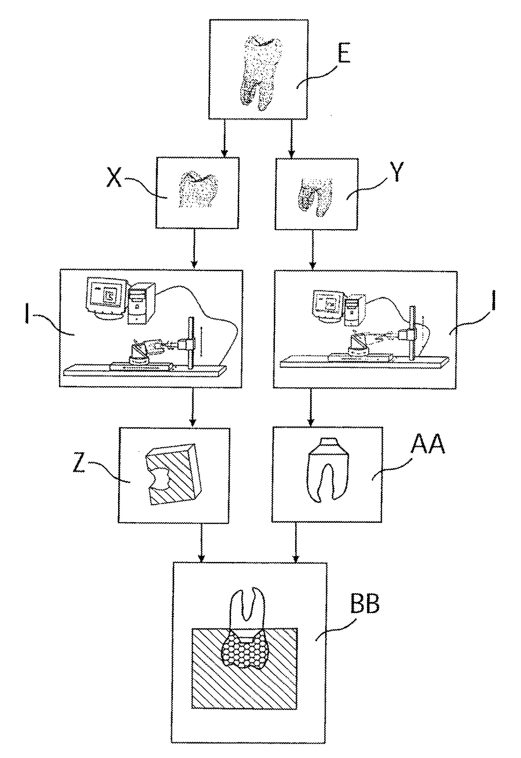
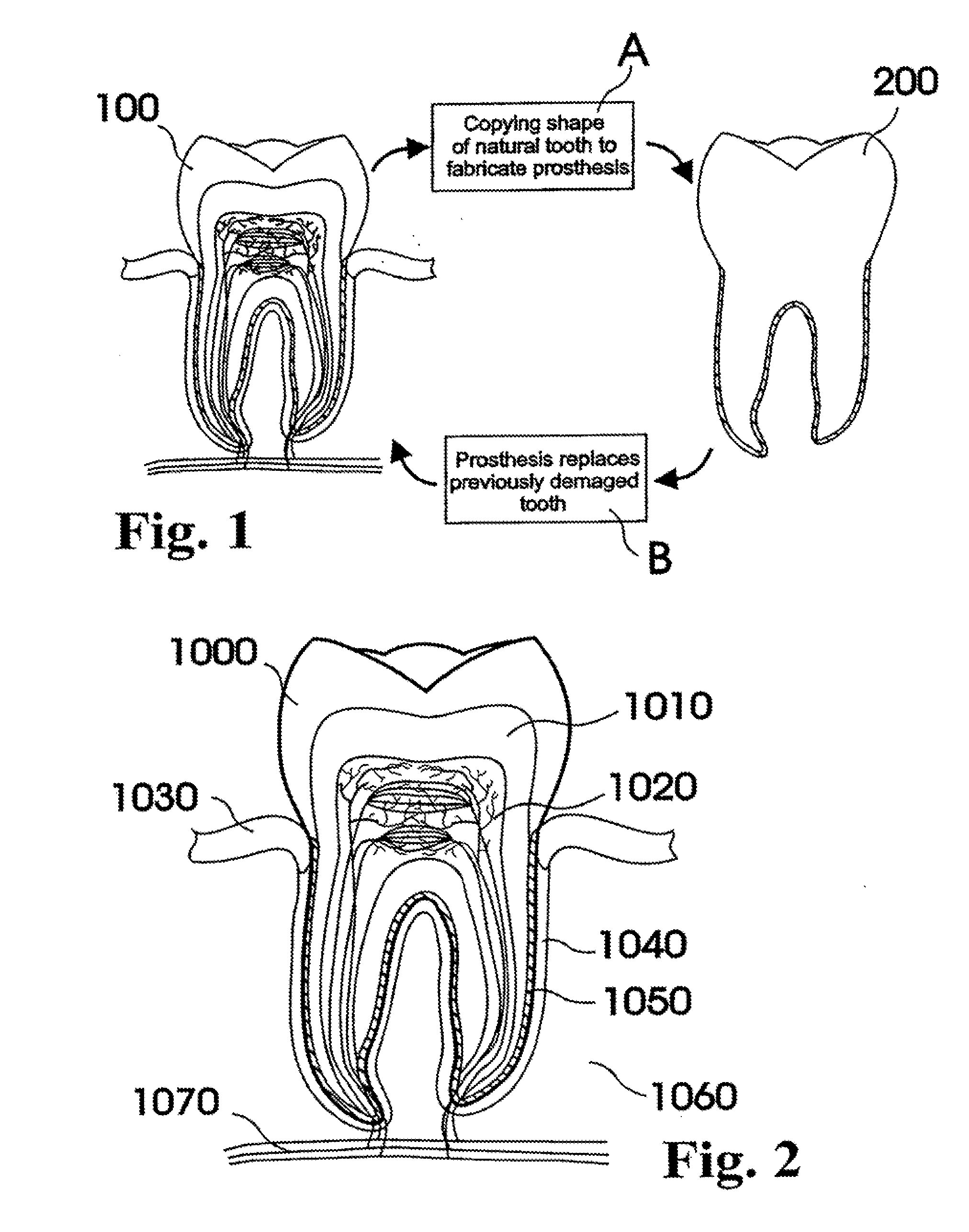
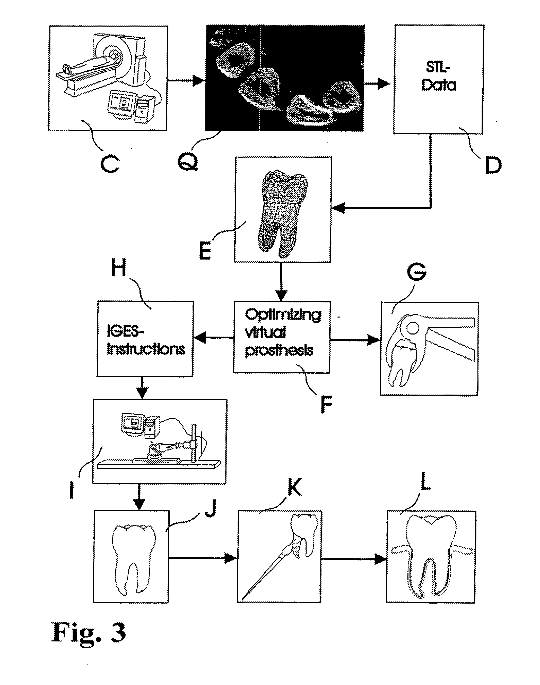
[0020]In view of the foregoing, various embodiments of the present invention beneficially provide a customized dental prosthesis and
implant in various embodiments based on a process that includes
copying a significant portion of the original root geometry of a
human tooth to be integrated after extraction of the original tooth either in the existing
biological cell structure of the periodontal
ligament or as one piece into the embedding
bone structure of the respective jaw. According to various embodiments of the present invention, primary stability is favorably achieved by a custom made splint that connects the dental prosthesis with the adjacent tooth or teeth or other dental structures like existing implants, bridges and the like. The concept of periodontal integration of an artificial tooth uses the existing human periodontal
ligament for integration and is certainly
less invasive than the integration of osseointegrated implants. The concept of integrating a one-piece prosthesis that includes a root-shape part, an
abutment, and a crown, according to an exemplary configuration, combines the two clinical episodes of integrating the root-shaped part and adapting the crown into one clinical event. Even if such one-piece prosthesis would include an
assembly of two or more parts, the
assembly can be fabricated in the controlled environment of a
dental laboratory or an industrial fabrication. As a result, the quality of the interface sealing between such parts can be expected to be of higher quality as produced in the mouth of the patient. This may reduce the
infection rate so that the success rate of the one-piece prosthesis according to an embodiment of the present invention would be higher as achieved with implementations according to conventional systems. Further, the concept of a splint that is custom made in the laboratory in advance can serve at least two purposes: the correct positioning of the prosthesis, and the achievement of reasonable primary stability. The concept of using in-vivo
imaging data in order to design and fabricate the prosthesis prior to the extraction of the tooth or teeth of interest enables a laboratory
lead time prior to the invasive clinical event. The concept of using data to design a root-shaped portion or portions of the prosthesis not actually of the tooth or teeth extracted or to be extracted, but of the anatomical alveolar structure, allows an ability to adapt the prosthesis to the post-extraction or even post-surgical shape of the alveolar situation.
[0022]According to an embodiment of the present invention, an example of a method of manufacturing a dental prosthesis for implantation into a
jaw bone cavity of a pre-identified patient includes the steps of receiving
imaging data describing a three-dimensional shape of an outer surface portion of a root portion of a nonfunctional
natural tooth of a pre-identified patient, deriving digital
design data defining a three-dimensional shape of an outer surface of a root portion of a tooth prosthesis responsive to the received imaging data, and manufacturing the root portion of the tooth prosthesis at least partially responsive to the digital
design data to form at least a substantial portion of the root portion of the tooth prosthesis. According to an exemplary configuration, the three-dimensional shape of the outer surface of the root portion of the tooth prosthesis defined by the derived a digital
design data dimensionally matches the three-dimensional shape of a corresponding outer surface portion of the
natural tooth of the pre-identified patient. Correspondingly, the three-dimensional shape of the at least a substantial portion of the outer surface portion of the root portion of the tooth prosthesis substantially produced by the manufacturing step correspondingly can also advantageously dimensionally match the three-dimensional shape of the corresponding outer surface portion of the root portion of the nonfunctional
natural tooth described by the imaging data.
[0027]According to an embodiment of the present invention, the step of manufacturing the root portion of the tooth prosthesis can also or alternatively include the steps of applying a first layer of
biocompatible material to outer surface portions of a main body portion of the root portion of the tooth prosthesis, and applying a second layer of
biocompatible material atop the first layer of
biocompatible material, performed, according to an exemplary embodiment of the method, prior to
insertion of the root portion of the tooth prosthesis into a
jaw bone cavity of the pre-identified patient. According to a specific example of an embodiment of the method, the first layer includes a
calcium hydroxide (Ca(OH)2)
cement and a second layer includes a matrix
protein derivative. Further, according to this exemplary embodiment of the method, the portions of the root portion comprising the first and the second
layers of biocompatible material that correspond to the
jaw bone cavity in its entirety do not dimensionally exceed a natural pre-
insertion three-dimensional shape of portions of the jaw bone cavity receiving the tooth prosthesis when the tooth prosthesis is clinically positioned in the jaw bone cavity of the pre-identified patient. This advantageously can reduce occurrences of
atrophy caused by a chronic pressure exerted on the surrounding tissue inherent with prosthetic systems which employ a “press-fit” methodology of securing the tooth prosthesis within the jaw bone cavity.
[0028]According to an embodiment of the present invention, the step of manufacturing the root portion of the tooth prosthesis can also or alternatively include the steps of applying a gel adapted to form a
barrier membrane when sprayed with water to enhance tissue growth to outer surface portions of the root portion of the tooth prosthesis adjacent the permanent crown portion connected thereto, and / or applying a layer of silver to outer surface portions of the root portion of the tooth prosthesis adjacent a permanent crown portion connected thereto to reduce healing gum tissue growth during integration into and adoption by the periodontal
ligament structure.
[0032]According to an example of a configuration of the splint, the tooth-facing outer surface can include a first three-dimensional shape portion dimensioned to substantially match to a three-dimensional shape of a corresponding substantial surface portion of the crown portion of the tooth prosthesis, and a second three-dimensional shape portion dimensioned to substantially match to a three-dimensional shape of a corresponding substantial surface portion of the crown portion of the at least one adjacent tooth. The first and the second three-dimensional shape portions can have a geometrical relationship configured so that when the first three-dimensional shape portion is affixed to the substantial surface portion of the crown portion of the tooth prosthesis and when the second three-dimensional shape portion is affixed to the substantial surface portion of the crown portion of the at least one adjacent tooth, and when the dental prosthesis is clinically inserted into the embedding portion of the jawbone cavity of the patient, the first three-dimensional shape portion provides temporary primary stability to the tooth prosthesis to thereby maintain the tooth prosthesis in a final geometrical relation to the embedding portion of the jawbone cavity. Further, according to an exemplary configuration, the splint is shaped to not interfere with the
occlusal contact between the crown portion of the at least opponent tooth with the crown portion of the at least one adjacent tooth and the crown portion of the tooth prosthesis.
[0034]According to another embodiment of the method, the method includes the steps of receiving imaging data describing a three-dimensional shape of an outer surface portion of a root portion of a nonfunctional natural tooth of a pre-identified patient, a three-dimensional shape of an outer surface portion of a crown portion of the nonfunctional natural tooth of the pre-identified patient, and a three-dimensional shape and position of an outer surface portion of a crown portion of at least one adjacent tooth immediately adjacent the jawbone cavity of the pre-identified patient and geometrical relation thereto. The method can also include deriving
digital data defining a three-dimensional shape of an outer surface of the root portion of a tooth prosthesis responsive to the received imaging data, and manufacturing the tooth prosthesis at least partially responsive to the
digital data to form at least a substantial portion of the root portion of the tooth prosthesis, whereby the three-dimensional shape of the outer surface of the root portion of the tooth prosthesis dimensionally matches a corresponding three-dimensional shape of a corresponding outer surface portion of the natural tooth of the pre-identified patient. The method can further include deriving digital design data defining a custom-shaped splint design, the splint design having an elongate tooth-surface contoured body including a tooth-facing outer surface, a non-tooth-facing outer surface opposite the tooth-facing surface, and an outer perimeter surface extending therebetween, and manufacturing the custom-shaped splint at least partially responsive to the derived digital design data defining the custom-shaped splint to form at least a substantial portion of the splint. The tooth-facing outer surface can include a first three-dimensional shape portion dimensioned to substantially match to a three-dimensional shape of a corresponding substantial surface portion of the crown portion of the tooth prosthesis and a second three-dimensional shape portion dimensioned to substantially match to a three-dimensional shape of a corresponding substantial surface portion of the crown portion of the at least one adjacent tooth, with the first and the second three-dimensional shape portions having a geometrical relationship so that when the first three-dimensional shape portion is affixed to the substantial surface portion of the crown portion of the tooth prosthesis and when the second three-dimensional shape portion is affixed to the substantial surface portion of the crown portion of the at least one adjacent tooth, and the tooth prosthesis is clinically inserted into an embedding portion of the jawbone cavity of the pre-identified patient, the first three-dimensional shape portion provides temporary primary stability to the tooth prosthesis and, due to the positioning and orientation of the at least one adjacent tooth, the splint maintains the tooth prosthesis in a final geometrical relation to the embedding portion of the jawbone cavity.


