Method for improving cartilage repair and/or preventing cartilage degeneration in a joint

a cartilage repair and joint technology, applied in the field of medical treatment, can solve the problems of limited regeneration capacity of cartilage, enormous amount of intensive and repetitive forces on articular cartilage, etc., and achieve the effect of improving cartilage repair and/or slowing down cartilage degeneration, and preventing further cartilage degeneration

Inactive Publication Date: 2010-08-26
ACADEMIC HOSPITAL MAASTRICHT +1
View PDF0 Cites 36 Cited by
  • Summary
  • Abstract
  • Description
  • Claims
  • Application Information

AI Technical Summary

Benefits of technology

[0015]We discovered that an active pharmaceutical agent is retained at the application site when injected into the infrapatellar fat pad of a human or animal joint, resulting in a superior treatment of medical conditions of the joint.
[0035]Hence, the invention relates to a method wherein said agents may reduce pain and suffering as a consequence of degeneration or trauma of cartilage, or as a consequence of operations to the joints.
[0089]This study shows that implantation (e.g. by injection) of a drug release system in the fatpad which is situated intra-articular or the fatpad adjacent to the joint (in particular Hoffa's fatpad in the knee joint) gives superior release characteristics compared to currently used injections in the joint space.
[0090]In conclusion, we have shown that injection of a Bio-Gel in Hoffa's fatpad is achieved easily and in reproducible manner. Fluoroscopy showed in both human and rat knees that the gel is retained in the fatpad and gives a controlled release of contrast agent. The osteoarthritic changes after injection of MIA were significant partially inhibited and / or reversed after injection of a Bio-Gel with a COX-2 inhibitor, whereas injection of a Bio-Gel only did not inhibit progression of OA. Cartilage repair, using techniques such as MACI and microfracturing, is significantly improved by controlled release of a COX-2 inhibitor from Hoffa's fatpad.

Problems solved by technology

Articular cartilage is exposed to an enormous amount of intensive and repetitive forces during life.
The wear resistance and friction modus of articular cartilage are unique, however when damaged, cartilage has a limited capacity for regeneration.

Method used


Image

  • Method for improving cartilage repair and/or preventing cartilage degeneration in a joint
  • Method for improving cartilage repair and/or preventing cartilage degeneration in a joint
  • Method for improving cartilage repair and/or preventing cartilage degeneration in a joint

Examples


example 1

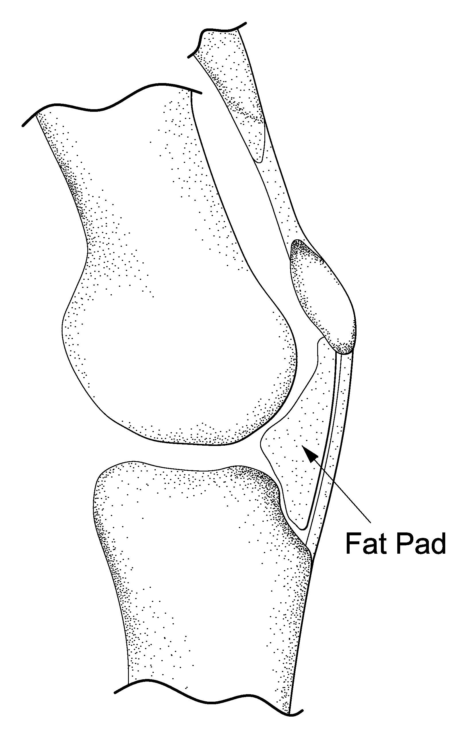
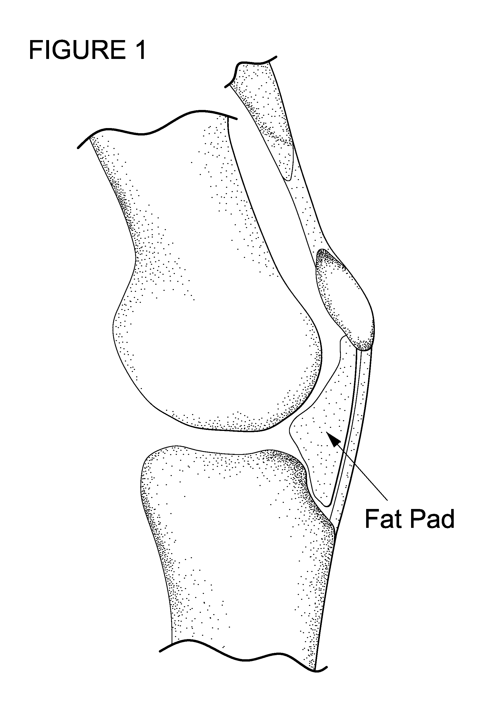
Injection of a Gel in Hoffa's Fat-Pad for Controlled Release of Pro Chondrogenic Factors

[0102]Rat model: Dutch laws on animal experimentation were strictly followed throughout the study and the experimental animal protocol was approved by the Maastricht University committee for animal experiments. After induction of general anaesthesia, both knees of Wistar rats were shaved and prepared for aseptic surgery. The centre of the patella tendon was identified. A 28 G needle with a 1 ml syringe was used to penetrate the centre patella tendon. Directly after the patella tendon was penetrated, approximately between 0.1 and 0.2 ml of the Bio-Gel containing iodine based contrastagent (Visipaque®) was injected in Hoffa's fat-pad (HFP). Since the gel was mixed with X-ray contrast, the side of injection was examined by fluoroscopy.

example 2

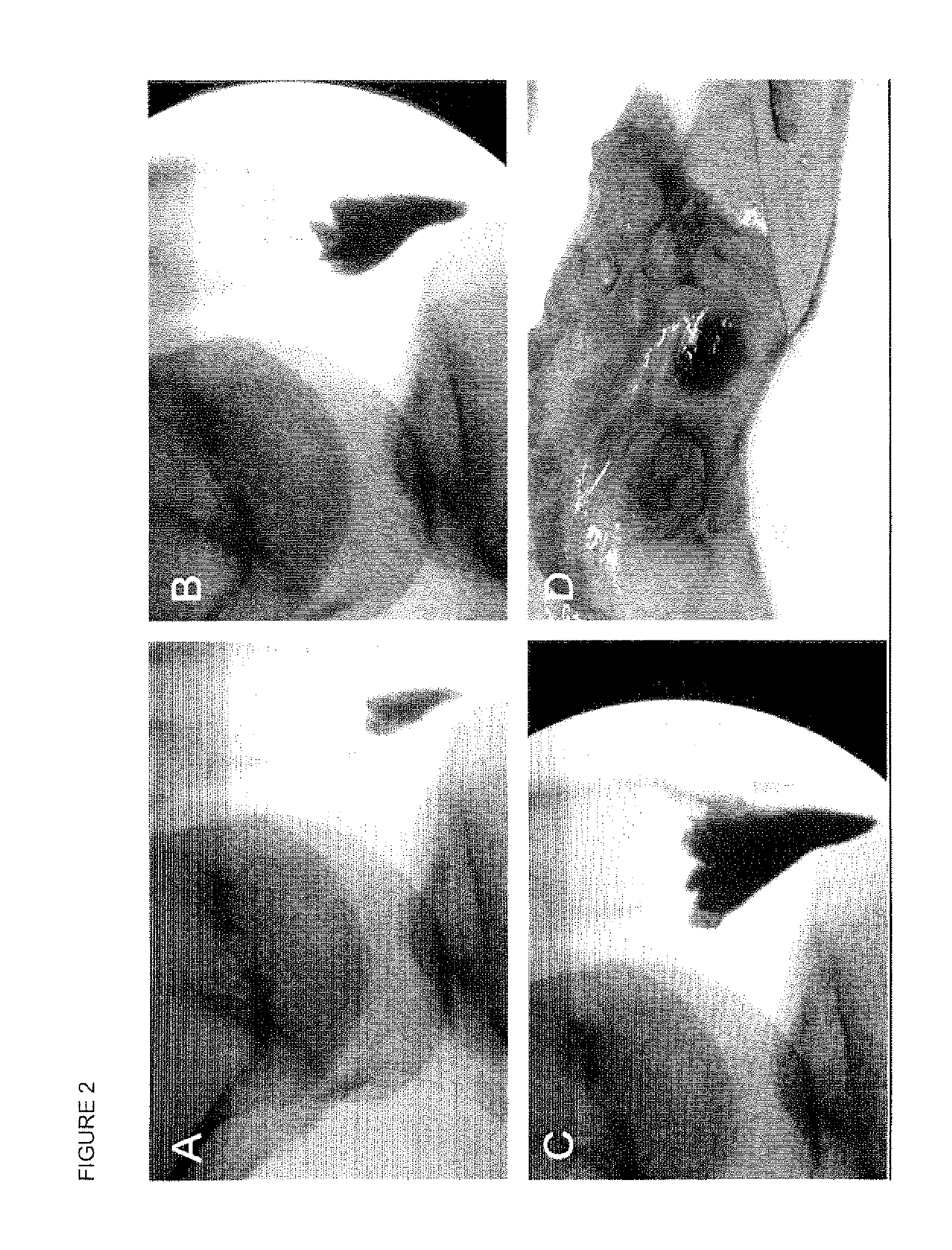
Ex Vivo Injection and Release from Hoffa's Fatpad

[0103]It is advantageous that injection in HFP is feasible, a certain amount can be injected, the injected gel is contained in Hoffa's fatpad, and a certain release is maintained in time. For this purpose we injected different amounts of a radiopaque contrast agent in human HFPs. The HFP could easily be reached through the patellar tendon. An amount of 10 cc could be injected in HFP. Within HFP the contrast agent was not contained in one depot but distributed equally through the fatpad (FIG. 2). For an ex vivo approach HFPs were collected after TKA. The freshly harvested HFPs were injected with bromophenol blue in InGell™. These studies show that at 37° C. gel formation was instantaneous, and the dye is maintained in InGell™ (FIG. 2D). In contrast, injections of the colour dye or contrast agent without the Bio-Gel showed immediate and complete dispersion throughout the Hoffa, with no containment of the additives at all (FIG. 2A-C). Di...

example 3

Histological Evaluation

[0104]Five days and 30 days post-injection the rats were sacrificed and whole knee joints were fixed in 4% buffered formalin and decalcified in 10% EDTA for histology. After embedding in paraffin 7 μm sections were cut and stained with hematoxylin / eosin or with safarin 0 / fast green.

[0105]Both fluoroscopy and histology showed that after penetration of the patella tendon a Bio-Gel could easily and reproducibly be injected in Hoffa's fatpad. Histology and fluoroscopy showed retention of the Bio-Gel in Hoffa's fatpad even after 30 days follow-up. The human cadaver study confirmed this finding; after 1000 flexion / extension cycles the Bic-Gel remained in Hoffa's fatpad.


PUM

PropertyMeasurementUnit
pore sizeaaaaaaaaaa
pore sizeaaaaaaaaaa
pore sizeaaaaaaaaaa

Abstract

The invention is in the field of methods for medical treatment. It provides an improved method for repairing damaged cartilage and / or preventing cartilage degeneration in tissue, in particular in a joint by administering a pharmaceutically active agent directly into the fat pad of a joint. The pharmaceutically active agent is preferably selected from the group consisting of agents that stimulate chondrogenic differentiation and / or cartilage matrix synthesis; agents that inhibit osteogenesis and / or hypertrophy, anti-inflammatory agents, agents that inhibit apoptosis of chondrocytes, agents that inhibit senescence of chondrocytes and agents that enhance lubrication of a joint.

Description

FIELD OF THE INVENTION[0001]The invention is in the field of methods for medical treatment. It provides an improved method for repairing damaged cartilage and / or preventing cartilage degeneration and / or managing pain in tissue, in particular in a joint.BACKGROUND OF THE INVENTION[0002]During our life moving, walking, sport, etc. is essential for our health and quality of life. We fully depend on joint motion for these abilities. The presence of healthy articular cartilage is essential for joint motion. Articular cartilage is exposed to an enormous amount of intensive and repetitive forces during life. The wear resistance and friction modus of articular cartilage are unique, however when damaged, cartilage has a limited capacity for regeneration. Articular cartilage lesions which fail to heal spontaneously eventually evolve in osteoarthritis (OA)3, 6, 7.[0003]OA is among the most frequent forms of musculoskeletal disorders affecting over 10% of the adult population9. While primary os...

Claims


Application Information

Patent Timeline
Patent Type & Authority Applications(United States)
IPC IPC(8): A61K38/19A61K9/00A61K9/127A61K38/39A61K38/17A61K39/395A61K31/728A61K38/45A61P19/10A61P19/02A61P19/00A61K31/42A61K38/02
CPCA61K9/0024A61K31/42A61K31/728A61L27/52A61L27/54A61K9/0019A61L2300/414A61L2300/426A61L2300/432A61L2300/452A61K38/2013A61L2300/252A61P19/00A61P19/02A61P19/10
Inventor EMANS, PETER JOSEPHVAN RHIJN, LODEWIJK WILLEMWELTING, TIM JOHANNES MARIADE LEEUW, MIKE
Owner ACADEMIC HOSPITAL MAASTRICHT
Why Eureka
Try Eureka