
Method and apparatus for monitoring spatial fibrin clot formation

a technology of fibrin clot and monitoring method, which is applied in the direction of biochemistry apparatus, biochemistry apparatus and processes, enzymes, etc., can solve the problems of long-standing and unreal spatial model of blood coagulation in vitro for diagnostical and basic research purposes, enormous labor and time-consuming methods, etc., and achieves convenient practical use, prolonging or shortening the lag time, and increasing or decreasing the growth rate of clots

Inactive Publication Date: 2010-10-14
HEMACORE SA
View PDF6 Cites 7 Cited by
  • Summary
  • Abstract
  • Description
  • Claims
  • Application Information

AI Technical Summary

Benefits of technology

[0008]Hence, it is a general object of the invention to provide a high resolution method and in particular an apparatus which allows for a sensitive, reproducible in vitro modelling of blood coagulation (specifically, the processes of fibrin clot formation and / or dissolution), taking into account the role of spatial heterogeneity and diffusion, and which are apt for the simultaneous monitoring of multiple samples, particularly important in clinical monitoring.
[0009]The present innovation discloses inter alia a cuvette assembly 26, a cuvette holder 13, an apparatus 14 comprising the cuvette holder and an optical detection device, as well as a method, which constitute a further development of the spatial clotting method, much simpler to manufacture and to use, providing more information, allowing automatization, and being apt for the use in a clinical environment.

Problems solved by technology

The problem of modelling blood coagulation in vitro for diagnostical and basic research purposes is long-standing.
This is the simplest approach, but it is non-physiological, because blood coagulation in vivo is a spatially non-uniform phenomenon.
Although this is a sensitive method, its major disadvantage is that it is not truly spatial: although reactants are distributed heterogeneously, the observed signal is an integral characteristic of the coagulation process.
The method is immensely labor-intensive and time-consuming.
In the present form it is impossible to use for diagnostics, because the method cannot be used for plasma clotting without modifications since formation of fibrin clot will prevent possibility of sampling.
Despite the advantages, the method and the apparatus for spatial clotting used previously are inadequate for practical use.
The method does not allow simultaneous measurement of more than one sample, which is not convenient for basic research and completely prevents wide clinical use.
Activation is performed by either fibroblast monolayer or glass; the former method is poorly reproducible, expensive and time-, labor-, and material-consuming (cell culturing facility is needed), the latter method is non-physiological (activation by glass is via the intrinsic pathway, which does not function in vivo).
To keep the sample at 37° C., the cuvette is thermostabilized by keeping it in a water incubator with a stirring device; however, this results bubble formation impairing the signal quality.
Although the method allows for the use of standard spectrophotometric cuvettes and ability to monitor several samples simultaneously, its major disadvantage is that the method requires very large quantities of plasma (1500 μl).
Furthermore, it provides a low spatial resolution for experiments with several samples (100 μm instead of 10 μm in “Spatial clotting method”), and is performed by non-physiological activation (by glass only).
There is no automatization, and the method is time-consuming and labor-intensive.
The method was designed for material testing only, and its suitability for diagnostic purposes, basic and pharmacological research is limited.
In this method, the optical axis is perpendicular to the activator surface, which does not allow monitoring spatial propagation in this direction.
However, this is a narrow and specific problem.
Suitability of the method for other types of basic research, for diagnostic purposes and pharmacological research is limited.

Method used


Image

  • Method and apparatus for monitoring spatial fibrin clot formation
  • Method and apparatus for monitoring spatial fibrin clot formation
  • Method and apparatus for monitoring spatial fibrin clot formation

Examples


Embodiment Construction

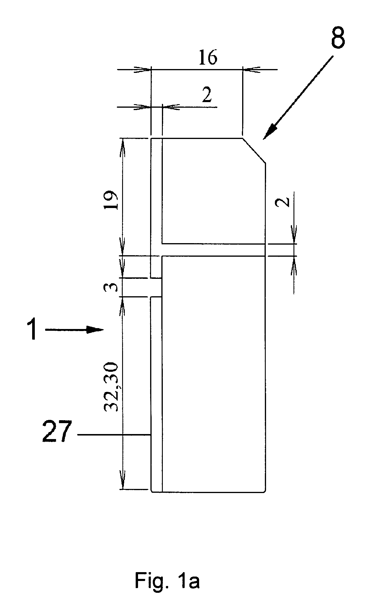
[0058]FIG. 1a shows a front-view of the experimental cuvette 1. The cuvette 1 is of substantially cuboid shape.

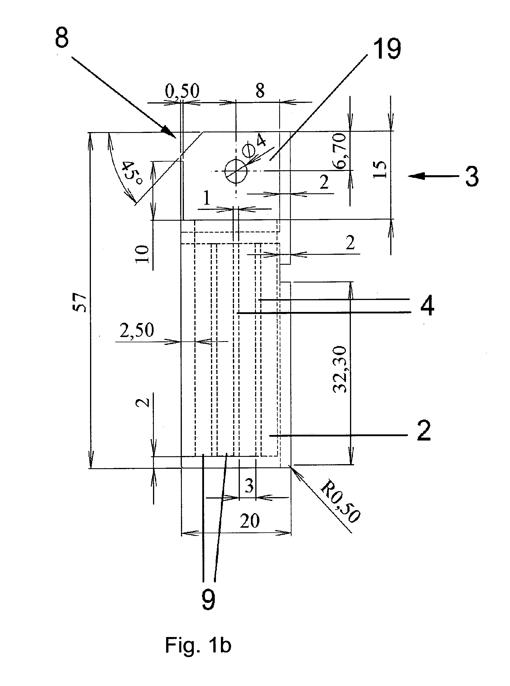
[0059]As can be seen in the cross-sectional view of FIG. 1b, the inner space of the cuvette 1 is divided by segmentation walls 4 into four wells 2 of equitable size. Each well 2 has a rectangular section of 3×1 mm2. However, the invention is not limited to the indicated dimensions; for example, more wells 2 with smaller rectangular sections are possible. In a preferred embodiment, the overall design of the cuvette 1 and the wells 2 are designed such that only a small sample volume is necessary to provide a reliable result. In the embodiment shown in FIGS. 1a and b, a sample volume of only 20 μl is necessary to achieve a reliable result.

[0060]Preferably, the segmentation walls 4 segment the cuvette 1 into a number of parallely and serially aligned wells 2 (see FIG. 1b). The width of the chambers or wells 2 is preferably equitable.

[0061]In this embodiment, the segmentation wa...


PUM

PropertyMeasurementUnit
distanceaaaaaaaaaa
distanceaaaaaaaaaa
timeaaaaaaaaaa

Abstract

The present invention relates to a method, a special designed cuvette (1) and an apparatus for the monitoring of spatial fibrin clot formation or dissolution in multiple samples.

Description

TECHNICAL FIELD[0001]The present invention relates to a method and an apparatus for the monitoring of spatial fibrin clot formation in multiple samples.BACKGROUND ART[0002]The problem of modelling blood coagulation in vitro for diagnostical and basic research purposes is long-standing. A number of methods and devices for measuring clotting parameters of plasma have been proposed; however, the vast majority of these methods (e.g. activated partial thromboplastin time test, thrombelastography, thrombin generation assay) monitor clotting times or other homogenous parameters. In other words, in these tests where the sample of blood (plasma) is uniformly mixed with the activator, coagulation of the sample as a whole is monitored. This is the simplest approach, but it is non-physiological, because blood coagulation in vivo is a spatially non-uniform phenomenon. Diffusion of reactants plays a critically important role in this process.[0003]There are few methods which take into consideratio...

Claims


Application Information

Patent Timeline
Patent Type & Authority Applications(United States)
IPC IPC(8): C12Q1/56C12N9/48C12M1/34
CPCG01N33/4905
Inventor ATAULLAKHANOV, FAZOILSARBASH, VASILIIOVANESOV, MIKHAILPANTELEEV, MIKHAIL
Owner HEMACORE SA
Learn More