
Rgd-modified alginate microparticles as a drug release system

a technology of rgd-modified alginate and microparticles, which is applied in the field of encapsulated cell systems, can solve the problems of capsules lacking high stability, progressive weakening of the structure of the microcapsule, and insufficient microenvironment for cell viability

Inactive Publication Date: 2010-10-28
UNIV DEL PAIS VASCO EUSKAL HERRIKO UNIBERTSITATEA
View PDF2 Cites 4 Cited by
  • Summary
  • Abstract
  • Description
  • Claims
  • Application Information

AI Technical Summary

Benefits of technology

The invention is about a new type of particle and microcapsule that can be used in medicine. These particles are made by modifying a polymer with a specific peptide and then attaching cells with specific receptors for that peptide to the modified polymer. The particles can also be coated with a membrane. The technical effect of this invention is that it provides a way to deliver peptides with biological activity to cells in a targeted and controlled way, which could be useful in the treatment of diseases. The particles can also be used as a device for the controlled release of peptides.

Problems solved by technology

The alginate microcapsules are stabilized by means of divalent cations which, after their implantation in the organism, tend to spread, resulting in a progressive weakening of the structure of the microcapsule.
However, this type of modified polymer does not provide a suitable microenvironment for cell viability.
However, the microcapsules are intended for the quick release of the cells, they are therefore capsules lacking high stability.
However, this study does not demonstrate that the encapsulated cells survive for more than 15 days from the moment of implantation.

Method used


Image

  • Rgd-modified alginate microparticles as a drug release system
  • Rgd-modified alginate microparticles as a drug release system
  • Rgd-modified alginate microparticles as a drug release system

Examples


example 1

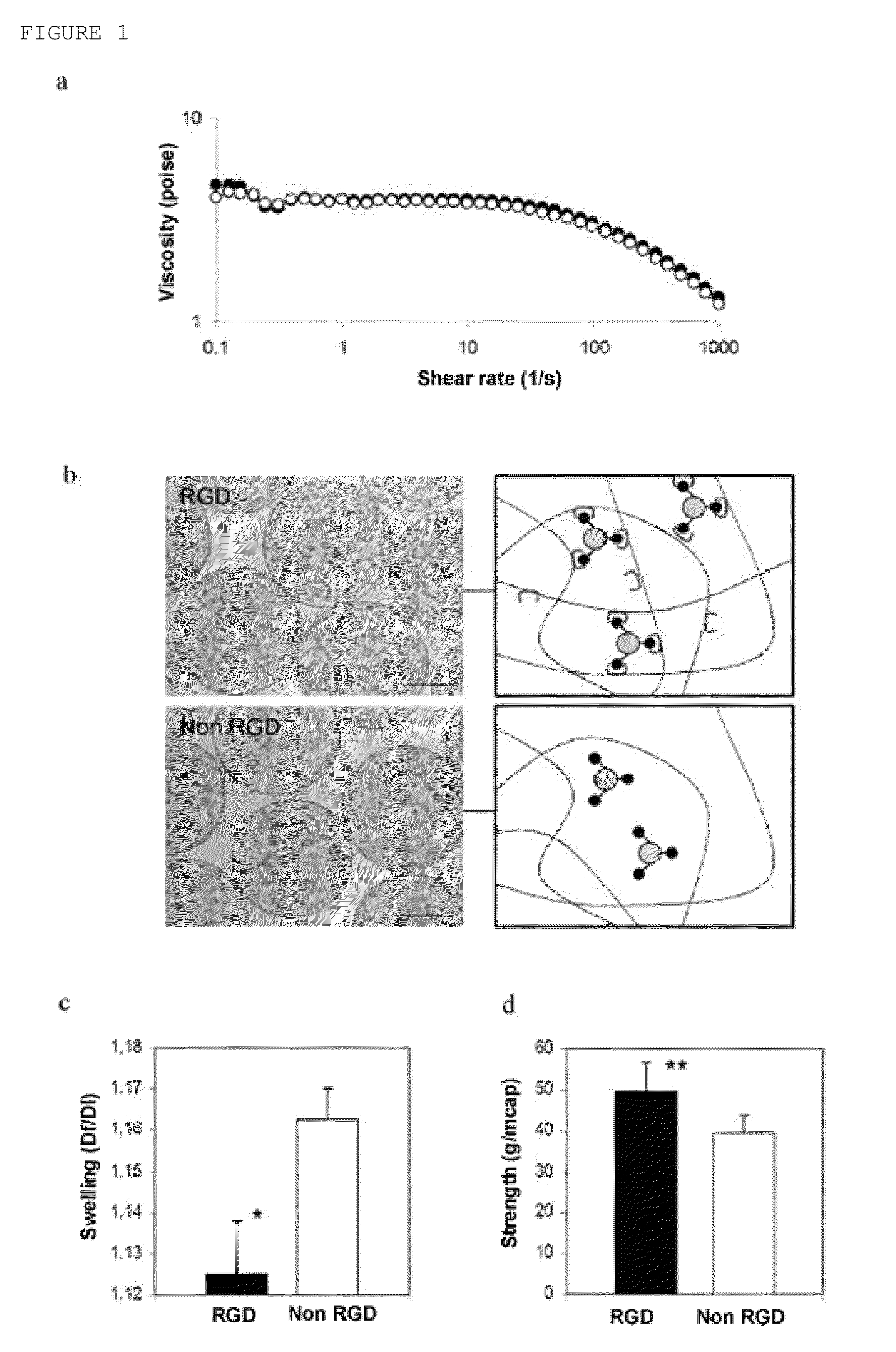
[0067]The molecular weight of the alginate molecules rich in guluronic acid residues (Protanal LF240 D, FMC Technologies) was adjusted by means of irradiation with a cobalt 60 source. The irradiation doses ranged from 2 to 5 Mrads. The final molecular weight of the alginate molecules was analyzed by means of gel filtration chromatography (Viscotek).

[0068]After irradiation, the alginate molecules were modified with synthetic oligopeptides containing the (glycine)4-arginine-glycine-aspartic acid-tyrosine sequences (GGGGRGDY, Commonwealth Biotechnology Inc.) using to that end aqueous carbodiimide reactions [1]. Briefly, alginate solutions were prepared in a 2-[N-morpholino]ethanesulfonic acid hydrate buffer (MES, Sigma) which were subsequently mixed with N-hydroxysulfosuccinimide (sulfo-NHS, Pierce Chemical), and 1-ethyl-3-[3-(dimethylamino)propyl]carbodiimide (EDC, Sigma) and the oligopeptides. After reacting for 24 hours, the modified alginate molecules were purified using filters (F...

example 2

Adhesive and Biomimetic Effects of RGD Alginates

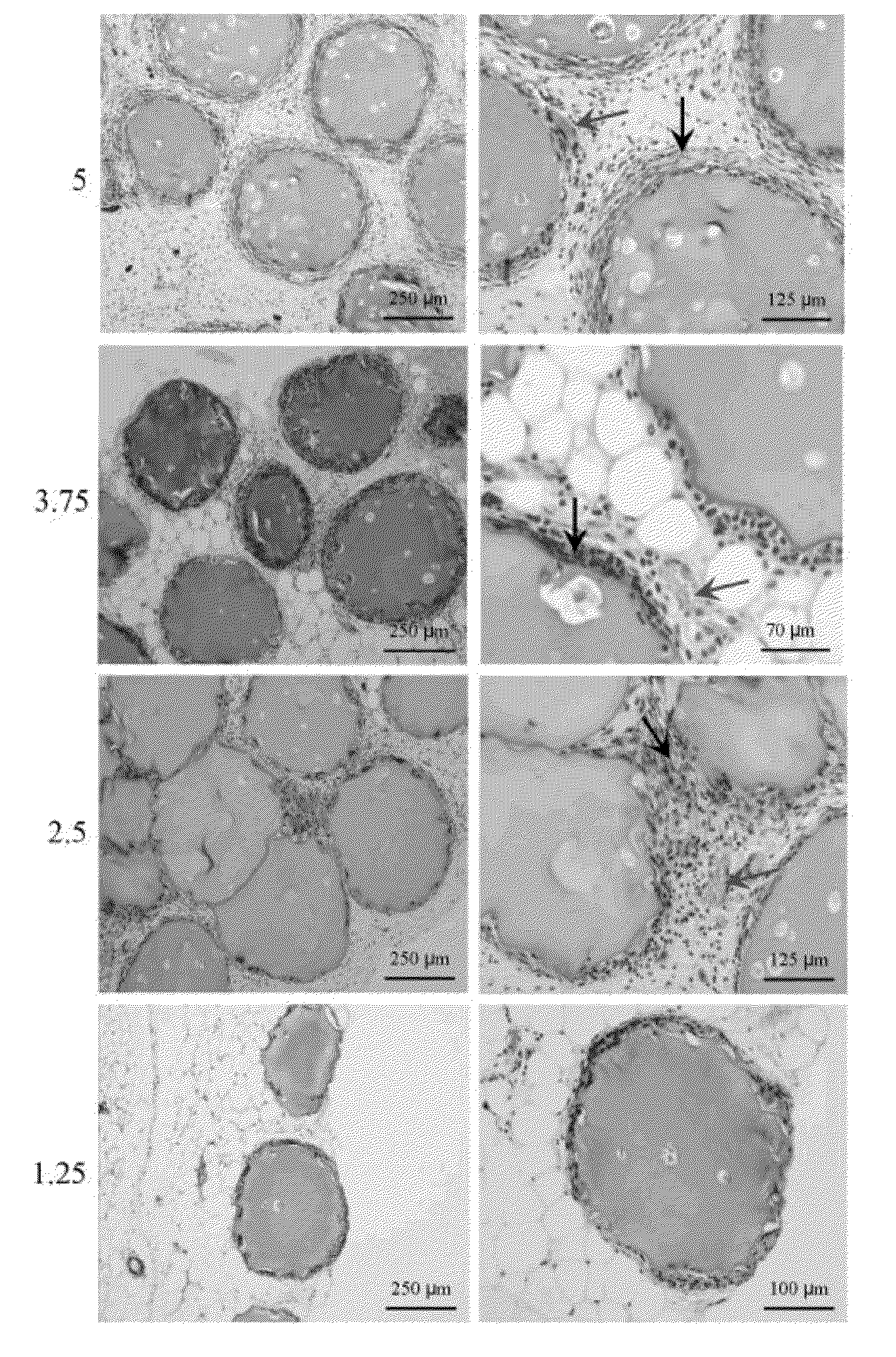
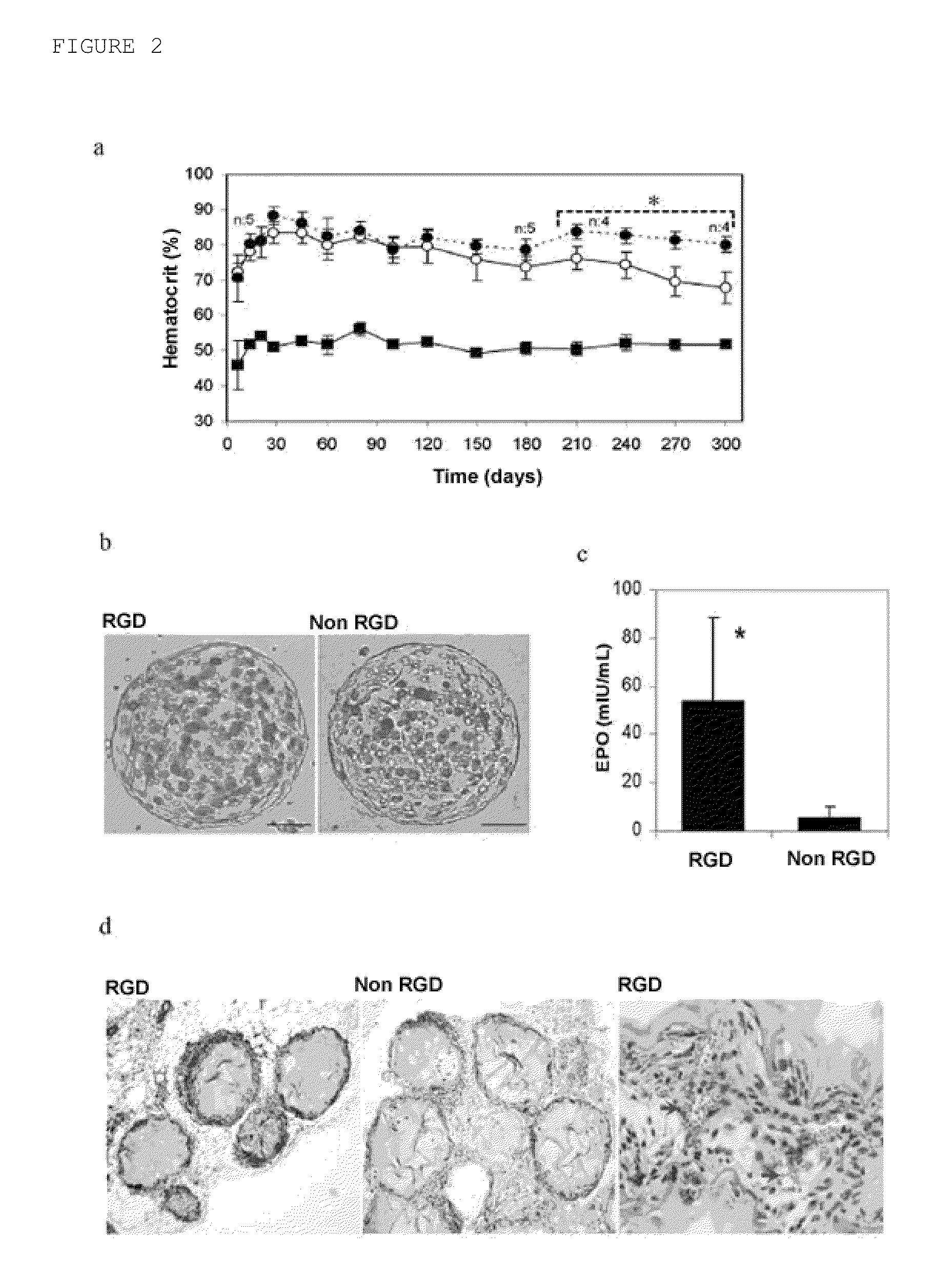
[0070]To evaluate the adhesive and biomimetic effects of RGD alginates, erythropoietin (EPO)-secreting mouse C2C12 skeletal myoblasts modified by genetic engineering were selected as candidate cells and mixed with the binary polymer solution. It was observed that both the RGD concentration and the alginate composition could control the phenotype of the myoblasts. These cells can be easily handled and once encapsulated and implanted in the host, they irreversibly come out of the cell cycle and fuse in multinucleated myofibrils. The latter is essential for controlling the therapeutic dose and for preventing possible biosafety risks. EPO was selected as model drug due to its resulting therapeutic effects and because it is easy to monitor its in vivo expression and bioactivity by following its hematocrit level. Furthermore, due to its weak pharmacokinetics, it is expected that cell encapsulation technology can prevent the continuous and re...

example 3

Viability and Functionality of the Myoblasts in the RGD Alginate Matrixes

[0073]To characterize the viability and functionality of the myoblasts in the matrixes, the cell metabolic activity and the release of EPO were studied in vitro during a 3-week period. These studies did not show major differences between the two types of microcapsules in cell viability or in the release of EPO in the short term. After 21 days in culture, the EPO secretion of RGD alginate microcapsules loaded with 100 cells measured was 71±9 IU of EPO / 24 h, whereas the production in non-modified alginate matrixes was 72±5 IU EPO / 24 h. These results suggest that the cells adapt satisfactorily to the new microenvironments. However, for the purpose of evaluating the possible impact of matrixes bioactivated with RGD on the survival and long-term functionality of encapsulated cells, 0.2 mL of both types of microcapsules (RGD alginate, 5×10⁶ cells / mL alginate) were implanted in subcutaneous tissue of immunocompetent B...


PUM

PropertyMeasurementUnit
diameteraaaaaaaaaa
diameteraaaaaaaaaa
cell adhesionaaaaaaaaaa

Abstract

The object of the present invention is to provide particles of a polymeric material containing cells therein, wherein said particles have a strength that is substantially greater than the microcapsules known in the state of the art. Said strength is achieved by means of functionalizing the polymeric material forming the microcapsule with a peptide which can bind to the adhesion proteins of the cell membrane, such that the cells act as cross-linking agents of the matrix resulting in an improvement of the mechanical properties of the matrixes

Description

TECHNICAL FIELD OF THE INVENTION[0001]The present invention relates to encapsulated cell systems and, more specifically, to microcapsules formed by a polymeric material which has been modified by a peptide which is capable of binding to the adhesion proteins found in the cell membrane such that the cells that are inside the microcapsule interact with said peptide. The invention also contemplates microcapsules in which the cells have been genetically modified to express a protein of therapeutic interest as well as the therapeutic applications of said microcapsules.BACKGROUND OF THE INVENTION[0002]Cell microencapsulation technology is based on the immobilization of cells within a polymeric matrix surrounded by a semi-permeable membrane. Said encapsulated cells are protected against the rejection of the cell and antibody-dependent immune system and have the potential of producing a wide range of therapeutically active substances (Orive, G, et al. 2003, Nat. Med. 9:104-107, Orive, G. Tr...

Claims


Application Information

Patent Timeline
Patent Type & Authority Applications(United States)
IPC IPC(8): A61K9/14C12N11/02C12N11/10A61K35/12B05D3/00A61P13/12A61P7/06A61P15/00A61P25/28A61P25/18
CPCC07K14/78C08B37/0084C08J3/126C08J2305/04C08L5/04C07K5/1019A61K9/5015A61K38/00C07K5/0817C07K5/1008A61K9/1652A61P13/12A61P15/00A61P25/18A61P25/28A61P7/06A61K9/50
Inventor ORIVE ARROYO, GORKAPEDRAZ MUNOZ, JOSE LUISHERNANDEZ, ROSA MARIA
Owner UNIV DEL PAIS VASCO EUSKAL HERRIKO UNIBERTSITATEA
Learn More