
In-Vitro Model of Blood-Brain Barrier, In-Vitro Model of Diseased Blood-Brain Barrier, and Drug Screening Method, Analysis Method for Functions of Diseased Blood-Brain Barrier, and Analysis Method for Pathogenesis Using the Same

a technology of bloodbrain barrier and in-vitro model, which is applied in the field of in-vitro model of bloodbrain barrier, can solve the problems of compound shortage as actual therapeutic drugs, the presence of bloodbrain barrier becomes a major obstacle to some artificial treatment of living organisms, and the development of drugs must be abandoned

Inactive Publication Date: 2010-10-28
PHARMACO CELL
View PDF0 Cites 11 Cited by
  • Summary
  • Abstract
  • Description
  • Claims
  • Application Information

AI Technical Summary

Benefits of technology

[0027]The present invention provides an in-vitro BBB model most analogous to the in-vivo blood-brain barrier and as such, efficiently achieves the development of a central nervous agent (centrally acting drug transported across the blood-brain barrier) which is currently urgently needed.
[0028]Moreover, the present invention provides a screening system for a drug transferred into the brain without being expected to centrally act and as such, can contribute to the development of appropriate drug therapies or the development of novel drugs. Moreover, the present invention is useful in the screening of a drug acting on the blood-brain barrier itself.

Problems solved by technology

However, its presence becomes a major obstacle to some artificial treatment of living organisms for causes such as diseases.
This presence is a major obstacle to the development of preventive or therapeutic drugs for central nervous diseases (dementia, Alzheimer's disease, prion disease, stroke, etc.) which is urgently needed.
Thus, such compounds are not available as actual therapeutic drugs, and their development must be abandoned.
In this case, it is also expected that drugs considered to be hardly transferred into the brain are transferred into the brain through drug interaction and causes central side effects.
Nevertheless, the law of constant transfer into the brain is absent due to the unique function of BBB, and drug permeability cannot be determined in advance from the chemical structure or molecular weight of the drug.
Study on drug transfer into the brain in animal experiments requires enormous labors and expenses.
In addition, it is difficult to correctly determine only the complicated in-vivo drug transfer.
The securing of the amount of lead compounds that can bear animal experiments requires additional times and expenses and thus, is not realistic.
However, previously reported in-vitro BBB models are only incomplete models such as a monolayer culture system of endothelial cells or a coculture system of endothelial cells with astrocytes.
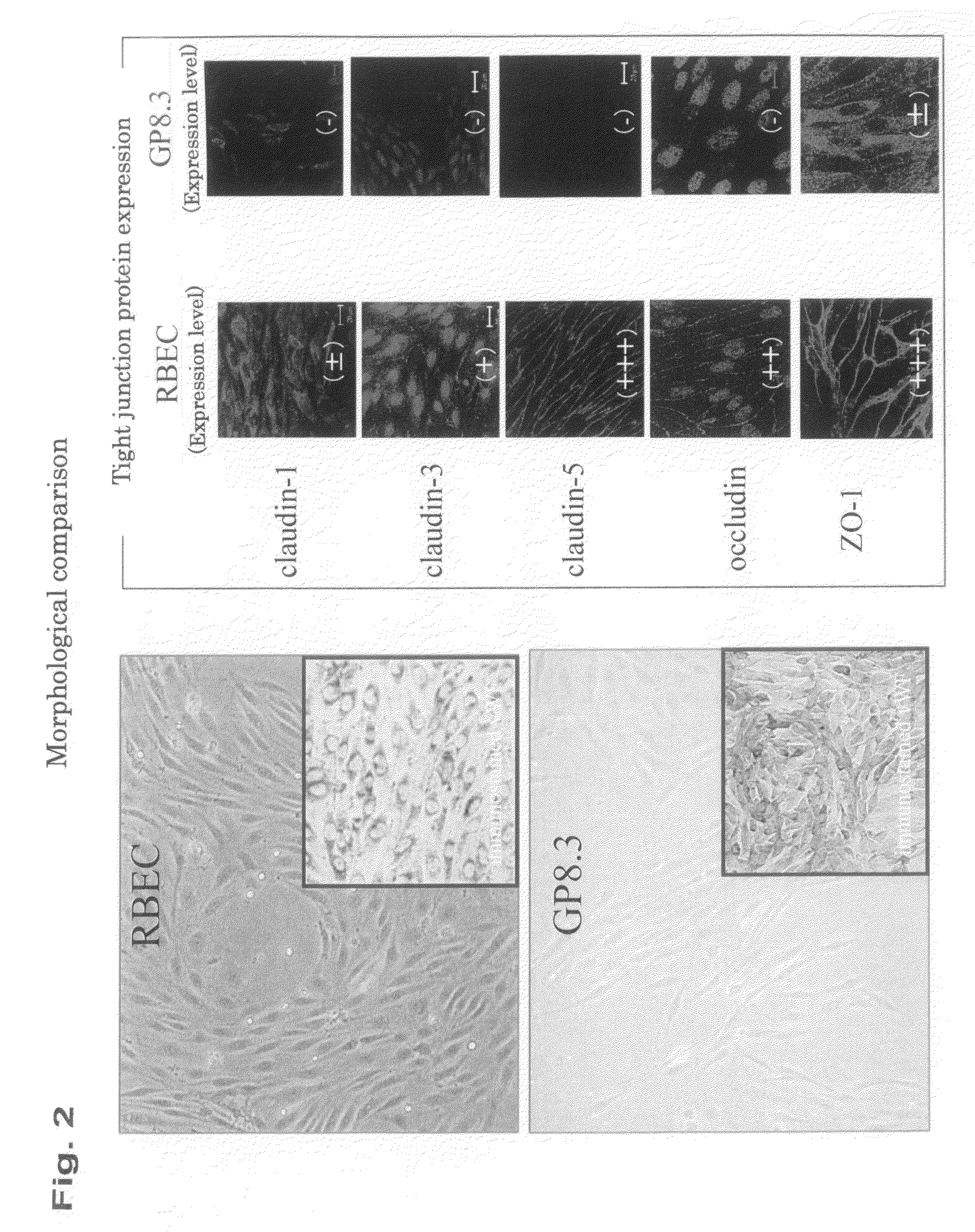
However, it is questionable whether immortalized brain capillary endothelial cells, which have been used previously because of their convenience, maintain this tight junction function.
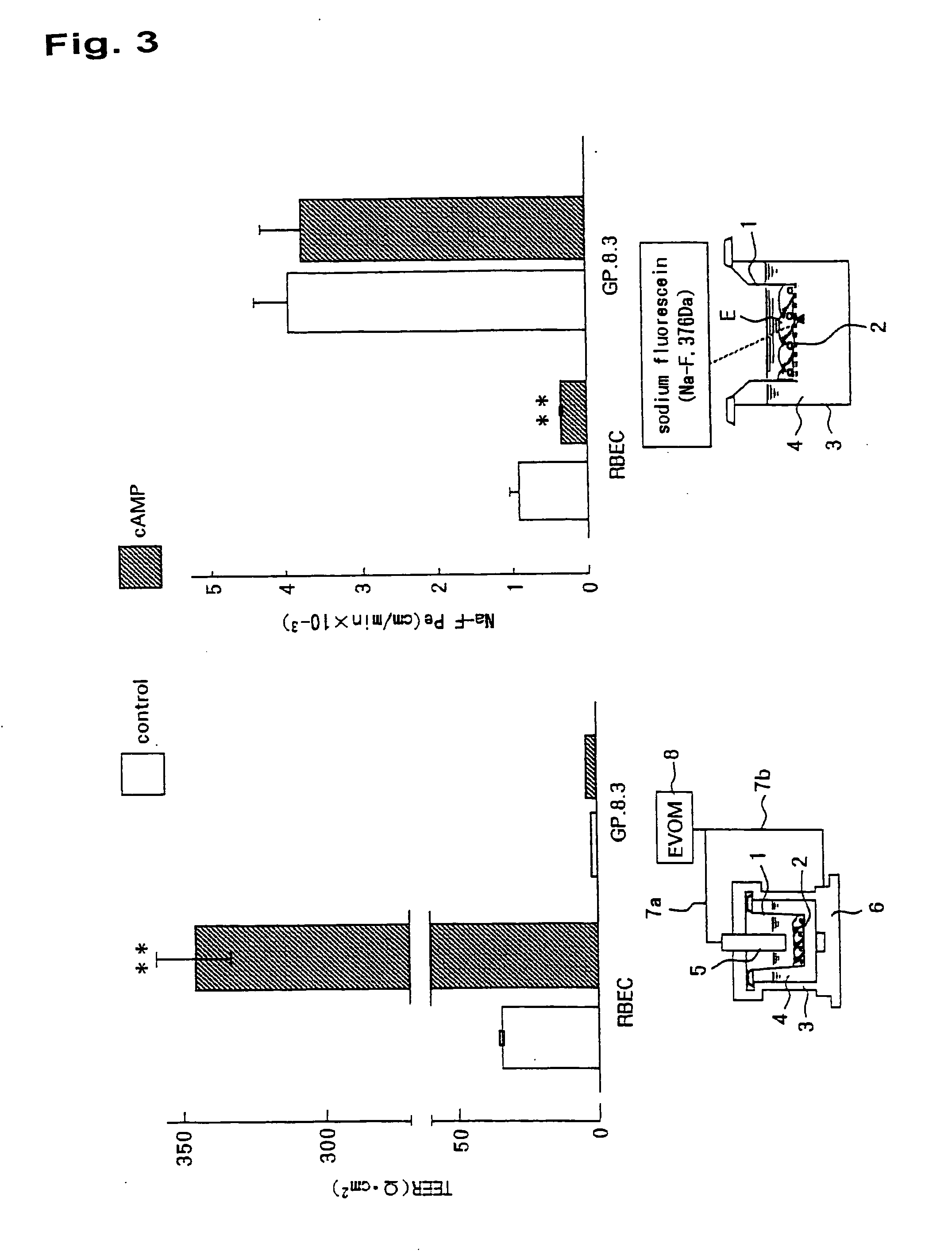
Particularly, such a model presents a problem in the screening of drugs that vary cAMP.
It is considered that incomplete BBB models having not all of BBB constituent cells, such as a culture system of endothelial cells alone or a coculture system of endothelial cells with astrocytes, do not sufficiently reproduce BBB in a diseased state.

Method used


Image

  • In-Vitro Model of Blood-Brain Barrier, In-Vitro Model of Diseased Blood-Brain Barrier, and Drug Screening Method, Analysis Method for Functions of Diseased Blood-Brain Barrier, and Analysis Method for Pathogenesis Using the Same
  • In-Vitro Model of Blood-Brain Barrier, In-Vitro Model of Diseased Blood-Brain Barrier, and Drug Screening Method, Analysis Method for Functions of Diseased Blood-Brain Barrier, and Analysis Method for Pathogenesis Using the Same
  • In-Vitro Model of Blood-Brain Barrier, In-Vitro Model of Diseased Blood-Brain Barrier, and Drug Screening Method, Analysis Method for Functions of Diseased Blood-Brain Barrier, and Analysis Method for Pathogenesis Using the Same

Examples


example 1

Isolation of Brain Capillary Slice

[0045]The brain of a three-week-old rat was excised within a clean bench and placed in an ice-cold Phosphate buffer saline (− / −) (PBS (− / −); Sigma). The dura mater, the cerebellum, the interbrain, the brain stem, and the like were removed on a sterilized filter paper to leave only the cerebral cortex. This cerebral cortex was placed in 3 ml of an ice-cold Dulbecco's Modified Eagle's Medium (DMEM; manufactured by Sigma) and finely cut into a size of approximately 1 mm3 with a surgical knife. 15 mL of DMEM containing enzymes (collagenase-class 2 (1 mg / ml; manufactured by Worthington) and 300 μL of DNase (15 μg / mL; manufactured by Sigma)) was further added thereto. The mixed solution was moved up and down 25 times with a 5-mL pipette to suspend a pellet. After shaking / incubation at 37° C. for 90 minutes, 10 mL of DMEM was added thereto, and the mixture was centrifuged. The precipitated pellet after centrifugation was centrifuged with 20% BSA (manufactu...

example 2

Brain Capillary Pericytes

[0046]The brain capillary slice of Example 1 contains a few percents of pericytes. Thus, the brain capillary slice was seeded onto a dish coated with collagen and cultured at 37° C. for 1 week in 5% CO2 / 95% atmosphere in a culture solution containing 10% fetal bovine serum (FBS)-DMEM supplemented with gentamicin (50 μg / mL) to promote pericyte growth. In this stage, the brain capillary endothelial cells are mixed with the pericytes. Next, the cells were detached with a 1×trypsin-EDTA solution (manufactured by Sigma) and seeded again onto a non-coated dish to isolate the pericytes (the endothelial cells cannot adhere to the dish, while only the pericytes grow therein).

example 3

Astrocytes

[0047]The brain was removed from a 1- or 2-day-old rat within a clean bench and placed in ice-cold PBS (− / −). The dura mater, the cerebellum, the interbrain, the brain stem, and the like were removed on a sterilized filter paper to leave only the cerebral cortex. This cerebral cortex was placed in a 50-mL tube, and 10 mL of ice-cold DMEM was added thereto. The mixed solution was slowly moved up and down with a 20-G syringe to break the tissue apart. The solution was left standing for a while. Then, 5 mL of the supernatant was placed in another 50-mL tube. 5 mL of DMEM was further added to 5 mL of the remaining cell suspension, and the mixed solution was moved up and down with a 20-G syringe. This procedure was repeated until the cell mass could not be observed visually. The last 5 mL of the cell suspension was also added thereto. Then, the cell suspension was passed through Cell Strainer (registered trademark; manufactured by Falcon) of 70 μm. The cells were collected by c...


PUM


Abstract

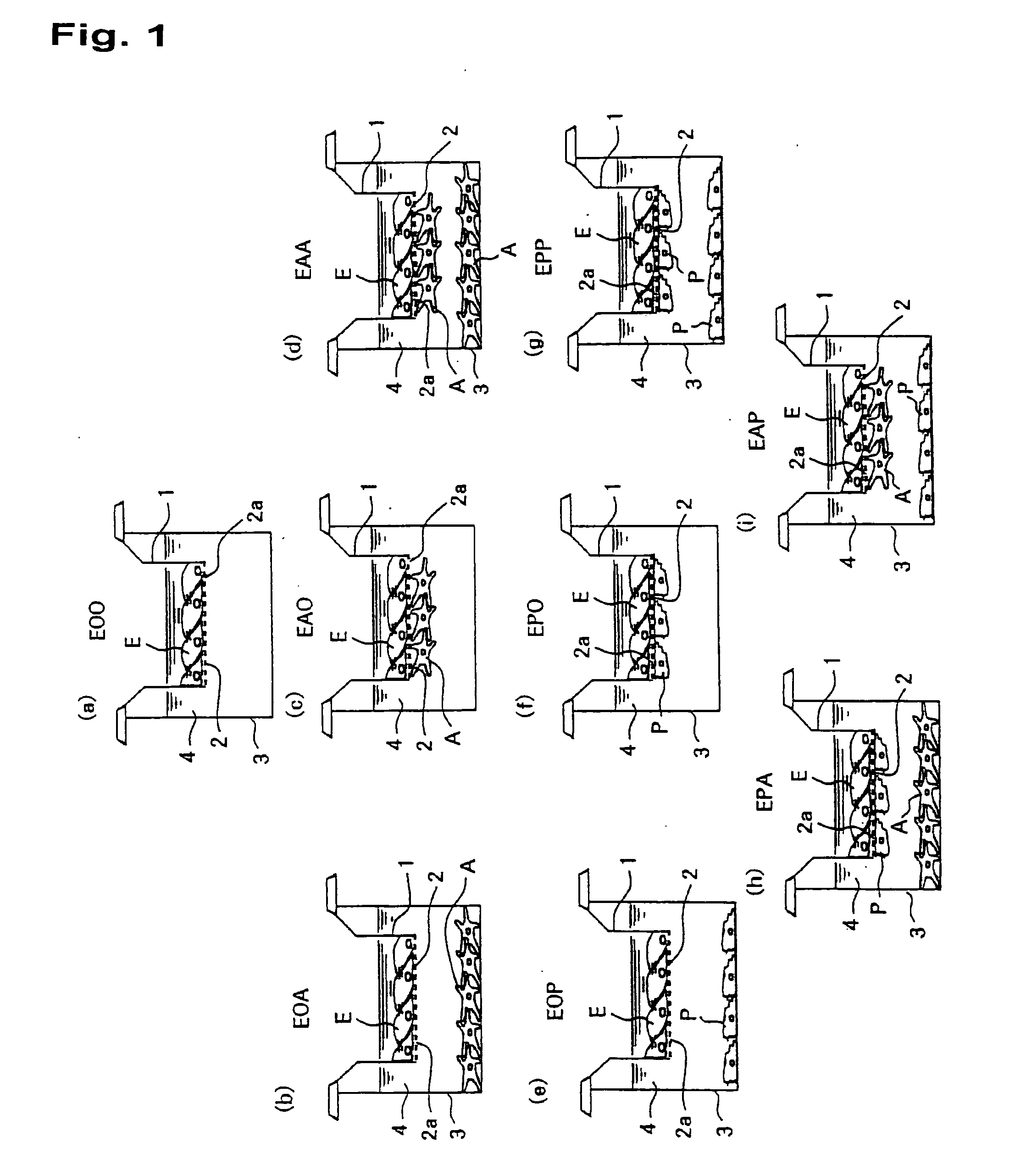
It is intended to provide a screening system for a centrally acting drug transported across the blood-brain barrier, a drug acting on the blood-brain barrier itself, or a drug transferred into the brain without being expected to centrally act. Moreover, another object of the present invention is to achieve pathogenesis analysis study or the screening in a diseased state by applying various diseased environments to this screening system. The present invention provides an in-vitro model of blood-brain barrier obtained by using a three-dimensional culture apparatus comprising: a culture solution; a plate holding the culture solution; and a filter immersed in the culture solution and placed in no contact with the inside bottom of the plate, the filter having plural pores of 0.35 to 0.45 μm in diameter, and by comprising: seeding primary cultured brain capillary endothelial cells onto the upper surface of the filter; seeding primary cultured brain pericytes onto the under surface of the filter; seeding primary cultured astrocytes onto the inside surface of the plate; and coculturing these cells in a normal culture solution.

Description

TECHNICAL FIELD[0001]The present invention relates to an in-vitro model of blood-brain barrier (generally called “BBB” as an abbreviation) and an in-vitro model of diseased blood-brain barrier.[0002]“In vitro” is a Latin term meaning “in glass” and refers to, as a biological term, a state in which a portion of a living organism is extracted and found free “outside the living organism” for various study purposes (see Iwanami Biological Dictionary, Iwanami Shoten, Publishers, Nov. 10, 1998).[0003]Specifically, the present invention relates to a screening system for a centrally acting drug transported across the blood-brain barrier, a drug acting on the blood-brain barrier itself, or a drug transferred into the brain without being expected to centrally act.[0004]Moreover, the present invention relates to a drug screening method using the in-vitro model of blood-brain barrier and the in-vitro model of diseased blood-brain barrier. Specifically, the present invention achieves pathogenesi...

Claims


Application Information

Patent Timeline
IPC IPC(8): C12Q1/02C12N11/00C12N5/071
CPCC12M35/08C12N5/0691C12N2502/28G01N33/5091C12N2533/54G01N33/5058G01N33/5064C12N2533/52
Inventor NIWA, MASAMINAKAGAWA, SHINSUKEDELI, MARIA ANNA
Owner PHARMACO CELL
Learn More