Method for Differentiating of Human Embryonic Stem Cells Into the Osteoblastic Lineage

a human embryonic stem cell and osteoblastic technology, applied in the field of differentiating human embryonic stem cells into osteoblastic lines, can solve the problems of limited cell division cycle, small number, and inability to collect by bone marrow aspiration, and achieve the effect of improving stem cells

Inactive Publication Date: 2010-11-25
KOREA RES INST OF BIOSCI & BIOTECH
View PDF2 Cites 0 Cited by
  • Summary
  • Abstract
  • Description
  • Claims
  • Application Information

AI Technical Summary

Benefits of technology

[0019]The composition for inducing stem cells to differentiate into an osteoblastic lineage and the method for differentiating stem cells into an osteoblastic lineage using the composition are useful in the development of cell therapeutics for metabolic bone diseases. Also, the present invention contributes to molecular biological research into the signaling pathway for the differentiation of human embryonic stem cells into an osteoblastic lineage, thus being useful in the discovery of other novel bone-related factors. Providing terminally differentiated mature cells, in addition, the present invention is applied to surgical and pharmaceutical research into metabolic bone diseases. The differentiated cells produced according to the method of the present invention find applications in various medical and pharmaceutical fields, including biological assay systems for medical efficacy using stem cells and assay systems for the medical efficacy of osteogenic inducers and bone tissue enhancements in stem cells.

Problems solved by technology

However, none of the therapies developed thus far ensure complete recovery from metabolic bone diseases.
The mesenchymal stem cells of adult tissues may be a useful approach to bone regeneration with autogenous bone grafts, but are disadvantageous in that they are very small in number and difficult to collect by bone marrow aspiration.
However, the fact that a limitation on the possible number of cell division cycles exists and that there is a high possibility of inducing cell modification during the large number of cycles of cell division act as a bottleneck for the use of mesenchymal stem cells.
The understanding of the mechanism by which osteoblasts are differentiated from embryonic stem cells and the techniques of inducing the differentiation have not advanced to a level sufficient to develop cell therapeutics having excellent clinical functions.

Method used


Image

  • Method for Differentiating of Human Embryonic Stem Cells Into the Osteoblastic Lineage
  • Method for Differentiating of Human Embryonic Stem Cells Into the Osteoblastic Lineage
  • Method for Differentiating of Human Embryonic Stem Cells Into the Osteoblastic Lineage

Examples


example 2

Preparation of Embryoid Bodies and Single Cells from Human Embryonic Stem Cells

[0034]In order to prepare consistent sizes of embryoid bodies, human embryonic stem cell colonies were sliced into cell clumps having dimensions of about 500×500 μm using a tool made from a glass pipette, and a suspension of the cell clumps was collected and transferred into bacterial culture dishes. The resulting embryoid bodies were dissociated into single cells by washing with phosphate buffered saline (PBS), adding a 1× trypsin-EDTA solution (TrypLE Express, Invitrogen), incubating at 37° C. for 1˜5 min, washing twice with PBS, and plating on Matrigel (BD biosciences, USA)-coated culture dishes.

example 3

Differentiation of Human Embryonic Stem Cells and Embryoid Bodies into Osteoblasts

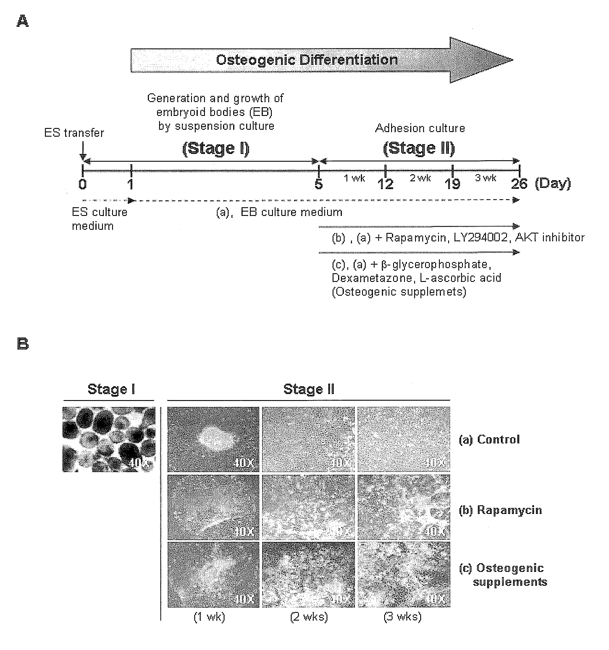
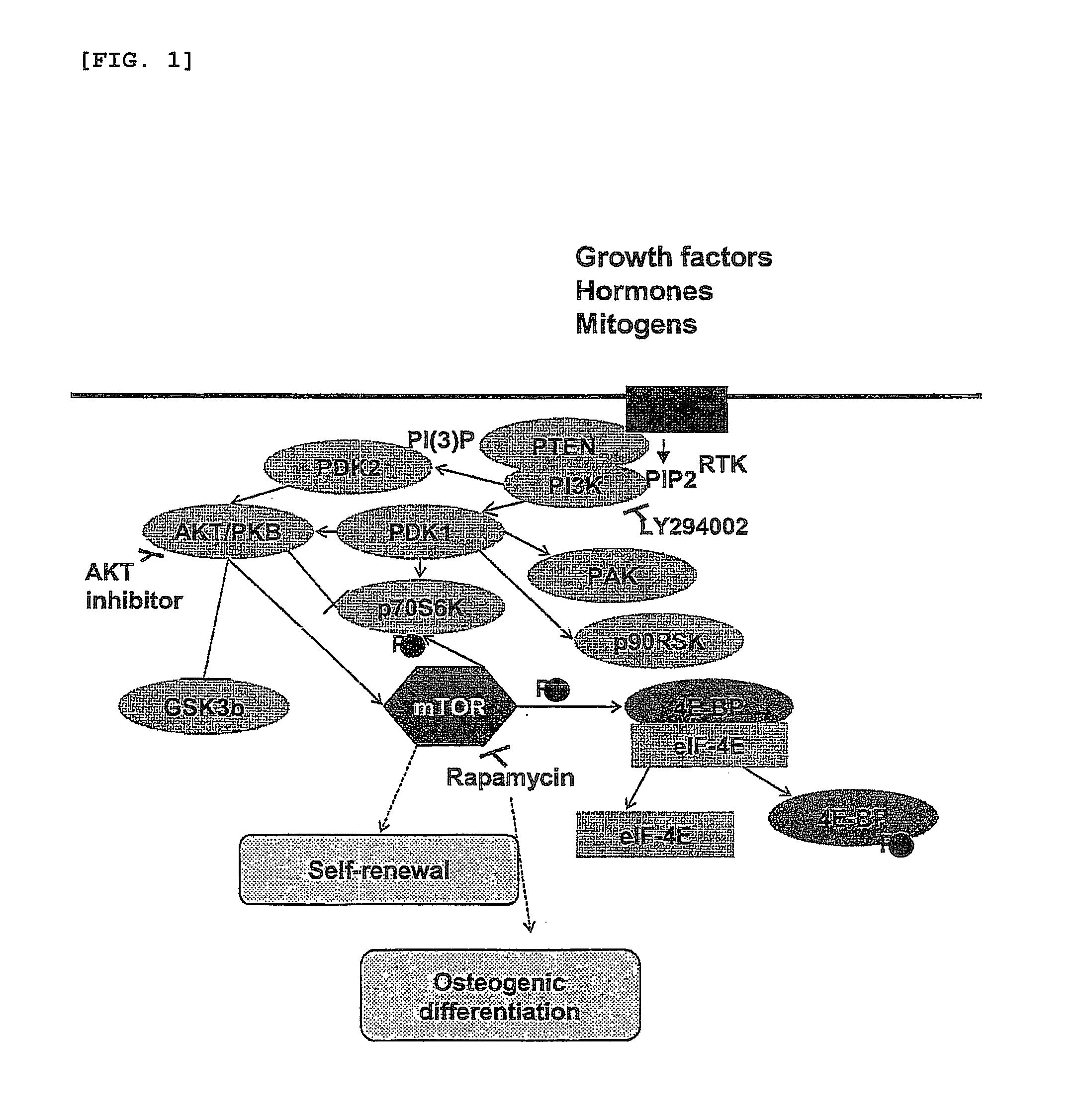
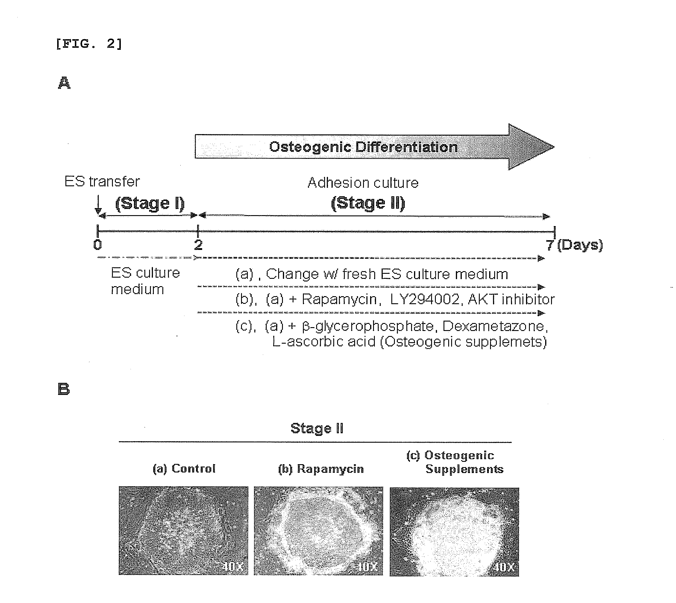
[0035]For osteogenic differentiation by adhesion culturing, the human embryonic stem cells obtained in Example 1, after passage, were cultured in the culture medium of Example 1 for 2 days, and then in a culture medium, free of recombinant human FGF basic, containing 0.1˜200 nM rapamycin, 0.01˜50 μM LY294002, 0.01˜20 μM AKT inhibitor, or conventional osteogenic supplements (10 mM β-glycerophosphate, 0.1 mM Dexamethasone, 0.1 mM L-ascorbic acid) for 7 days, with daily replacement with a fresh embryonic stem cell culture medium (FIG. 2A).

[0036]For osteogenic differentiation by embryoid body formation, as shown in FIG. 3A, the human embryonic stem cells obtained in Example 1 were first cultured in the stem cell culture medium of Example 1 for one day after passage, and the stem cell colony was sliced into cell clumps having dimensions of 500×500 μm. After being collected as a suspension, the cell clumps w...

example 4

Detection of Osteoblastic Marker

[0038] Reverse Transcriptase PCR (RT-PCR) and Quantitative Real-Time PCR

[0039]The differentiation of human embryonic stem cells into osteoblasts in Example 3 was examined by detecting the expression of osteoblast-specific genes through RT-PCR and quantitative real-time PCR. For this, after treatment or non-treatment with rapamycin, LY294002, an AKT inhibitor or osteogenic supplements (10 mM β-glycerophosphate, 0.1 mM Dexamethasone, and 0.1 mM L-ascorbic acid), cells were collected according to differentiation stages and subjected to total RNA isolation using a TRIzol reagent (Invitrogen, USA). The RNA was used as a template to synthesize cDNA with a oligo(dT) primer in the presence of a Superscript II reverse transcriptase, followed by PCR with various primers listed in Table 1, below.

TABLE 1AccessionProductGenesNos.SequencesSize (bp)GAPDHNM_002046ForwardGAAGGTGAAGGTCGGAGTC226ReverseGAAGATGGTGATGGGATTTCOct4NM_002701ForwardGAAGGATGTGGTCCGAGTGT243Revers...


PUM

PropertyMeasurementUnit
sizeaaaaaaaaaa
densityaaaaaaaaaa
sizesaaaaaaaaaa

Abstract

Disclosed are a composition for introducing the osteogenic differentiation of human embryonic stem cells and a method for differentiating human embryonic stem cells into an osteoblastic lineage by inhibiting the mTOR signaling pathway. When cultured in the presence of an inhibitor of the mTOR signaling pathway, human embryonic stem cells are effectively induced to differentiate into an osteoblastic lineage. The osteogenic differentiation of human embryonic stem cells using the method and the composition is useful in examining the development and differentiation mechanism of osteoblasts and the cause of metabolic bone diseases, including osteoporosis. In addition, the method and the composition can be applied to the development of osteogenic differentiation techniques for generating clinically useful, terminally differentiated mature cells or progenitor cells.

Description

TECHNICAL FIELD[0001]The present invention relates to a method for differentiating human embryonic stem cells into an osteoblastic lineage, using an inhibitor against the mTOR signaling pathway, and a composition for inducing osteogenic differentiation.BACKGROUND ART[0002]Stem cells, especially human embryonic stem cells, have recently arisen as a promising cell therapeutic in the regenerative medicine and medical industries. In recognition of the economic and industrial use and added value of human embryonic stem cells, the development of technologies and materials for inducing human embryonic stem cells to differentiate into functionally specific cells has emerged as one of the most interesting topics. As embryonic stem cells were first derived from mouse embryos in 1981 by Evans and Kaufman, mouse embryonic stem cells have been used as basic materials for use in the study of embryonic stem cells and the development of differentiation inducing technologies.[0003]Embryonic stem cel...

Claims


Application Information

Patent Timeline
Patent Type & Authority Applications(United States)
IPC IPC(8): C12N5/0735C12N5/077
CPCA61K31/436C12N5/0654C12N2500/38C12N2502/02C12N2501/04C12N2501/39C12N2501/70C12N2500/42A61K31/445C12N5/00C12N5/0602
Inventor CHO, YEE SOOKLEE, KYU-WON
Owner KOREA RES INST OF BIOSCI & BIOTECH
Why Eureka
Try Eureka