Method for screening patient-specific Anti-cancer agent using limiting dilution assay

a screening method and anti-cancer technology, applied in the field of screening methods, can solve the problems of limiting the sensitivity of drugs according to gene information, increasing the risk and burden of patients, and affecting the effect of clinical trials,

Active Publication Date: 2015-04-09
SAMSUNG LIFE PUBLIC WELFARE FOUND
View PDF1 Cites 9 Cited by
  • Summary
  • Abstract
  • Description
  • Claims
  • Application Information

AI Technical Summary

Benefits of technology

[0023]According to the present invention, since the patient-specific anti-cancer agents reflecting individual genetic properties may be precisely and rapidly selected by using an extremely small amount of cancer cells, the screening method of the patient-specific anti-cancer agents according to the present invention is useful for development of novel anti-cancer agents and personalized medical field.

Problems solved by technology

A main cause that a normal cell is changed into a cancer cell is due to abnormality of gene, wherein the abnormality is caused by genetic factors inherited from parents; however, there are many cases that the abnormality is developed by acquired factors such as carcinogen, smoking, diet, virus infection, and the like.
Meanwhile, in the case where expected effectiveness of an anti-cancer agent known to have excellent effects is not shown due to patient-specificity of cancer, trial and error for an appropriate treatment is inevitable, risk and burdens of patients are increased, such that trial for reflecting personal characteristics in development and screening of an anti-cancer agent has been actively conducted, and it has been verified through various clinical tests that in the case where a customized targeted treatment having a target as a specific patient group is performed, treatment reaction of the patient is better than that of the existing standard anti-cancer treatment (Alterovitz G. et al., Oral Oncol., 47(10):951-5, 2011; Arnedos M. et al., Mol Oncol., 6(2):204-10, 2012; Black A. and Morris D., Curr Oncol., 19(Suppl 1):S73-85, 2012).
However, since the above-described existing drug development system performs a drug screening based on a single cell line cultured in vitro for a long time, there are many cases that an efficacy of a developed drug is different from that in the actual clinical test.
It is general that a proliferation assay constructing anti-cancer agent screening is useful for evaluating an efficacy to an abnormal proliferation ability of a cancer cell; however, has a limitation in reflecting sensitivity to drug according to gene information.
In particular, since a high throughput anti-cancer agent screening system constructed up to now demands an adherent culture of a cell onto an artifact surface, there is high probability to bring genotypic and phenotypic changes and it is difficult to represent in vivo environment (US Patent Application Publication No. 2012-0077698).
In order to overcome the above-described recent limitation, effort to apply a three-dimensional culture to the high throughput screening system has been conducted; however, there are problems in that many cells are demanded for an analysis, and determination depends on a subjective determination of an inspector.
In addition, in the case where the three-dimensional culture is a floating culture, since a focal distance for each area is different from each other, there are limitations in view of time and physical aspect in that each image should be taken from several thousands of wells for an automatic analysis (US Patent Application Publication No. 2009-0221441).
Meanwhile, genetically modified model and xenograft transplantation model based on cancer cell lines among animal models easily cause a change in the cell lines, such that there are many cases of losing original properties as the cancer cell.
However, about 90% of anti-cancer agents showing a remarkable anti-cancer effect in a preclinical model and entering a clinical test are not capable of reproducing effects in actual patients up to the present.

Method used


Image

  • Method for screening patient-specific Anti-cancer agent using limiting dilution assay
  • Method for screening patient-specific Anti-cancer agent using limiting dilution assay
  • Method for screening patient-specific Anti-cancer agent using limiting dilution assay

Examples


example 1

Isolation and Culture of Cells

[0060]Tumor tissues extracted from a surgical process of a brain glioblastoma patient was washed with PBS within 6 hours, and mechanically pulverized using surgical scissors or automatic pulverization device. The pulverized tumor tissues were subjected to enzymatic degradation by being treated with collagenase (Invitrogen, US) and trypsin (Invitrogen, US). The dissociated tissues were subjected to centrifugation under a condition of 400×g for 3 minutes, the obtained precipitate was washed with PBS several times and passed through poresizenylon mesh having a pore size of 100 μm, thereby obtaining a cell suspension dissociated at a level of a single cell.

[0061]For culture of cancer cells, the obtained reactant was subjected to suspension-culture at a concentration of 1˜3×10⁵ cells / ml using a nerve stem cell culture medium (Neurobasal-A) [Gibco, US] containing cell growth factor (bFGF / EGF) [R&D systems, US] and additive (L-glutamine / B27 / N2) (Gibco, US), th...

example 2

Coating of Micropillar Chip

[0062]A surface of ends of a micropillar chip (Samsung Electro-Mechanics Co., Korea) was modified so that hydrogel containing cells at the time of performing a three-dimensional culture of a patient-derived cancer cell was not desorbed. Each 60 nl of 0.01% poly-L-lysine (Sigma-Aldrich, US) was discharged onto the ends of the micropillar using a microarray spotter (Samsung Electro-Mechanics Co., Korea), followed by incubation in a humid chamber for 1 hour. After incubation, PLL-coated pillarchip was washed with Dulbecco's phosphate buffered saline buffer (Invitrogen, US), and dried at room temperature. Then, 60 nl of 0.05% BaCl2 solution (Sigma-Aldrich, US) was discharged onto the PLL-coated pillarchip so that alginate droplet was not desorbed.

example 3

Three-Dimensional Culture of Patient-Derived Cancer Cell

[0063]The suspension-cultured neurosphere obtained in Example 1 above were collected and treated with 0.025% trypsin / EDTA for 3 minutes, thereby obtaining cell suspensions at a level of a single cell. The cell suspensions were prepared at a concentration of 4 cells / 30 nl, 10 cells / 30 nl, 20 cells / 30 nl, 40 cells / 30 nl, 100 cells / 30 nl and 200 cells / 30 nl, respectively.

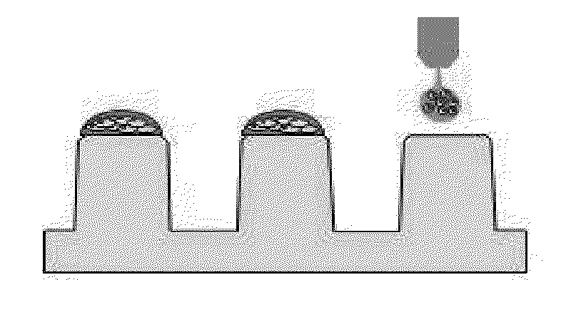
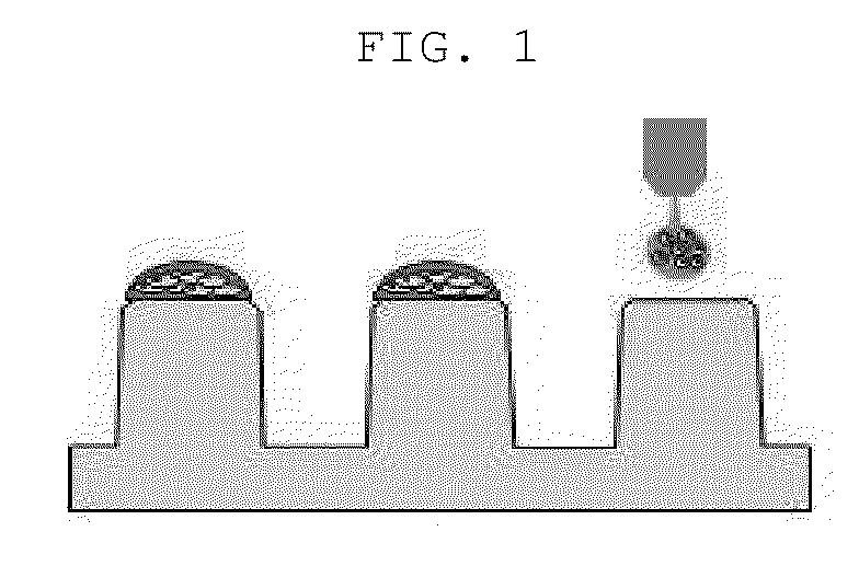
[0064]50 μL of 3% alginate (Sigma-Aldrich, US) and 100 μμL distilled water were mixed to prepare 1% alginate, and then the cell suspensions and 1% alginate as prepared above were mixed together in the same amount. The prepared alginate / cell mixture was discharged onto the surface-modified micropillar using a microarray spotter (Samsung Electro-Mechanics Co., Korea), followed by gelation for 2 minutes so that an alginate gel containing 2, 5, 10, 20, 50 or 100 cells was fixed onto each micropillar (FIG. 1).


PUM

PropertyMeasurementUnit
sizeaaaaaaaaaa
pore sizeaaaaaaaaaa
concentrationaaaaaaaaaa

Abstract

A screening method is described for selecting patient-specific anti-cancer agents reflecting individual genetic properties, in a precise and rapid manner, using an extremely small amount of cancer cells. Such screening method is useful for development of novel anti-cancer agents and the personalized medical field.

Description

CROSS-REFERENCE TO RELATED APPLICATION[0001]This application claims priority under the provisions of 35 USC 119 to Korean Patent Application No. 10-2013-0120118 filed Oct. 8, 2013. The disclosure of Korean Patent Application No. 10-2013-0120118 is hereby incorporated herein by reference, in its entirety, for all purposes.TECHNICAL FIELD[0002]The present invention relates to a screening method for precisely and efficiently selecting patient-specific anti-cancer agents, and more specifically, to a screening method of patient-specific anti-cancer agents using a limiting dilution assay (LDA) characterized by three-dimensional culturing an small amount of patient-derived cancer cells together with anti-cancer agents, automatically analyzing the sphere formed therefrom, and selecting the patient-specific anti-cancer agent.BACKGROUND ART[0003]Cancer is a term commonly known as a disease occurred by indiscreet proliferation of cells due to damage to mechanism of regulating cell proliferatio...

Claims


Application Information

Patent Timeline
Patent Type & Authority Applications(United States)
IPC IPC(8): G01N33/50
CPCG01N33/5011G01N2800/52G01N2500/10A61P35/00C12Q1/68G01N33/48G01N33/53G01N33/574
Inventor NAM, DO HYUNJOO, KYEUNG MINCHOI, YEONSOOK
Owner SAMSUNG LIFE PUBLIC WELFARE FOUND
Why Eureka
Try Eureka