Method for evaluating blush in myocardial tissue

a tissue and myocardial technology, applied in image enhancement, instruments, angiography, etc., can solve the problems of the inability to accurately evaluate the inability to accurately assess the intensity of the myocardium, so as to reduce the variance of intensity between pixels, reduce the risk of myocardial infarction, and improve the effect of myocardial perfusion

Active Publication Date: 2010-12-02
STRYKER EUROPEAN OPERATIONS LIMITED
View PDF1 Cites 32 Cited by
  • Summary
  • Abstract
  • Description
  • Claims
  • Application Information

AI Technical Summary

Benefits of technology

[0010]The present invention is directed to a method for evaluating myocardial blush in tissue from images recorded following injection of fluorescent dyes using a static ROI (Region-of-Interest) that is fixed in position on the image while the heart (or other tissue of interest) moves under it in the image sequence. The static ROI uses a statistical technique to eliminate intensity outliers and to evaluate only those pixels that have less inter-pixel intensity variance. The technique is highly robust, and the results depend only insignificantly on changes to the ROI size and position, providing the ROI is placed in the same general region of the anatomy.

Problems solved by technology

However, substantial evidence has grown over the years showing that distortion of microvasculature and myocardial perfusion is often present despite epicardial artery patency.
The consequences of microvascular damage are extremely serious.
This approach is quite time-consuming and is difficult to perform on a beating heart and to conclude within a reasonable time.
However, this approach is computationally intensive and not reliable with low contrast images.

Method used


Image

  • Method for evaluating blush in myocardial tissue
  • Method for evaluating blush in myocardial tissue
  • Method for evaluating blush in myocardial tissue

Examples


Embodiment Construction

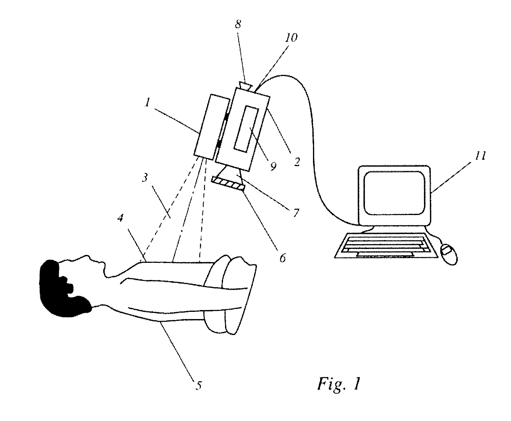
[0033]FIG. 1 shows schematically a device for non-invasively determining blush of myocardial tissue by ICG fluorescence imaging. An infrared light source, for example, one or more diode lasers or LEOs, with a peak emission of about 780-800 nm for exciting fluorescence in ICG is located inside housing 1. The fluorescence signal is detected by a CCD camera 2 having adequate near-IR sensitivity; such cameras are commercially available from several vendors (Hitachi, Hamamatsu, etc.). The CCD camera 2 may have a viewfinder 8, but the image may also be viewed during the operation on an external monitor which may be part of an electronic image processing and evaluation system 11.

[0034]A light beam 3, which may be a divergent or a scanned beam, emerges from the housing 1 to illuminate an area of interest 4, i.e. the area where the blush of myocardial tissue is to be measured. The area of interest may be about 10 cm×10 cm, but may vary based on surgical requirements and the available illumin...


PUM


Abstract

Vessel perfusion and myocardial blush are determined by analyzing fluorescence signals obtained in a static region-of-interest (ROI) in a collection of fluorescence images of myocardial tissue. The blush value is determined from the total intensity of the intensity values of image elements located within the smallest contiguous range of image intensity values containing a predefined fraction of a total measured image intensity of all image elements within the ROI. Vessel (arterial) peak intensity is determined from image elements located within the ROI that have the smallest contiguous range of highest measured image intensity values and contain a predefined fraction of a total measured image intensity of all image elements within the ROI. Cardiac function can be established by comparing the time differential between the time of peak intensity in a blood vessel and that in a region of neighboring myocardial tissue both pre and post procedure.

Description

CROSS-REFERENCE TO RELATED APPLICATIONS[0001]This application is a continuation of prior filed copending PCT International application no. PCT / CA2009 / 000073, filed Jan. 23, 2009, which designated the United States and has been published as International Publication No. WO 2009 / 092162 in the English language, and which claims the benefit of U.S. Provisional Application Ser. No. 61 / 023,818, filed Jan. 25, 2008. This application also claims the benefit of prior filed U.S. provisional Application No. 61 / 243,688, filed Sep. 18, 2009, the entire contents of which are incorporated herein by reference.BACKGROUND OF THE INVENTION[0002]The invention relates to a method for evaluating myocardial blush in tissue from images recorded following injection of fluorescent dyes.[0003]TIMI (Thrombolysis In Myocardial Infarction) studies initially suggested that successful restoration of flow in an infarcted artery was the major goal of reperfusion. However, substantial evidence has grown over the year...

Claims


Application Information

Patent Timeline
Patent Type & Authority Applications(United States)
IPC IPC(8): A61B6/00
CPCA61B5/0059G06T7/90A61B6/504A61B5/0261A61B5/489A61B5/7225A61M5/007A61B6/507G06T7/0012A61B2576/02A61B5/0275G06T2207/10064G06T2207/30104G06T7/12G06T7/248A61B6/503
Inventor DVORSKY, PETERGOYETTE, DAVID MARK HENRI
Owner STRYKER EUROPEAN OPERATIONS LIMITED
Why Eureka
Try Eureka