Detection methods of proteins on polyacrylamide gels using gel background staining and organic dye compositions for the same

a polyacrylamide gel and protein technology, applied in the direction of benz-azabenzanthrone dyes, isotope separation, electrolysis, etc., can solve the problems of long time required for cbbr staining and destaining, low sensitivity, and complicated multi-step processes, and achieve the effect of improving sensitivity

Active Publication Date: 2011-03-03
IND FOUND OF CHONNAM NAT UNIV
View PDF6 Cites 4 Cited by
  • Summary
  • Abstract
  • Description
  • Claims
  • Application Information

AI Technical Summary

Benefits of technology

[0012]In order to solve the above described problems of the prior arts and to provide a novel staining method with improved sensitivity, the present inventors had performed extensive studies. As a result, th...

Problems solved by technology

However, CBBR staining requires a long time (8-12 hours) to perform staining and destaining, and provides a relatively low sensitivity [sensitivity of 50 ng for bovine serum albumin (BSA)].
Although the silver staining method provides such a high sensitivity that the method allows detection of up to 0.1 ng, i.e., femtogram scales, the method requires complicated and multi-step processes.
Although the fluorescence staining method provides a high sensitivity, the method requires complicated process, ultraviolet (UV) irradiation, and precise instruments for quantification, and thus is not cost effective.
Although...

Method used


Image

  • Detection methods of proteins on polyacrylamide gels using gel background staining and organic dye compositions for the same
  • Detection methods of proteins on polyacrylamide gels using gel background staining and organic dye compositions for the same
  • Detection methods of proteins on polyacrylamide gels using gel background staining and organic dye compositions for the same

Examples


example 2

Detection of BSA by Phloxine B Background Staining Method

[0057](1) Preparation of Gels and Electrophoresis

[0058]First, SDS-containing slab gels were prepared according to Laemmli's method. Protein concentrations were measured by Bradford's method using Bio-Rad Standard I for quantification of protein (see, Anal. Biochem. 151, 369-374). Before electrophoresis, protein samples, i.e., 250 ng, 125 ng, 62 ng, 31 ng, 15 ng, 8 ng, 4 ng, 2 ng, 1 ng and 0.5 ng of BSA, were heated in a sample buffer (70 mM Tris-HCl, pH 6.8, 11.4% glycerol, 3% SDS, 0.01% bromophenol blue, and β-mercaptoethanol) to 100° C. for 5 minutes. The developing buffer contained 0.025 M Tris, 0.2 M glycine and 0.1% SDS, and had a pH of 8.3. The gel had a thickness of 1 mm, and stacking gels (4.5%) with a length of 1.5 cm were stacked on separating gels (10%) with the ratio of acrylamide:bisacryamide of 30:0.8.

[0059]Then, 250 ng, 125 ng, 62 ng, 31 ng, 15 ng, 8 ng, 4 ng, 2 ng, 1 ng and 0.5 ng of BSA were loaded onto wells ...

examples 3 and 4

Detection of Protein Standards by Eosin and Phloxine B Background Staining Methods

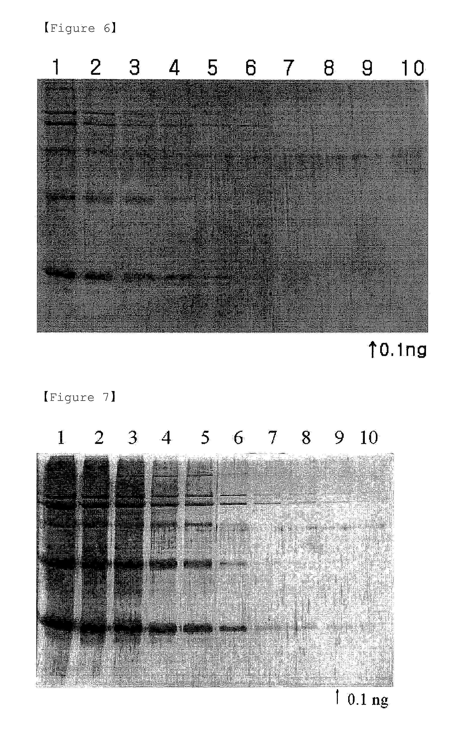
[0064]The substantially same procedures as described in Examples 1 and 2 were performed, except that protein standards containing equal amounts of myosin (205 kDa), β-galactosidase (116 kDa), phosphorylase b (97.4 kDa), BSA (66 kDa), ovalbumin (45 kDa) and carbonic anhydrase (29 kDa) were loaded onto wells, at amounts of 200 ng, 80 ng, 32 ng, 12 ng, 5 ng, 2 ng, 0.8 ng, 0.3 ng, 0.1 ng and 0.04 ng, in order from the left.

[0065]The results are shown in FIGS. 6 and 7. As shown in FIGS. 6 and 7, background staining methods with eosin Y and phloxine B showed similar staining patterns to those of CBBR, zinc-imidazole and Sypro® Ruby staining methods (see, FIGS. 8-10).

[0066]However, the background staining methods with eosin Y and phloxine B allowed detection of the protein standards up to 0.1 ng. Therefore, it can be seen from the above results that the background staining methods using eosin Y and phloxine B...


PUM

PropertyMeasurementUnit
Fractionaaaaaaaaaa
Sensitivityaaaaaaaaaa

Abstract

Disclosed are a method for detecting proteins on a polyacrylamide gel by background staining method using an organic dye composition containing eosin Y or phloxine B, and the organic dye composition for use in the method, which enables the rapid and simple detection of the protein on the polyacrylamide gel with a high sensitivity.

Description

TECHNICAL FIELD [0001]The present invention relates to the detection of proteins on polyacrylamide gels using gel background staining.[0002]More specifically, it relates to a method for detecting proteins on a polyacrylamide gel through gel background staining using an organic dye composition containing eosin Y or phloxine B, and to the organic dye composition for use in the method.BACKGROUND ART [0003]Polyacrylamide gel electrophoresis (PAGE) is protein analysis technique useful for separation, identification, and purity and size determination of proteins. Particularly, in sodium dodecyl sulfate-PAGE (SDS-PAGE), proteins are strongly bound to SDS, an anionic surfactant, in a ratio of 1.4:1, so that the protein molecules have negatively charged surfaces and are separated only based on their sizes. The SDS-PAGE is simple and has a high resolution, and thus is widely used as a method for protein analysis (see, A. T. Andrews, Electrophoresis, pp. 1-58).[0004]Most proteins to be analyze...

Claims


Application Information

Patent Timeline
IPC IPC(8): G01N33/53
CPCY10T436/25375G01N33/6803G01N33/559G01N33/582
Inventor CHOI, JUNG-KAP
Owner IND FOUND OF CHONNAM NAT UNIV
Why Eureka
Try Eureka