
Eureka
For R&D, Eureka makes reading and utilizing patents & technical documents easy.
Eureka AIR
Designed for self-driven R&D workflows. Generate viable solutions, solve complex R&D challenges, empower your innovation with AI.

Eureka Materials
Designed for material experts only. Revolutionize your material R&D, from search, analyze, to developing new materials.

TechResearch
Generate reliable direction feasibility study reports for your R&D in just a few steps.

TechSeek
Discover and master advanced knowledge NOW. Basics, ideas, possibilities, all at once.

TechMind
As an expert in R&D Theories, TechMind can generates customized viable solutions instantly.

TechRisk
Analyze your overall solution with one click, know your potential R&D risks in advance.

TechMonitor
Get weekly tech updates, stay abreast of the latest tech innovations and key insights.
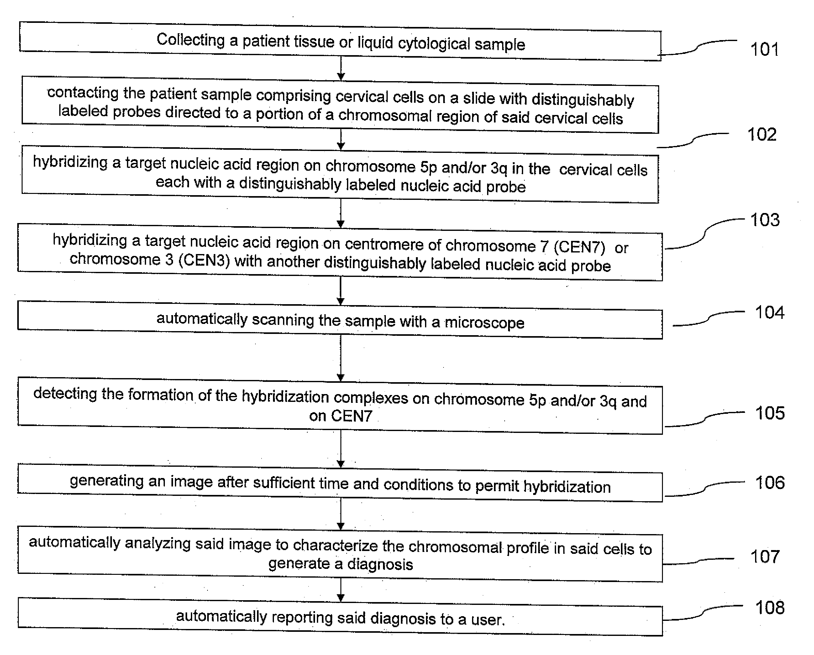
Method and system for automated image analysis in cancer cells
- Summary
- Abstract
- Description
- Claims
- Application Information
AI Technical Summary
Benefits of technology
Problems solved by technology
Method used
Image

Examples
example 1
Methods and Specimen Selection for Automated Analysis and Scoring
A total of twenty-seven (27) specimens were identified within the tested population that had negative results for cytology, HPV and two-color method testing. In addition, these 27 specimens all had 1000 total cells counted in order to minimize variation within the analysis. The triple negative specimens were considered disease negative and, therefore, any abnormal cells identified by FISH would be considered ‘false positives.’ Once the number of ‘false positive’ cells is determined for the 27 specimens, the described BETAINV calculation can be used to determine the threshold between normal and abnormal specimens.
The results of FISH testing for the 27 triple negative specimens were reviewed to identify the total number of ‘false positive’ cells per specimen at each chromosomal loci. A specimen had 5 cells identified by FISH to be abnormal across both loci, 3q26 and 5p15, the greatest number of observed ‘false positive’ ...
example 2
FISH was performed on previously prepared thin layer, liquid-based cytology samples (THINPREP®, Cytyc, Marlborough, Mass.). Slides were made from THINPREP® vials and then subject to a pretreatment protocol that includes protease digestion, formaldehyde fixation, washing, and dehydration. Hybridization was performed using a two-color multi-target interphase FISH probe kit (Abbot Molecular). The kit included directly labeled probes to CEN7-aqua and to the locus of 3q26 (3q-orange) and to the locus of 5p15 (5p-green). The cells were analyzed using fluorescence microscopy.
Samples had a minimum of 800 cells for analysis. Positive tests showed aneuploidy and (1) gains of either 3q copy number or 5p copy number in 1.0% or more of the analyzed cells; (2) gains of only 3q copy number of 0.9% or more of the analyzed cells; or (3) gains of only 5p copy number in 0.7% or more of the analyzed cells. Negative tests showed normal ploidy and (1) less than 1.0% of analyzed cells with an increase in ...
example 3
Evaluation of FIG. 7 this specimen has revealed an abnormal copy number of the TERC gene on 3q26 and the Cri du Chat locus 5p15. Of 1000 cells analyzed, 986 cells were normal while 8 were found abnormal for extra copies of TERC (3q), 2 were found abnormal for extra copies of Cri du Chat (5p), and 4 were found abnormal for extra copies of TERC and Cri du Chat (3q and 5p) for an abnormal cell percentage of 1.40%.
PUM
| Property | Measurement | Unit |
|---|---|---|
| Mass | aaaaa | aaaaa |
| Molar density | aaaaa | aaaaa |
| Molar density | aaaaa | aaaaa |
Abstract
Description
Claims
Application Information
 - R&D Engineer
- R&D Manager
- IP Professional
- Industry Leading Data Capabilities
- Powerful AI technology
- Patent DNA Extraction
Browse by: Latest US Patents, China's latest patents, Technical Efficacy Thesaurus, Application Domain, Technology Topic, Popular Technical Reports.
© 2024 PatSnap. All rights reserved.Legal|Privacy policy|Modern Slavery Act Transparency Statement|Sitemap|About US| Contact US: help@patsnap.com