
Isolation of Anti-Desmoglein 1 Antibodies by Phage Display of Pemphigus Foliaceus Autoantibodies

Active Publication Date: 2011-04-21
THE TRUSTEES OF THE UNIV OF PENNSYLVANIA
View PDF0 Cites 12 Cited by
  • Summary
  • Abstract
  • Description
  • Claims
  • Application Information

AI Technical Summary

Benefits of technology

[0014]The invention also includes an inhibitor of an anti-desmoglein autoantibody or fragment thereof, wherein the inhibitor inhibits the expression of a nucleic acid sequence selected from the group consisting of SEQ ID NOs: 1-43, 87-129, 130-172, and any combination thereof.
[0015]In one embodiment, the inhibitor is selected from the group consisting of a is small interfering RNA (siRNA), a microRNA, an antisense nucleic acid, a ribozyme, a polyamide, a triple-helix-forming agent, a synthetic peptide nucleic acids (PNAs), a

Problems solved by technology

Polyclonal anti-Dsg1 antibodies from PF patients are pathogenic in organ culture of normal human skin and by passive transfer to neonatal mice, which result in blisters with the typical histology of PF from loss of cell-cell adhesion.
Study of PF serum-derived polyclonal antibodies has limitations for addressing many of the remaining questions regarding the pathophysiology of the autoantibodies in this disease.

Method used


Image

  • Isolation of Anti-Desmoglein 1 Antibodies by Phage Display of Pemphigus Foliaceus Autoantibodies
  • Isolation of Anti-Desmoglein 1 Antibodies by Phage Display of Pemphigus Foliaceus Autoantibodies
  • Isolation of Anti-Desmoglein 1 Antibodies by Phage Display of Pemphigus Foliaceus Autoantibodies

Examples


example 1

Immunochemical Properties of the Anti-Dsg 1 scFv are Associated with their Heavy-Chain Gene Usage

[0246]ELISA Analysis

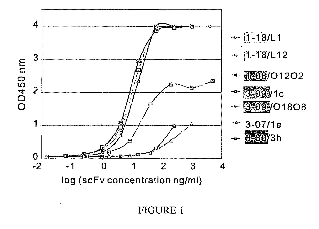
[0247]Representative soluble scFvs were tested for binding to Dsg1 (FIG. 1) and Dsg 3 by ELISA. All scFvs bound Dsg1 as expected, and did not bind Dsg3, except for two 3-07 / 1e clones (differed from each other only in their light chain VJ recombination) that showed weak binding to Dsg3 at very high concentrations (data not shown). Six VH1-18 clones bound strongly by ELISA, giving an OD450=1 values at concentrations from 0.4 to 2.8 ng / ml (2 clones shown in FIG. 1, light green lines). VH3-09 clones also showed good binding; 9 of 12 clones tested gave OD450=1 values at 0.5-20 ng / ml (i.e. dark green lines in FIG. 1). The VH1-08 and VH3-07 clones showed much weaker binding, and the VH3-30 clone showed intermediate binding (FIG. 1).

[0248]Indirect Immunofluorescence on Mouse Tail and Human Skin

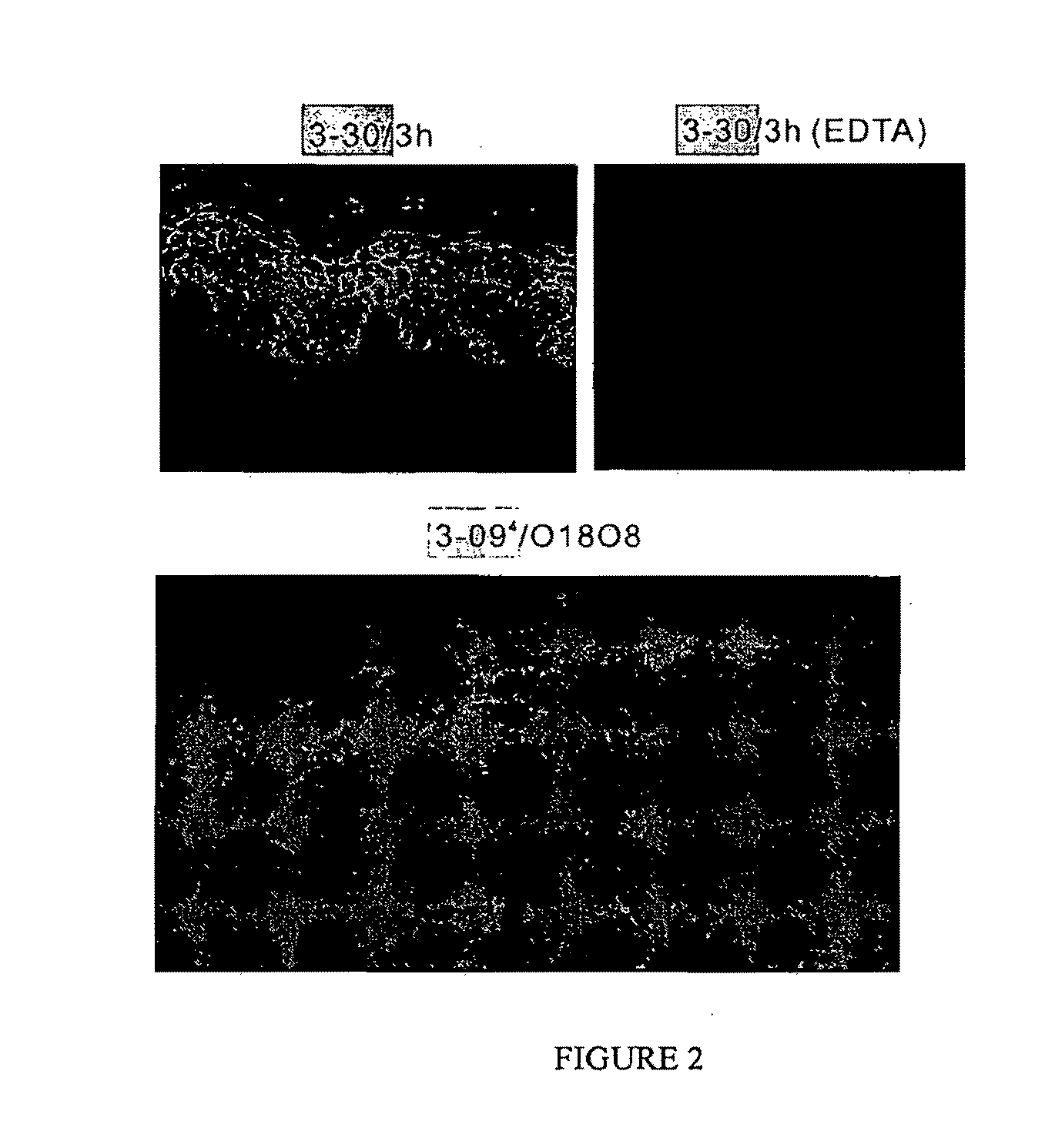
[0249]All VH1-18 clones stained the cell surface of keratinocytes throughout human ...

example 2

Light Chain Suffling Shows that Only Certain Light Chains can Pair with the Restricted Heavy Chains to Allow Dsg1 Binding

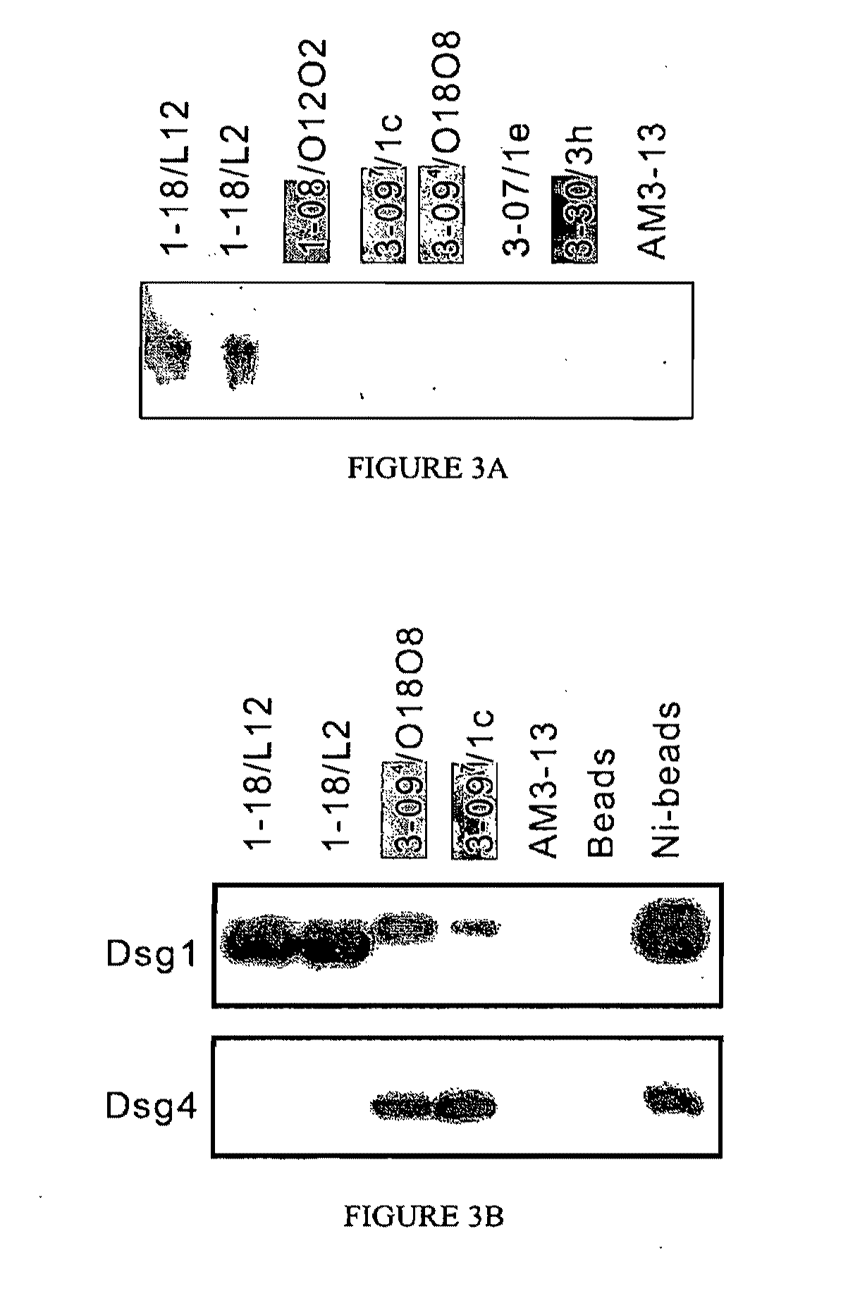
[0257]Although the heavy chain gene usage is generally associated with the immunochemical properties of these scFvs, and the light chain gene usage is much less restricted, light chain shuffling experiments showed that the pairing of light and heavy chains is not entirely random, as only certain light chains allow binding to Dsg1. To demonstrate the light chain contribution to Dsg1 binding, a derivative phage display library was constructed using the heavy chain of scFv 3-098 / L11 paired with the entire original light chain repertoire from the PF patient. Analysis of 16 clones from this derivative library before panning showed that none bound to Dsg1 by ELISA, even though all had the VH3-09 heavy chain. The clones did, however, bind an anti-hemagglutinin (HA) tag-coated ELISA plate, showing that the scFv (which is engineered to express the HA tag on its carboxy-ter...

example 3

Monovalent, Monoclonal Anti-Dsg1 ScFv can Cause the Pathology of PF

[0258]Initially pathogenicity was tested in the neonatal mouse model of pemphigus. Because VH1-18 and VH3-09 clones did not bind well to mouse skin by indirect immunofluorescence, it was believed seemed that theses antibodies would not bind epidermis or induce pathology, which turned out to be the case (Table 1). However, clones 1-08 / O12O2 and 3-07 / 1e did not induce pathology either, even though they did bind mouse epidermis (Table 2, FIG. 5A). Clone 3-30 / 3h, on the other hand, caused extensive gross blisters with the typical histology and direct immunofluorescence of PF (FIG. 5A).

TABLE 2Code table for PFl-scFv clonesCommonOriginal name ofVH geneVDJD segmentJ segmentVL geneclonesname in paperVH1VH1-18VDJ1D3-10 / DXP′1JH4bB3PF1-2-71-18 / B3L8PF1-2-11-18 / L8L1PF1-2-51-18 / L1L12PF1-2-61-18 / L12PF1-3-K111-18 / L2LFVK431PF1-2-151-18 / LFVK431VH1-08VDJ2D3-3 / DXP4JH6bO12 / O2PF1-2-221-08 / O12O2VH3VH3-9VDJ3D2-2JH6bO12 / O2PF1-2-113-093 / O12O2...


PUM

PropertyMeasurementUnit
Fractionaaaaaaaaaa

Abstract

This invention relates to compositions and methods for the use of anti-autoimmune reagents that specifically bind to anti-desmoglein antibodies, which are responsible for pemphigus foliaceus. In addition, the invention relates to methods and compositions for inhibiting the expression or function of a variable region of an anti-desmoglein (anti-Dsg) pathogenic autoantibody.

Description

BACKGROUND OF THE INVENTION[0001]Pemphigus foliaceus (PF) is a tissue-specific autoimmune disease in which antibodies against the keratinocyte cell surface cause skin blisters. These blisters are due to loss of cell adhesion in the superficial living epidermis (i.e. the granular layer) as shown by the diagnostic histology from biopsies of the skin lesions of these patients. The autoantibodies bind desmoglein 1 (Dsg1), a desmosomal cadherin found predominantly in the superficial layers of stratified squamous epithelia. Desmogleins are thought to function as cell-cell adhesion molecules which, in epidermis, maintain its integrity. Polyclonal anti-Dsg1 antibodies from PF patients are pathogenic in organ culture of normal human skin and by passive transfer to neonatal mice, which result in blisters with the typical histology of PF from loss of cell-cell adhesion.[0002]In most PF patients the predominant antibody response is directed against the amino terminal 161 amino acids of the 496 ...

Claims


Application Information

Patent Timeline
IPC IPC(8): A61K39/395C07K16/18C07K16/42C07K2/00C07H21/00C07H21/02A61K38/02A61K31/7088A61K31/713G01N33/566G01N33/53A61K31/7105A61P17/00A61P29/00
CPCA61K2039/505C07K16/28C07K2316/96C07K2319/30C07K2317/565C07K2317/622C07K2317/21A61P17/00A61P29/00
Inventor PAYNE, AIMEE S.STANLEY, JOHN R.SIEGEL, DONALD L.ISHII, KEN
Owner THE TRUSTEES OF THE UNIV OF PENNSYLVANIA
Learn More