Method of motion compensation for trans-catheter aortic valve implantation

a technology of aortic valve and motion compensation, applied in the field of cardiac imaging, can solve the problems of static fluoroscopic overlay techniques, inability to distinguish soft tissue in 2d x-ray projection images, and increased cost compared to image-based localization

Active Publication Date: 2011-07-07
SIEMENS HEALTHCARE GMBH
View PDF3 Cites 39 Cited by
  • Summary
  • Abstract
  • Description
  • Claims
  • Application Information

AI Technical Summary

Benefits of technology

[0006]There is a need for image-based localization for both cardiac and respiratory motion compensation during minimal invasive AVI procedures. There is also a need for such techniques that do not utilize specialized catheters and control systems.

Problems solved by technology

Unfortunately, 2D X-ray projection images generally cannot distinguish soft tissue.
Consequently, the current fluoroscopic overlay techniques are usually static.
However, the proposed method is based on a magnetic tracking system for device localization using specialized catheters and, therefore, is more expensive compared to image-based localization.

Method used


Image

  • Method of motion compensation for trans-catheter aortic valve implantation
  • Method of motion compensation for trans-catheter aortic valve implantation
  • Method of motion compensation for trans-catheter aortic valve implantation

Examples


Embodiment Construction

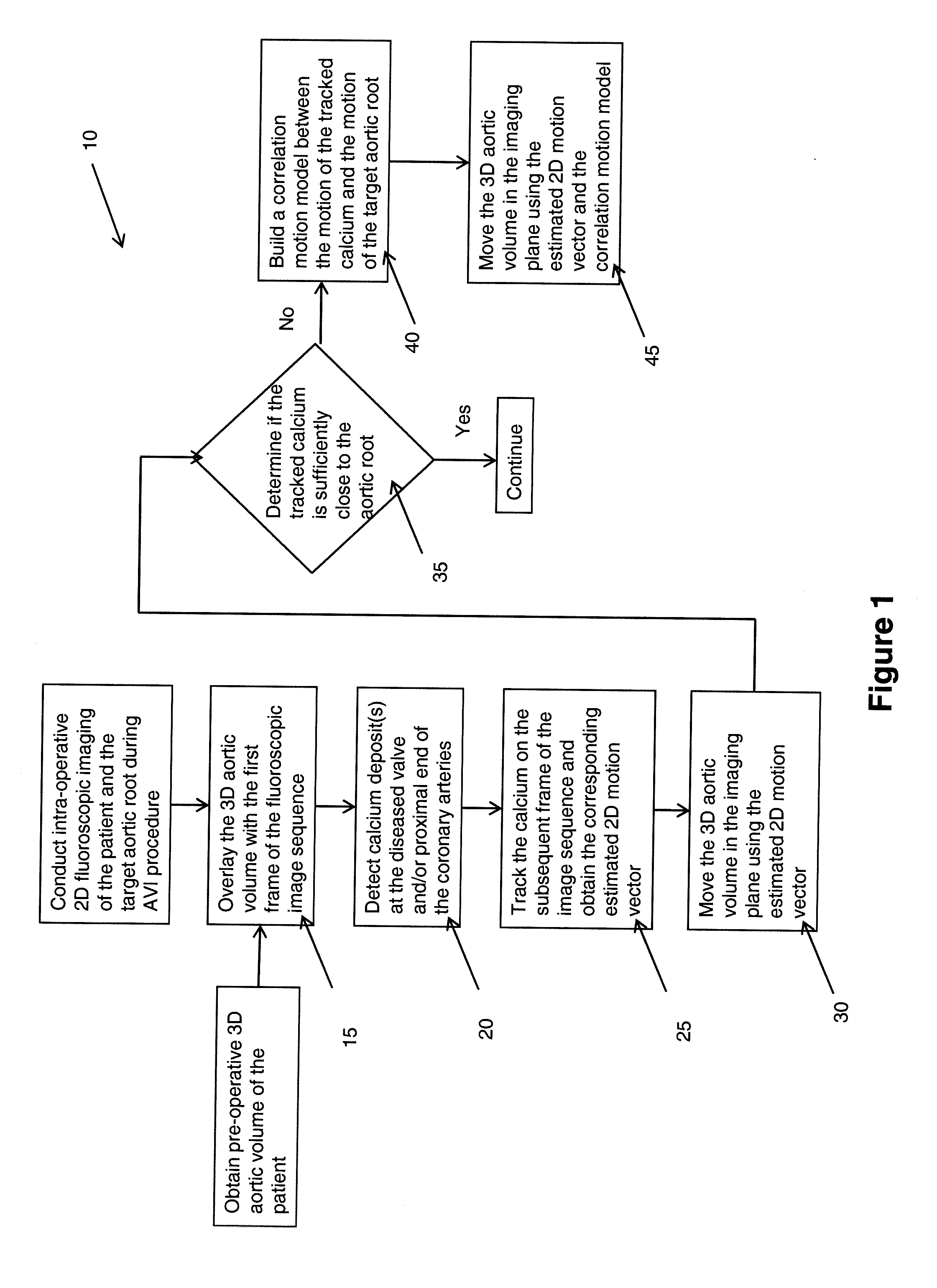
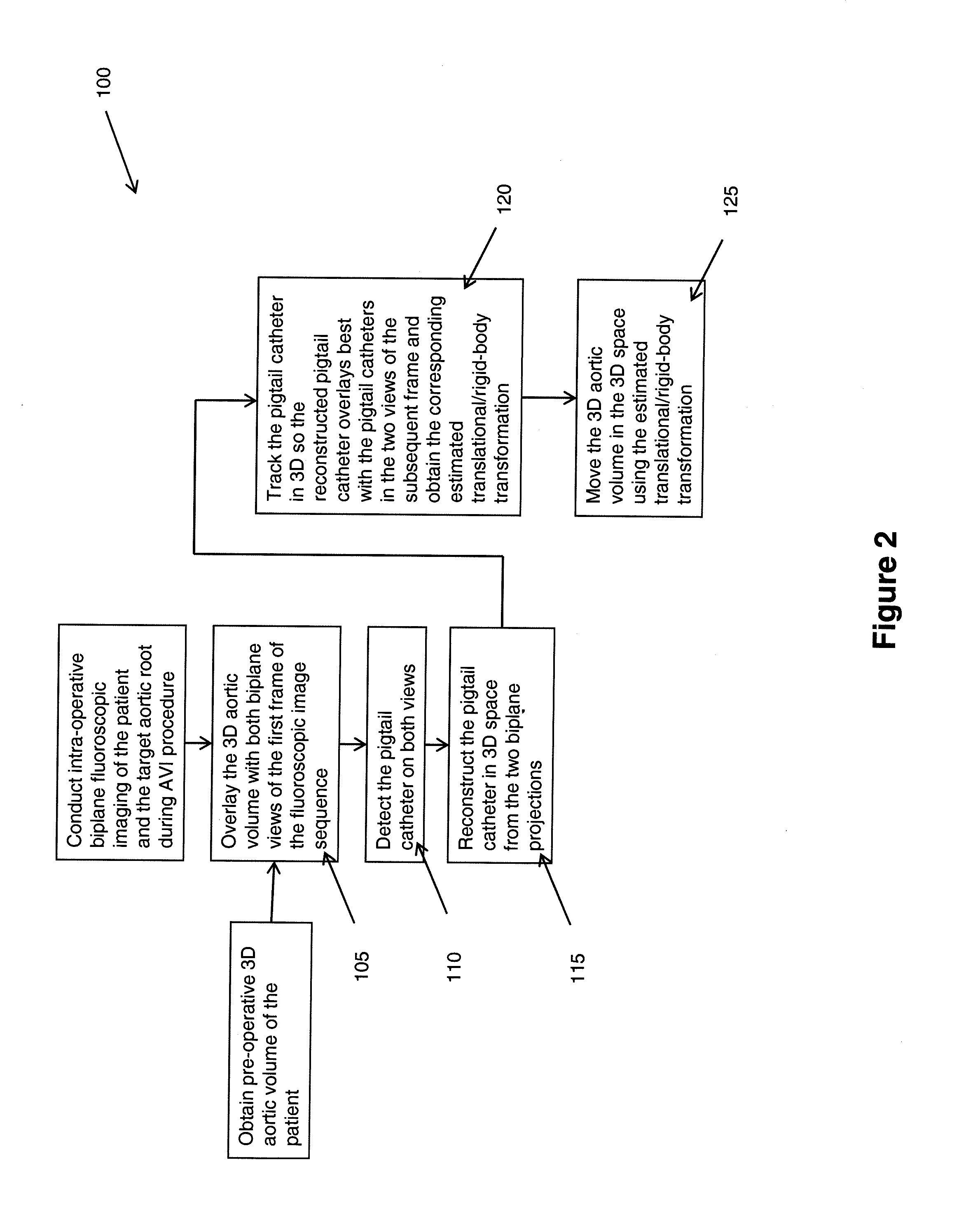
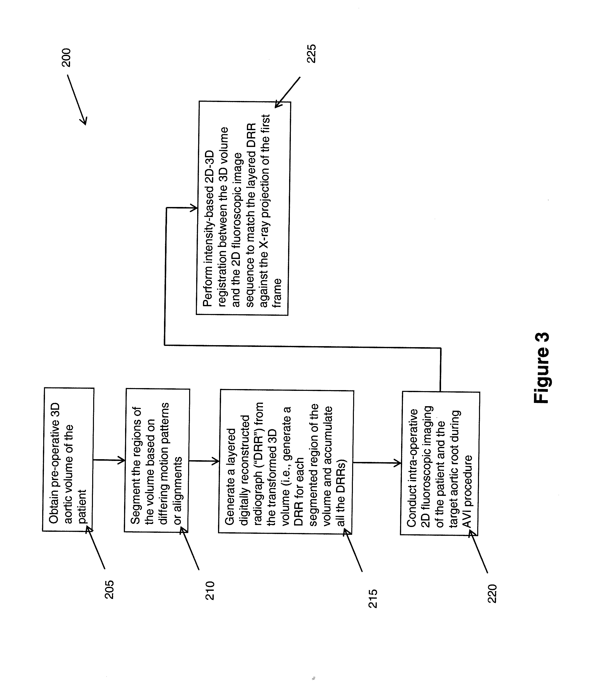
[0021]The present invention provides several methods to compensate for cardiac and respiratory motion for cardiac imaging. The methods are particularly directed to compensate for cardiac and respiratory motion during minimal invasive (e.g., trans-catheter) AVI procedures by image-based tracking on fluoroscopic images. The methods are implemented with the assistance of the imaging system and its visualization and computing capabilities. As noted above, minimal invasive AVI procedures, like other complex non-invasive cardiac interventional procedures, are routinely guided by an X-ray coronary angiography system. Generally, a medical professional inserts an appropriate catheter into a blood vessel of the subject patient and advances the catheter to the target site (i.e., the aortic root, which is the outflow tract from the left ventricle to the aorta) to perform the desired interventional actions. To assist in the visualization for the AVI procedure, the medical professional utilizes t...


PUM


Abstract

A method (10) to compensate for cardiac and respiratory motion in cardiac imaging during minimal invasive (e.g., trans-catheter) AVI procedures by image-based tracking (20, 25) on fluoroscopic images.

Description

CROSS-REFERENCE TO RELATED APPLICATIONS[0001]This application claims the benefit of Provisional U.S. Patent Application Ser. No. 61 / 292,892, entitled, “Motion Compensation for Trans-Catheter Aortic Valve Implantation by Image-Based Tracking on Fluoroscopy”, filed in the name of Rui Liao, Ali Kamen, Matthias John, Alois Noettling, Jan Boese, and Marily Sarmiento, on Jan. 7, 2010, the disclosure of which is also hereby incorporated herein by reference.FIELD OF INVENTION[0002]The present invention relates to cardiac imaging. More particularly, the present invention relates to respiratory motion compensation in certain cardiac imaging.BACKGROUND OF THE INVENTION[0003]Aortic valve disease (such as regurgitation or stenosis) affects over 100 million people globally, with half of them requiring implantation of a mechanical or biological aortic valve to replace the diseased valve. In 2007, there were about seventy to eighty thousand aortic valve implantation (AVI) procedures in the United S...

Claims


Application Information

Patent Timeline
Patent Type & Authority Applications(United States)
IPC IPC(8): G06T15/00
CPCG06T7/0024G06T7/0028G06T7/2033G06T2207/30101G06T2207/10121G06T2207/10124G06T2207/30048G06T2207/10072A61B6/12A61B6/4266A61B6/481A61B6/487A61B6/503A61B6/5264A61B6/541G06T7/246G06T7/30G06T7/33
Inventor LIAO, RUIKAMEN, ALIJOHN, MATTHIASNOTTLING, ALOISBOESE, JANSARMIENTO, MARIA LYDIA
Owner SIEMENS HEALTHCARE GMBH
Why Eureka
Try Eureka