Diagnosis/Therapeutic Strategy For Gynecological Cancer by Utilizing Micro-RNA as Biomarker

a technology of gynecological cancer and expression profile, which is applied in the field of diagnostic/therapeutic strategy for gynecological cancer by utilizing micro-rna as biomarkers, can solve the problems of difficult to differentiate cancer from benign disease, lack of biomarkers in both sensitivity and specificity, and avoid the removal of the uterine lining, etc., to achieve rapid and accurate determination, efficient screening, and rapid and accurate determination

Inactive Publication Date: 2011-07-07
KEIO UNIV
View PDF3 Cites 5 Cited by
  • Summary
  • Abstract
  • Description
  • Claims
  • Application Information

AI Technical Summary

Benefits of technology

[0033]The method for the determination of gynecological cancer and the method for using a microRNA as a biomarker for gynecological cancer according to the present invention enable the gynecological cancer to be rapidly and accurately determined. The kit for the determination of gynecological cancer and the method for using a microRNA for the determination of gynecological cancer according to the present invention also enable the rapid and accurate determination of the presence of the gynecological cancer. In addition, the method for screening therapeutic agents for gynecological cancer enables the therapeutic agents for gynecological cancer to be efficiently screened. Particularly, a microRNA whose concentration in the blood of a subject also changes when a subject suffers from gynecological cancer is excellent especially in terms of speeding-up and simplifying the method for use as a biomarker for gynecological cancer, the method for the determination of gynecological

Problems solved by technology

Thus in the case of gynecological cancer, imaging tests cannot qualitatively diagnose the cancer and in many instances make it difficult to differentiate the cancer from benign disease.
Although several factors for prediction of onset and prognosis of the cancer are reported (see Patent Documents 4, 5 and 6), there currently exists no biomarker sufficient in both sensitivity and specificity.
For cervical cancer and endometrial cancer, it cannot be sometimes avoided that the uterine is removed by surgery; however, this poses serious problems because it causes the loss of fecundity in a young woman who has never borne a child (see Non-Patent Document 5).
Ovarian cancer produces poor subjective symptoms and promptly becomes worse; thus, the cancer has often already made progress when it is detected.
It is also difficult to early detect the cancer because an e

Method used


Image

  • Diagnosis/Therapeutic Strategy For Gynecological Cancer by Utilizing Micro-RNA as Biomarker
  • Diagnosis/Therapeutic Strategy For Gynecological Cancer by Utilizing Micro-RNA as Biomarker
  • Diagnosis/Therapeutic Strategy For Gynecological Cancer by Utilizing Micro-RNA as Biomarker

Examples


example 1

[0077][Collection of Sample Tissue-1]

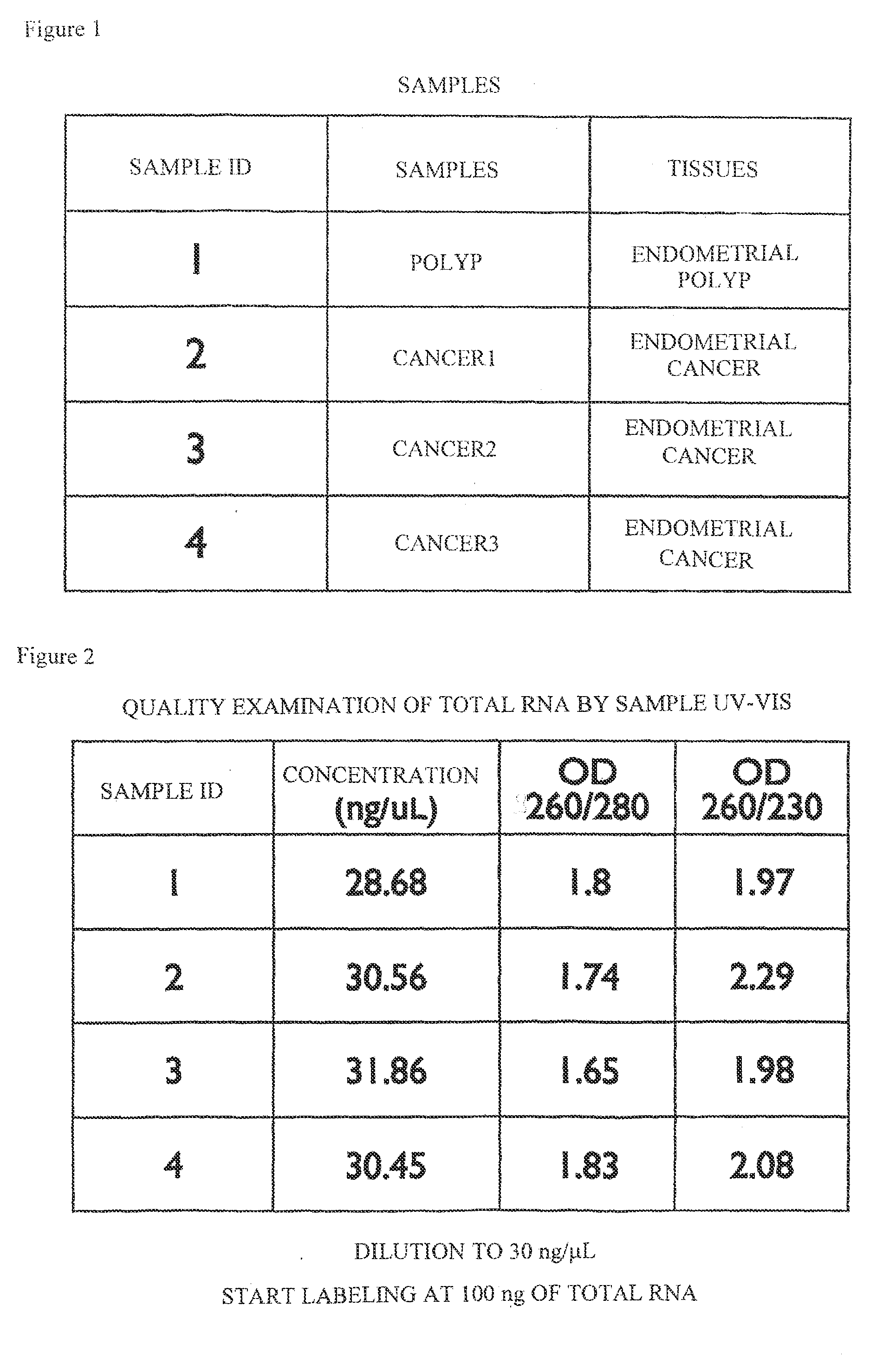
[0078]Under the approval of the ethics committee of Keio University School of Medicine, patients visiting the gynecology clinic of Keio University Hospital were selected and used as persons to be asked for sample donation. Then, after obtaining informed consent from the persons, 4 sample tissues (“SAMPLE IDS 1 to 4” in FIG. 1) were collected from 4 of the persons. The tissue types of these 4 sample tissues (the article “TISSUES” in FIG. 1) consisted of two types: endometrial polyp and endometrial cancer; and the tissue conditions (the article “SAMPLES” in FIG. 1) consisted of 2 conditions: polyp and cancer conditions.

[0079]A portion of each of the collected tissues was placed in a tube into which RNAlater (trade name) (from Applied Biosystems) was dispensed and subjected to frozen storage to stabilize RNA in each tissue. The condition (cancer, polyp, or the like) of each sample tissue was subjected to definite diagnosis by performing the patholog...

example 2

[0080][Extraction of RNA from Sample Tissue and Qualitative Evaluation Thereof-1]

[0081]RNA was extracted from each tissue frozen in Example 1 in order to use in a microarray to be described later. Specifically, total RNA comprising microRNAs was extracted from each of the above tissues using mirVana miRNA Isolation Kit (from Applied Biosystems) according to the appended protocol.

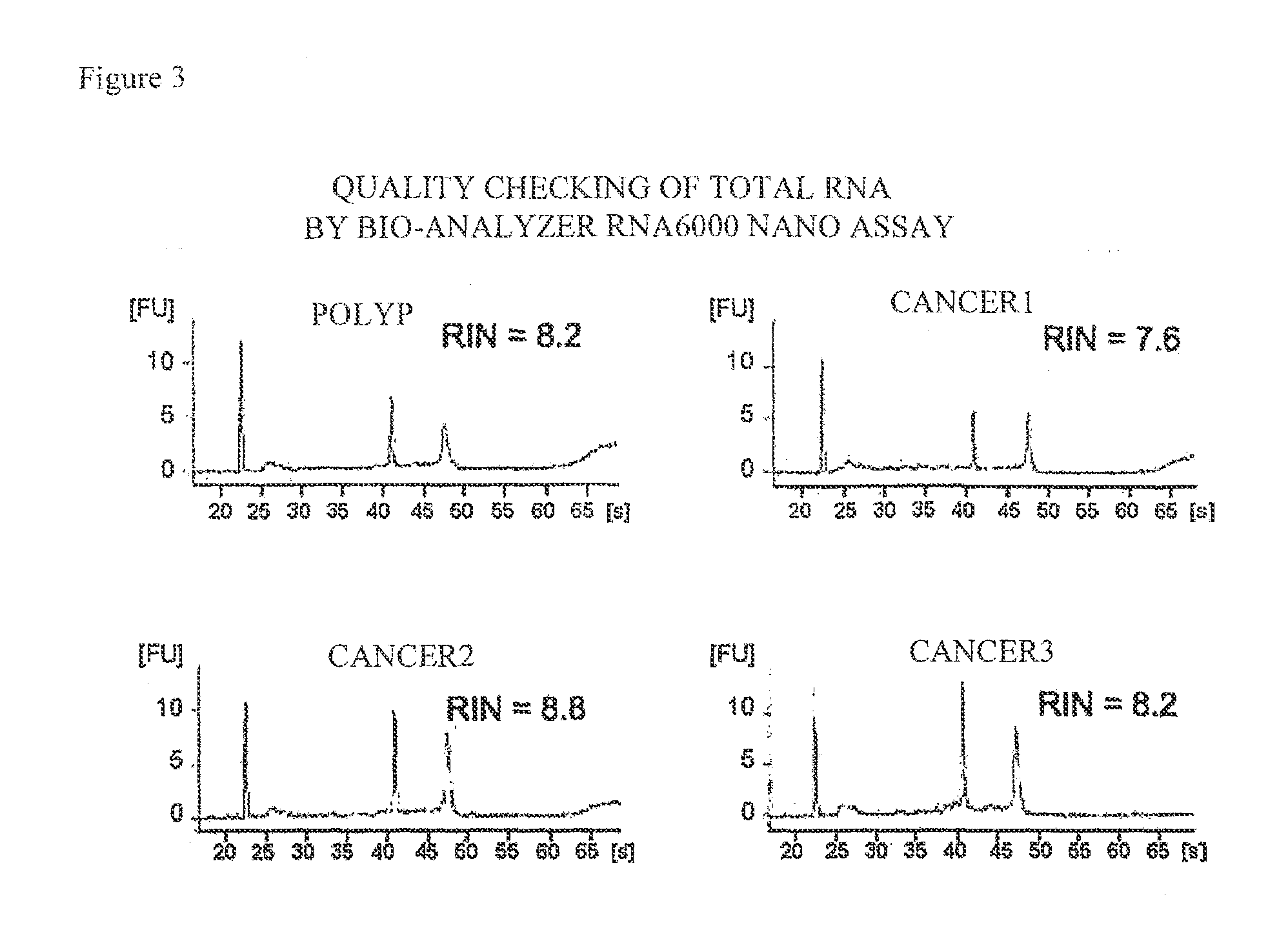
[0082]Then, the extracted RNA was subjected to qualitative evaluation in order to make sure that sufficient accuracy would be obtained in the microarray to be described later. Specifically, the resultant RNA was adjusted to a concentration of about 30 ng / μL using distilled water, and OD260 / 280 (the numerical value obtained by dividing the measured value of OD260 by the measured value of OD280) and the like were measured using a spectrophotometer for calculation. The results are shown in FIG. 2. As shown in FIG. 2, each OD260 / 280 fell in the range of about 1.6 to 2.0, indicating that each RNA was little conta...

example 3

[0083][Expression Analysis of microRNAs in Each Tissue-1]

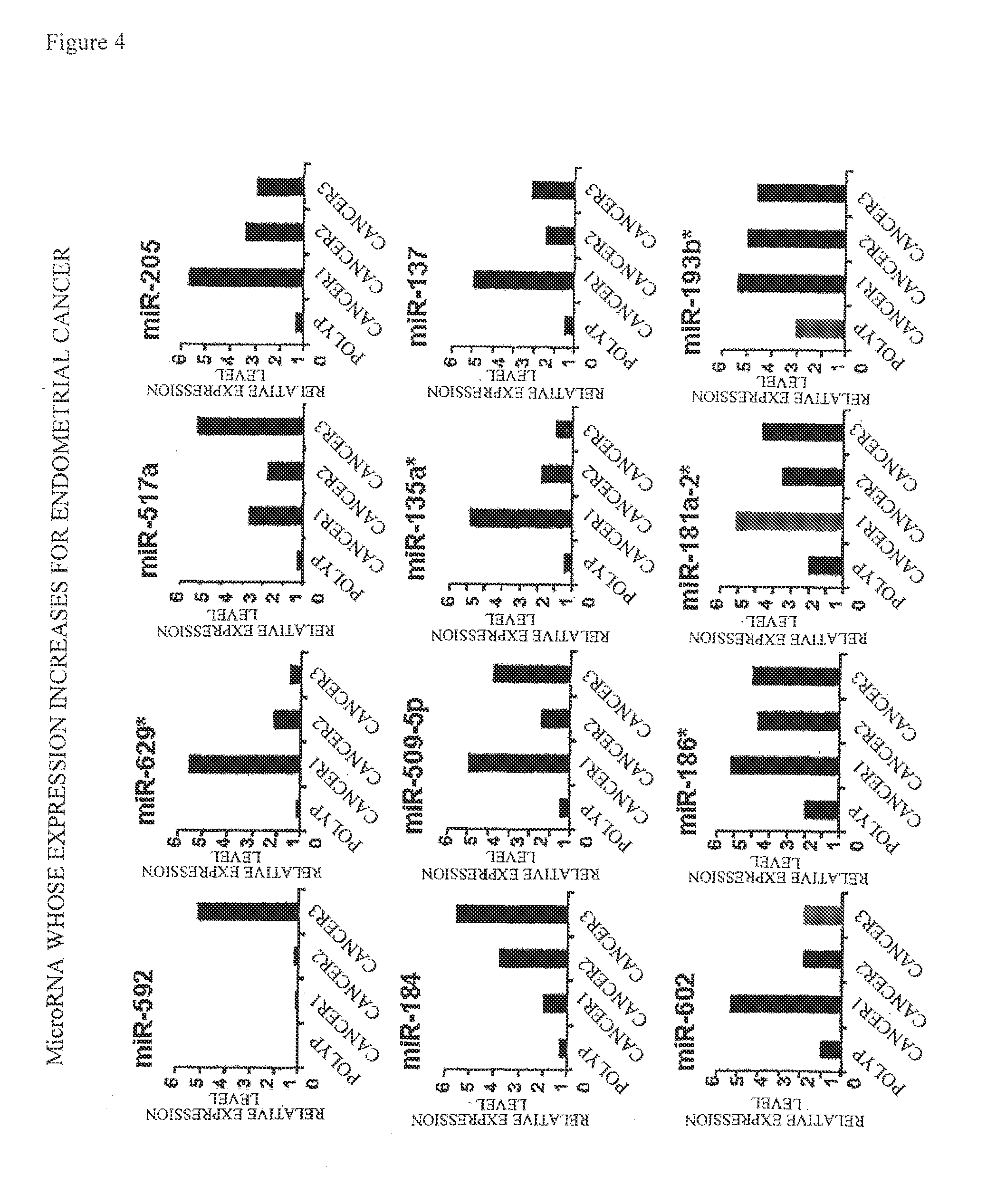
[0084]Using Agilent Human miRNA V2 (from Agilent Technologies), 723 human microRNAs were subjected to exhaustive analysis. The above-described Agilent Human miRNA V2 microarray comprises DNA sequences complementary to nucleotide sequences represented by SEQ ID NOS: 1 to 23 and 25 to 27 as probes for miR-592, miR-629*, miR-517a, miR-205, miR-184, miR-509-5p, miR-135a*, miR-137, miR-602, miR-186*, miR-181a-2*, miR-193b*, miR-377*, miR-449b, miR-449a, miR-369-3p, miR-323-3p, miR-329, miR-299-5p, miR-34b, miR-411, miR-34c-5p, miR-376b, miR-337-3p, miR-337-5p, and miR-127-3p, respectively. The method for analysis was according to the method described in Agilent Technologies' miRNA Microarray Protocol Version 1.5. Specifically, the analysis was performed by the following method. The amount of a reagent is described as an amount for one sample.

[0085]First, the total RNA obtained in Example 2 was diluted to about 25 ng / μL with DNase / R...


PUM


Abstract

Disclosed are: a method for using a particular microRNA as a biomarker for gynecological cancer; a method for the determination of gynecological cancer; a kit for the determination of gynecological cancer, and the like. The present invention is characterized in that at least one microRNA selected from the group of microRNAs consisting of miR-592, miR-629*, miR-517a, miR-205, miR-184, miR-509-5p, miR-135a*, miR-137, miR-602, miR-186*, miR-181a-2*, miR-193b*, miR-377*, miR-449b, miR-449a, miR-369-3p, miR-323-3p, miR-329, miR-299-5p, miR-34b, miR-411, miR-34c-5p, miR-376b, miR-885-5p, miR-337-3p, miR-337-5p, miR-127-3p, miR-138, miR-203, miR-488, miR-489, miR-643, miR-888, miR-424, miR-432, miR-433, miR-450a, miR-503, miR-542-3p, miR-542-5p and miR-584 is used as a biomarker for gynecological cancer.

Description

TECHNICAL FIELD[0001]The present invention relates to an expression profile of a microRNA (miRNA) in gynecological cancer and a use thereof. More specifically, the present invention relates to a method using a specific microRNA as a biomarker for the gynecological cancer, a method for the determination of the gynecological cancer, a kit for the determination of the gynecological cancer, and the like.BACKGROUND ART[0002]Gynecological cancer is a cancer occurring in the uterine cervix, uterine corpus, ovary, uterine tube, vagina, or external genitalia, and accounts for over ¼ the malignant tumor of women. The main gynecological cancers can include cervical cancer, endometrial cancer, and ovarian cancer (see Patent Document 1 and Non-Patent Document 1).[0003]In recent years, basic and clinical studies on cancer have been dramatically advanced and demonstrated that cancer is a disease caused by abnormal genes. As a result, therapies are attempted which use various genes or proteins rela...

Claims


Application Information

Patent Timeline
IPC IPC(8): C40B40/06C12Q1/68
CPCC12N15/113C12N2310/113C12N2310/141C12Q2600/178C12Q1/6886C12Q2600/136C12Q2600/158C12N2330/10
Inventor ZAMA, TAKERUHIRASAWA, AKIRASAITO, KOICHIROAKAHANE, TOMOKO
Owner KEIO UNIV
Why Eureka
Try Eureka