
Medical Device, Method And System For Temporary Occlusion Of An Opening In A Lumen Of A Body

a technology of a medical device and a lumen is applied in the field of medical devices, systems and procedures, which can solve the problems of not being able to perform acute surgery, major bleeding may occur, and the supply vessel is sometimes difficult to reach for the surgeon, so as to achieve the effect of reducing the risk of major bleeding

Inactive Publication Date: 2011-07-14
AEEG
View PDF23 Cites 46 Cited by
  • Summary
  • Abstract
  • Description
  • Claims
  • Application Information

AI Technical Summary

Benefits of technology

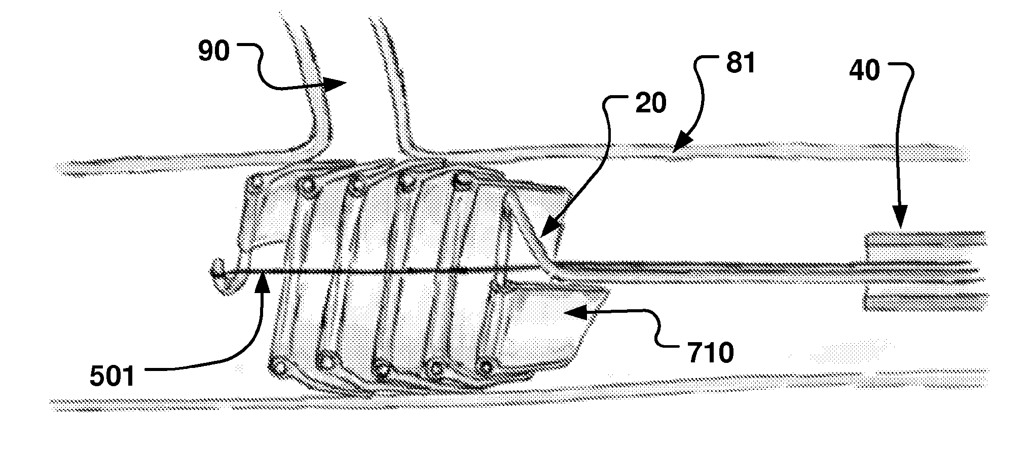
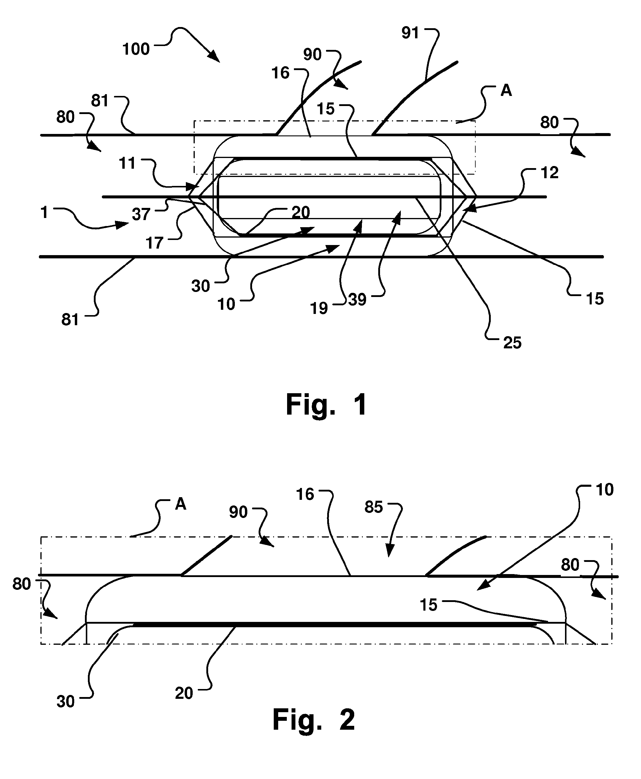
[0023]Fluid communication through the opening is temporary prevented, e.g. a fluid flow from a main lumen of a body through an opening in the main lumen at a site inside the body is prevented.
[0046]In embodiments, the device may also be provided and used as a balloon for aortic valvuloplasty and post stent dilatation of percutaneous aortic valves, eliminating the need for rapid pacing, avoiding its negative consequences.

Problems solved by technology

However, this may not be possible during acute surgery.
Also, supply vessels are sometimes difficult to reach for the surgeon.
Thus, major bleeding may occur.
Moreover, the clamping of vessels leads to vascular trauma, which is undesired.
A background is that a total occlusion of a coronal vessel for instance may cause a heart thump or skip and may lead to angina akin to a heart attack because the vessel is completely blocked while the balloon is inflated.
Another issue is that, the pressure from the blood flow on the proximal part of a standard dilation balloon may cause it to displace in the vessel.
When trying to compensate this, e.g. with higher inflation pressure or larger balloon diameter in relation to the vessel inner diameter, this may cause vascular trauma.
When applied in lumen of the urinal tract, trauma and displacement of occlusion balloons is also an issue.
However, the prior disclosed inflatable balloons having a through channel are not suitable for use in applications for occluding lateral openings in vessels, such as ostia of side branch vessel occlusion.
For instance, these known balloons would not have sufficient stability.
Also, these known balloons may easily harm the surrounding tissue due to high inflation pressures resulting in rock-like inflated balloons.
This results also in a reduced flexibility of the balloons, which is not advantageous when aiming at sealing a side branch lumen fluid tight.
Occasionally, the angioplasty balloon fails to deflate, after the relatively short inflation time, which may cause serious injuries or even death to patients due to the blood flow blockage.
Furthermore, such existing balloons may be difficult to maneuver into some lumen, as e.g. side branch vessels, for instance due to small size thereof, and / or anatomy restrictions as e.g. narrow bifurcations from main vessels.
Therefore, existing inflatable dilation balloons are not intended or suitable for long term occlusion of lumina.
Moreover, existing dilation balloons are not intended or suitable for reliable long-term occlusion of vessels, as e.g. during a surgical procedure.
Besides the aforementioned risk of inducing ischemia distal to the occlusion, total occlusion of major blood vessels has other drawbacks.
For instance, when occluding peripheral vessels, once blood flow is restored, the release of toxic components, which built up during the occluded phase, into the circulation may cause severe side effects, such as vasodilatation that can be clinically difficult to handle sometimes. In addition, embolic material may be released into a blood vessel, in particular when removing the aforementioned clamps or clips.
Embolic particles, which may include thrombus, atheroma and lipids, may become dislodged by surgical or catheter manipulations and enter the bloodstream, embolizing in the brain or other vital organs downstream.
Cerebral embolism can lead to neuropsychological deficits, stroke and even death.
However, the devices disclosed in U.S. Pat. No. 6,183,492 are difficult to position in lumen of high flows, e.g. in vessels close to the heart.
This is partly due to the fact that the flow isolator disclosed in U.S. Pat. No. 6,183,492 is not suited for applications were a large inner diameter is necessary for a high flow through the primary conduit.
Another issue with the inflatable device disclosed in U.S. Pat. No. 6,183,492 is that it released from a catheter and then positioned in a fluid flow to a vessel intersecting position.
In high flow vessels, this would make a reliable sealing of secondary conduits difficult, as the device could not be quickly enough positioned before tumbling and generating turbulent flow in the primary conduit.
Also, inflating the balloon in such an environment would make the aforementioned difficulties worse, increase pressure upstream the device, and influence cardiac output substantially.

Method used


Image

  • Medical Device, Method And System For Temporary Occlusion Of An Opening In A Lumen Of A Body
  • Medical Device, Method And System For Temporary Occlusion Of An Opening In A Lumen Of A Body
  • Medical Device, Method And System For Temporary Occlusion Of An Opening In A Lumen Of A Body

Examples


Embodiment Construction

[0077]Specific embodiments of the invention will now be described with reference to the accompanying drawings. This invention may, however, be embodied in many different forms and should not be construed as limited to the embodiments set forth herein; rather, these embodiments are provided so that this disclosure will be thorough and complete, and will fully convey the scope of the invention to those skilled in the art. The terminology used in the detailed description of the embodiments illustrated in the accompanying drawings is not intended to be limiting of the invention. In the drawings, like numbers refer to like elements.

[0078]The following description focuses on embodiments of the present invention applicable to body lumen in form of blood vessels and in particular to a side branch blood vessel branching from a main blood vessel, or other openings in blood vessels, e.g. ruptures, or openings into aneurysms. However, it will be appreciated that the invention is not limited to ...


PUM


Abstract

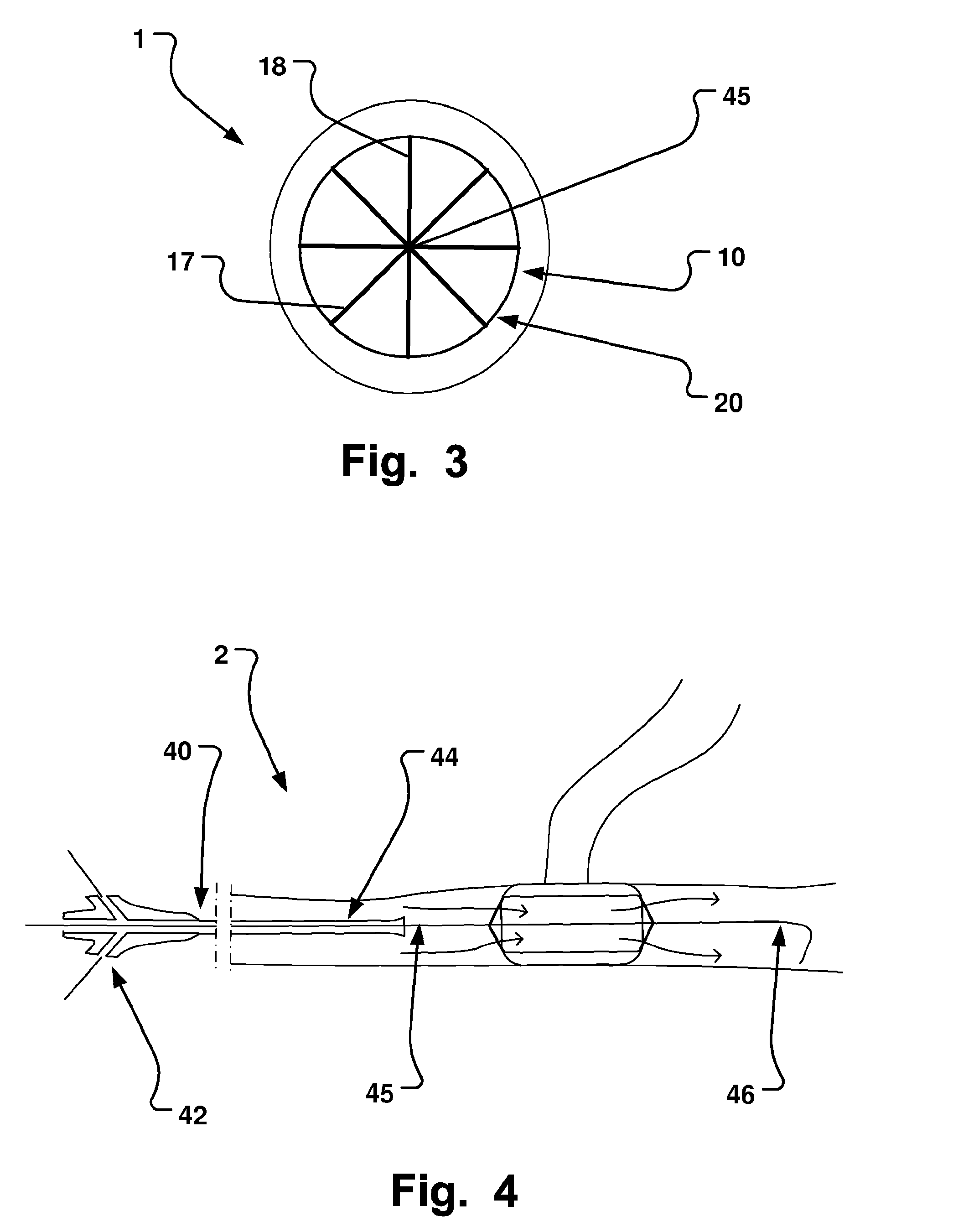
A medical system is disclosed that has three basic components; a retractable sheet, a first balloon that has a centrally arranged hollow, and a collapsible / expandable support structure at the hollow. The first balloon is for instance mounted / molded onto the exterior surface of the support structure. The aggregate of support structure and the first balloon is positioned, and once the sheet has been retracted from the first balloon, the first balloon is inflated. The support structure may be self-expandable or expandable by an expansion unit, such as a further balloon arranged at its inside. The lumen of the support structure is chosen to be smaller than that of a main lumen. The outside diameter of the inflated first balloon is chosen to be larger than the interior diameter of the main lumen. This procedure may be done using standard Seldinger technique and fluoroscopy. This makes the system user friendly and increases patient safety, as a well established clinical method may be used with some modifications according to the invention.

Description

RELATED APPLICATIONS[0001]This application claims priority to International Patent Application No. PCT / EP2009 / 061982, International Filing Date 15 Sep. 2009, entitled Medical Device, Method And System For Temporary Occlusion Of An Opening In A Lumen Of A Body, which claims priority to Swedish Patent Application No. 0850015-9 filed 15 Sep. 2008 entitled Medical Device And System For Temporary Occlusion Of An Opening In A Lumen Of A Body And Method Of Temporary Occluding A Fluid Flow Through Such Opening and to U.S. Provisional Application Ser. No. 61 / 097,315 filed 16 Sep. 2008 entitled Medical Device And System For Temporary Occlusion Of An Opening In A Lumen Of A Body And Method Of Temporary Occluding A Fluid Flow Through Such Opening, all of which are hereby incorporated herein by reference in their entireties.FIELD OF THE INVENTION[0002]This invention pertains in general to the field of medical devices, systems and procedures. More particularly the invention relates to temporary o...

Claims


Application Information

Patent Timeline
Patent Type & Authority Applications(United States)
IPC IPC(8): A61M29/00A61F2/958
CPCA61B17/12036A61M2039/2466A61B17/12045A61B17/12104A61B17/12109A61B17/12118A61B17/12136A61B17/12145A61B17/1215A61B17/12172A61B17/12177A61B2017/00597A61B2017/0061A61B2017/00783A61B2017/00867A61B2017/22054A61F2/013A61M25/1002A61M25/104A61M29/02A61M2025/0024A61M2025/1052A61M2025/1097A61M2039/2406A61M2039/246A61B17/1204
Inventor JONSSON, ANDERS
Owner AEEG
Learn More