
Programmed-release, nanostructured biological construct for stimulating cellular engraftment for tissue regeneration

a technology of cellular engraftment and biological construct, which is applied in the direction of prosthesis, coating, pharmaceutical delivery mechanism, etc., can solve the problems of affecting affecting the cytocompatibility of the implant, so as to promote positive tissue remodeling and organ function, improve the biocompatibility of the biological construct, and optimize the cytocompatibility surface characteristics

Inactive Publication Date: 2011-11-03
SCHAPIRA JAY N +1
View PDF0 Cites 45 Cited by
  • Summary
  • Abstract
  • Description
  • Claims
  • Application Information

AI Technical Summary

Benefits of technology

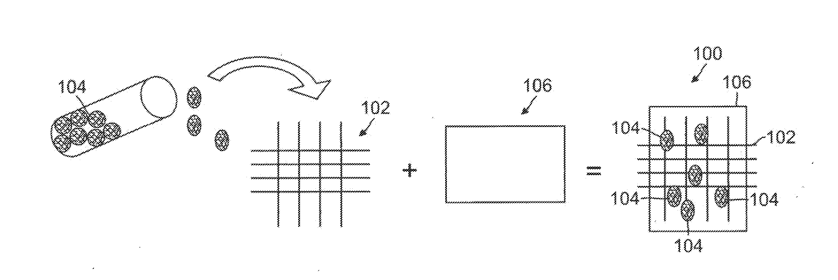
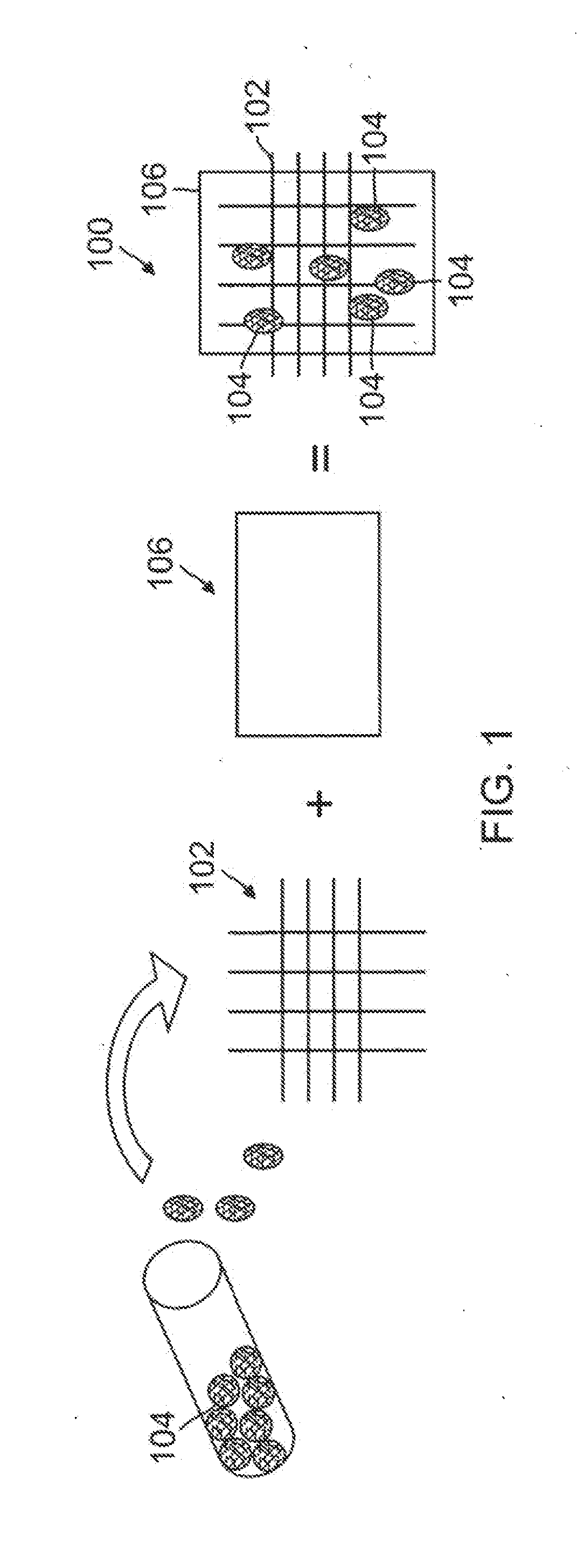
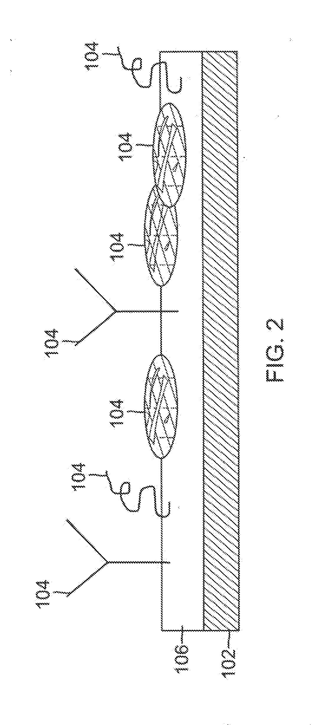
[0015]The unique biological construct for improved, timed-release drug delivery and tissue remodeling following implantation, comprises a layered polymeric biomatrix, either with or without a polymeric bioscaffold having a nanophase surface texture designed to mimic the specific extracellular matrix of a tissue into which the polymer is implanted to improve the biocompatibility of the biological construct; and various therapeutic agents seeded within the polymeric biomatrix to promote positive tissue remodeling and organ function through controlled drug delivery, optimized cyto-compatible surface characteristics, favorable protein adsorption, and improved cellular interaction. The therapeutic agent may be a therapeutic substance such as a drug, chemical compound, biological compound, or a living cell.

Problems solved by technology

The cyto-compatibility of these implants is still imperfect, however.
Implantation is often accompanied by a risk of biological rejection, cellular migration, impaired, undesirable, and excessive tissue healing, clot development on the device surface, or infection.
This problem has limited the application of the currently available implantable biomaterials, drug-delivery technology, and cell therapy strategies.
Implantation upsets the organic systems physiology of the host tissue.
Following device placement, the tissue becomes a hostile environment for cellular function and subsequent tissue regeneration.
Regardless of the organ or tissue type, these injuries inevitably disrupt the fine balance of cellular signaling, differentiation, proliferation, and death.
These chemical signals are crucial for cellular survival, and are greatly disrupted by the introduction of a therapeutic device.
The natural healing process is impeded and can often be further complicated by age and disease state.
In the field of tissue engineering, physicians and scientists have encountered numerous problems with poor osteoblast adhesion and osteointegration following bone implant surgeries.
Similarly, bladder and tissue implants have been problematic, as the un-seeded, or bare polymeric scaffolds used to regenerate “new” tissue, while promising, have demonstrated issues with cyto-compatibility, toxicity, and infection following placement.
This response essentially “indigenizes” the device, but, in 25-30% of situations, smooth muscle cell proliferation becomes excessive, and results in re-stenosis of the vascular device.
These complications invariably extend to any organ system following device implantation, as they are perceived as foreign bodies by the human immune system.
These polymers have shown some success in large arteries, bone, and dental applications, but their surface features are not optimal and, as they degrade, they are known to be thrombogenic in applications such as small diameter vessel grafts.
Considering this, the existing implants designed to improve both biocompatibility and healing demonstrate promise, but they fail to address the critical design issues of the device: 1) the need for surface topography that mimics the native biological environment of the tissue and 2) an implant that is able to recreate the spatial and temporal aspects of the physiological healing process of specific tissues.
Surface features on existing implantable medical devices having micro-scale resolution, and not nano-scale resolution, have proven to be inadequate, and those applications that have attempted nano-topography are generally directed at texturing the non-polymeric portion of the construct, which in many cases, is not exposed.
As a result, the surface topography of the currently available implantable medical devices and / or polymers does not mimic a natural environment, limits organic bio-interaction, and does not create a suitable cellular environment for tissue regeneration.

Method used


Image

  • Programmed-release, nanostructured biological construct for stimulating cellular engraftment for tissue regeneration
  • Programmed-release, nanostructured biological construct for stimulating cellular engraftment for tissue regeneration
  • Programmed-release, nanostructured biological construct for stimulating cellular engraftment for tissue regeneration

Examples


Embodiment Construction

[0023]The detailed description set forth below in connection with the appended drawings is intended as a description of presently-preferred embodiments of the invention and is not intended to represent the only forms in which the present invention may be constructed and / or utilized. The description sets forth the functions and the sequence of steps for constructing and operating the invention in connection with the illustrated embodiments. It is to be understood, however, that the same or equivalent functions and sequences may be accomplished by different embodiments that are also intended to be encompassed within the spirit and scope of the invention.

[0024]The present invention provides a biological construct and method for tissue remodeling and / or drug delivery following medical device implantation by utilizing a cyto-compatible, layered, bio-compatible polymeric biomatrix optimally constructed with a specialized surface texture of grain sizes up to 100 nm seeded with various ther...


PUM

PropertyMeasurementUnit
grain sizesaaaaaaaaaa
grain sizeaaaaaaaaaa

Abstract

A biologically engineered construct comprising of a polymeric biomatrix, designed with a nanophase texture, and a therapeutic agent for the purpose of tissue regeneration and / or controlled delivery of regenerative factors and therapeutic substances after it is implanted into tissues, vessels, or luminal structures within the body. The therapeutic agent may be a therapeutic substance or a biological agent, such as antibodies, ligands, or living cells. The nanophase construct is designed to maximize lumen size, promote tissue remodeling, and ultimately make the implant more biologically compatible. The nano-textured polymeric biomatrix may comprise one or more layers containing therapeutic substances and / or beneficial biological agents for the purpose of controlled, physiological, differential substance / drug delivery into the luminal and abluminal surfaces of the vessel or lumen, and the attraction of target molecules / cells that will regenerate functional tissue. The topographic and biocompatible features of this layered biological construct provides an optimal environment for tissue regeneration along with a programmed-release, drug delivery system to improve physiological tolerance of the implant, and to maximize the cellular survival, migration, and integration within the implanted tissues.

Description

CROSS-REFERENCES TO RELATED APPLICATIONS[0001]This application is a continuation-in-part of U.S. application Ser. No. 12 / 150,329, filed Apr. 25, 2008, which claims the benefit of U.S. Provisional Application Nos. 60 / 926,306, filed Apr. 25, 2007; 60 / 931,749, filed May 25, 2007; 60 / 935,021, filed Jul. 20, 2007; and 60 / 963,290, filed Aug. 3, 2007. This application is also a continuation of U.S. application Ser. No. 12 / 221,139, filed Jul. 31, 2008, which claims the benefit of U.S. Provisional Application Nos. 60 / 935,021, filed Jul.20, 2007; and 60 / 963,290, filed Aug. 3, 2007. Each of the foregoing applications is incorporated herein by this reference.FIELD OF THE INVENTION[0002]The present invention relates to the use of a biologically engineered construct that will be used for tissue regeneration and controlled drug delivery after it is implanted into tissues, vessels, or luminal structures within the body.BACKGROUND OF THE INVENTION[0003]Each year, millions of patients undergo the imp...

Claims


Application Information

Patent Timeline
Patent Type & Authority Applications(United States)
IPC IPC(8): A61K9/00B82Y5/00
CPCA61L27/50A61L27/54A61L2300/252A61L2300/256A61L2300/412A61L2400/18A61L2300/426A61L2300/432A61L2300/602A61L2300/61A61L2400/12A61L2300/414
Inventor SCHAPIRA, JAY N.MAKKAR, RAJ
Owner SCHAPIRA JAY N
Learn More