Cytological method for analyzing a biological sample by raman spectroscopy

a cytological method and biological sample technology, applied in the direction of radiation pyrometry, light diagnosis, instruments, etc., can solve the problems of high cost, high degree of technical knowledge, and high cost of high resolution analysis,

Inactive Publication Date: 2011-12-29
DUBLIN INST OF TECH
View PDF25 Cites 26 Cited by
  • Summary
  • Abstract
  • Description
  • Claims
  • Application Information

AI Technical Summary

Benefits of technology

[0025]In some embodiments, aspects of the invention relate to cytological methods for analyzing a biological sample, the method comprising the steps of: (a) consolidating a biological sample into a mass; (b) obtaining a Raman spectrum for the mass; and (c) comparing the Raman spectrum of the mass with one or more reference spectra, each of the reference spectra corresponding to a known abnormality to determine whether the contents of the mass contain one of the known abnorma...

Problems solved by technology

Raman scattering is a basic property of the interaction of light with molecules; however, Raman scattering is not simply a direct alternative to other techniques, such as InfraRed (IR) spectroscopy.
Exp and Mol Pathol...

Method used


Image

  • Cytological method for analyzing a biological sample by raman spectroscopy
  • Cytological method for analyzing a biological sample by raman spectroscopy
  • Cytological method for analyzing a biological sample by raman spectroscopy

Examples


example 1

Comparison of Low and High Resolution Raman Spectroscopy in Normal and Abnormal Tissue Sections

[0105]Formalin-fixed paraffin preserved (FFPP) tissue samples were obtained from the National Maternity Hospital, Holles St., Dublin. Two parallel 10 μm FFPP sections were cut from each block using a microtome, mounted on glass slides and dried. Samples were dewaxed by immersion in hexane. One section from each sample (the reference section) was stained with hematoxylin and eosin and the other kept unstained for spectroscopic examination. FFPP cervical tissue sections were characterized by a consultant pathologist at the National Maternity Hospital, Holles St., Dublin, and the samples consisted of 20 normal and 20 invasive carcinoma sections from 40 patients. Of the 20 carcinoma samples, 10 samples were identified as having various grades of cervical intraepithelial neoplasia (CIN), which were also marked for examination.

[0106]An Instruments S.A. (now Horiba Jobin Yvon) Labram 1B Raman spe...

example 2

Comparison of Low and High Resolution Raman Spectroscopy in Normal and Abnormal Smears

[0111]Cervical cytology samples were obtained from the National Maternity Hospital, Holles St., Dublin and the Coombe Women and Infants University Hospital, Dublin. Samples were collected by scraping of the cervix using the THINPREP® Pap Test Cervex-Brush protocol. Cervical cells were fixed in PRESERVCYT® solution (Cytyc Corporation, Marlborough, USA).

[0112]The cells were transferred onto a microscopic slide using a CYTOSPIN® centrifuge (Cytospin3, Shandon, USA). The samples were left to air dry and were analyzed unstained. After Raman analysis, the samples were stained with the Papanicolaou stain and coverslipped. The cells from which Raman spectra were acquired were re-visited and assessed by a cytologist.

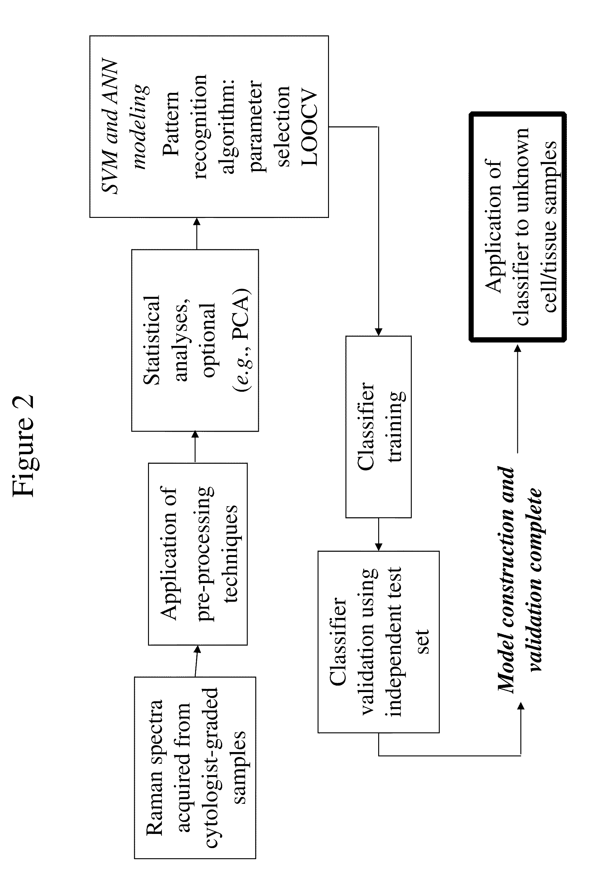
[0113]Raman spectra were acquired and subjected to data analysis, as described in Example 1. For the low resolution spectroscopy of the smear samples, a support vector machine (SVM) model was tr...

example 4

Cell Mass Consolidation

[0115]A mixed population of normal and abnormal cells (held in a liquid preservative) were consolidated into a solid mass by centrifugation at a speed of 1200 RPM for 8 minutes. The supernatant was then removed. The pellet was placed on a slide, and the slide was placed in the system described above in Examples 1 and 2. The pellet was aligned, and a representative spectrum for the pellet (rather than an individual cell) was obtained using low resolution Raman spectroscopy. The representative spectra was then compared with a library of reference spectra of abnormal cells, as described above in Examples 1 and 2. Approximately 30% of the cells were identified as abnormal.


PUM


Abstract

Provided herein are systems and methods that permit low resolution Raman spectroscopy to be used for detection of biological components within cells in order to classify the cells, for example, as premalignant, malignant, or benign.

Description

RELATED APPLICATIONS[0001]This application is a continuation-in-part of International Application Number PCT / EP2009 / 067595, filed Dec. 18, 2009, which claims priority from GB 0823071.6, filed Dec. 18, 2008, both of which are hereby incorporated herein in their entireties.FIELD OF THE INVENTION[0002]Aspects of the invention relate to cytological analyses using low resolution Raman spectroscopy.BACKGROUND[0003]Cancer is a class of diseases in which a group of cells displays uncontrolled growth and is responsible for approximately 13% of all deaths. Significant research has been conducted in the area of diagnosis because early detection of cancer leads to improved survival rates and less drastic treatments.[0004]For cervical cancer, the second deadliest cancer in women, a currently used screening method is a smear test (a Papanicolaou smear, or Pap smear). In a smear test, cell / tissue samples are collected from the outer opening of the cervix using, for example, a spatula or brush. The...

Claims


Application Information

Patent Timeline
IPC IPC(8): G01J3/44
CPCG01N15/1463G01N2035/0091G01N2015/1006G01N21/65A61B5/0059G01J3/44
Inventor LYNG, FIONA M.BYRNE, HUGHO FAOLAIN, EOGHANCLARKE, COLINOSTROWSKA, KAMILA
Owner DUBLIN INST OF TECH
Why Eureka
Try Eureka