Transscleral delivery

a transscleral and ocular technology, applied in the direction of biocide, drug composition, extracellular fluid disorder, etc., can solve the problems of fibrovascular scarring of the macular region, no treatment, and current treatment that is not optimal,

Inactive Publication Date: 2012-02-09
SANTEN PHARMA CO LTD
View PDF5 Cites 8 Cited by
  • Summary
  • Abstract
  • Description
  • Claims
  • Application Information

AI Technical Summary

Benefits of technology

[0122]Generally the therapeutic agent and compositions containing the therapeutic agent may be delivered using any delivery system capable of transscleral delivery of a therapeutically effective amount of the therapeutic agent. Delivery systems and routes of administration that may be used include but are not limited to delivery by injection, solid polymer implant, backed solid polymer implant, solid bioadhesive implant; solid implant with anchoring surface, coated suture, coiled fiber, and solid therapeutic agent.

Problems solved by technology

There are a variety of retinal disorders for which there is currently no treatment or for which the current treatment is not optimal.
Retinal disorders such as uveitis (an inflammation of the uveal tract: iris, ciliary body, and choroid), macular degeneration, macular edema, proliferative diabetic retinopathy, and retinal detachment generally are all retinal disorders that are difficult to treat with conventional therapies.
Organization of serous or hemorrhagic exudates escaping from these vessels results in fibrovascular scarring of the macular region with attendant degeneration of the neuroretina, detachment and tears of the retinal pigment epithelium, vitreous hemorrhage and permanent loss of central vision.
Currently there is no optimal treatment for wet AMD.
Unfortunately, AMD subjects with subfoveal lesions subjected to laser treatment experienced a rather precipitous reduction in visual acuity (mean 3 lines) at 3 months follow-up.
Another drawback of the procedure is that vision after surgery is immediately worse.
Unfortunately, even with successful conventional laser photocoagulation, the neovascularization recurs in about 50-70% of eyes (50% over 3 years and >60% at 5 years).
In addition, many subjects who develop CNV are not good candidates for laser therapy because the CNV is too large for laser treatment, or the location cannot be determined so that the physician cannot accurately aim the laser.
Photodynamic therapy, although utilized in up to 50% of new cases of subfoveal CNV has only marginal benefits over natural history, and generally delays progression of visual loss rather than improving vision which is already decreased secondary to the subfoveal lesion.
PDT is neither preventive or definitive.
Several PDT treatments are usually required per subject and additionally, certain subtypes of CNV fare less well than others.
Although there is currently some off label use of intravitreal triamcinolone acetate, there are no other widely accepted therapies for subfoveal CNV.
Uveitis is another retinal disorder that has proven difficult to treat using existing therapies.
Ocular complications of uveitis may produce profound and irreversible loss of vision, especially when unrecognized or treated improperly.
Visual loss may progress over a period of months, and can be very annoying because of the inability to focus clearly.
ME is a common cause of severe visual impairment.
There are currently no approved devices to deliver therapeutic agents to the posterior segment of the eye from a location external to the eye.
Additionally, therapeutic agents are likely to have undesirable side effects when delivered systemically to treat posterior segment disease.
Interior placement, however, will result in a composition or device being present in the interior of the eye, which may have adverse effects on the proper functioning of the eye.
Although transscleral delivery of therapeutic agents to the eye is advantageous, there are many difficulties in developing such delivery mechanisms.

Method used


Image

  • Transscleral delivery
  • Transscleral delivery
  • Transscleral delivery

Examples


example 1

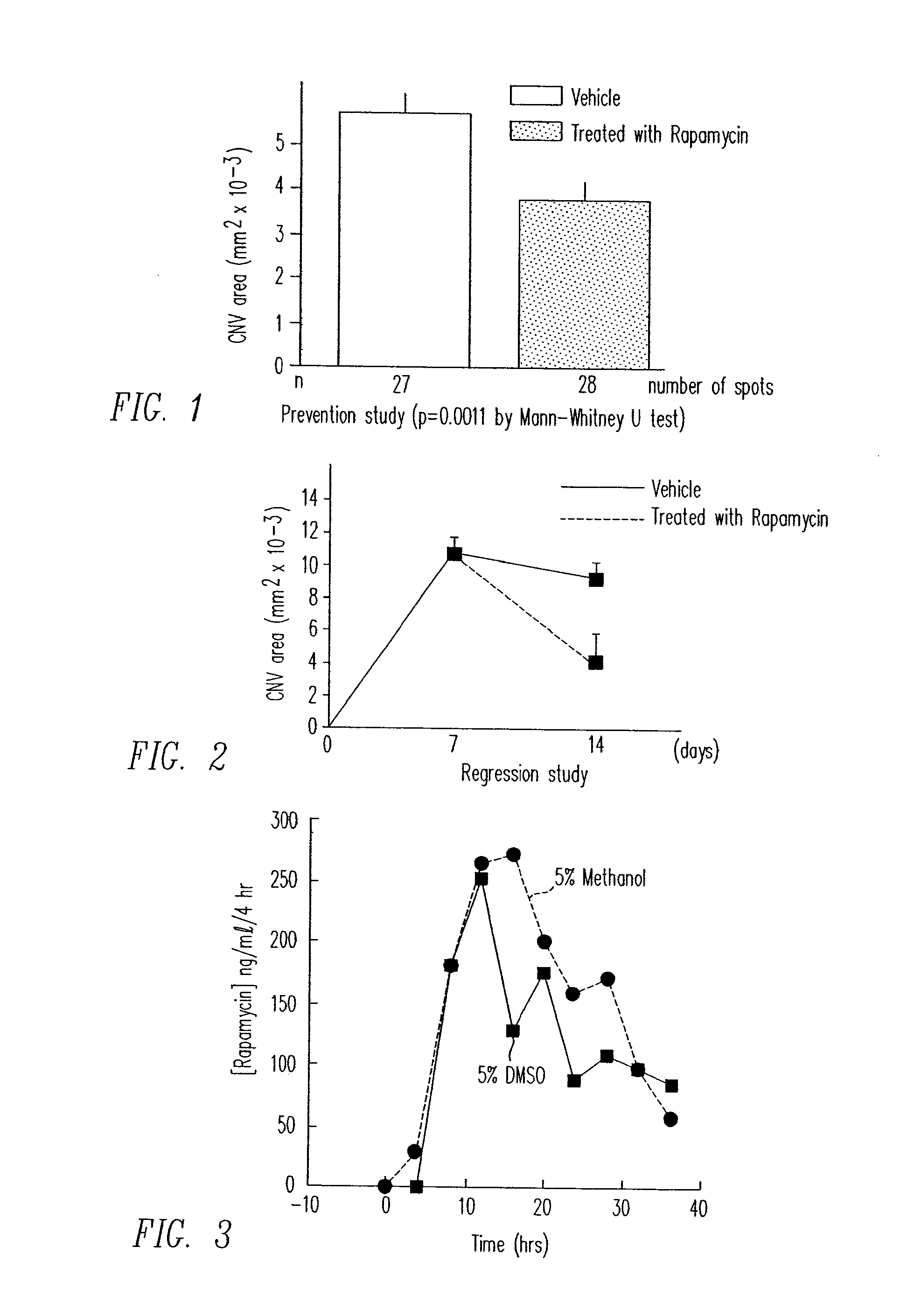
Inhibition of Laser Induced CNV by Rapamycin

[0196]CNV may be induced by rupturing Bruch's membrane with laser. Fifteen mice were given daily periocular injections of 5 micrograms of rapamycin in corn oil vehicle in one eye. After two days of treatment, Bruch's membrane was ruptured by laser at three sites in each eye. The fellow eye served as control and was treated with periocular injections of corn oil only. Two weeks after laser rupture, 10 mice were perfused with fluorescein-labeled dextran and CNV areas were measured in each eye on choroidal flat mounts. The remaining mice had retinas and RPE / choroid dissected and stored at −70° C. for tissue therapeutic agents level measurement.

[0197]As depicted in FIG. 1, rapamycin treatment led to a statistically significant reduction in CNV size versus vehicle alone (p=0.0011 by Mann-Whitney U test). The mean CNV area for rapamycin treated eyes was 0.00381 mm2 with a standard deviation of 0.00197 and the vehicle eyes showed a CNV area of 0....

example 2

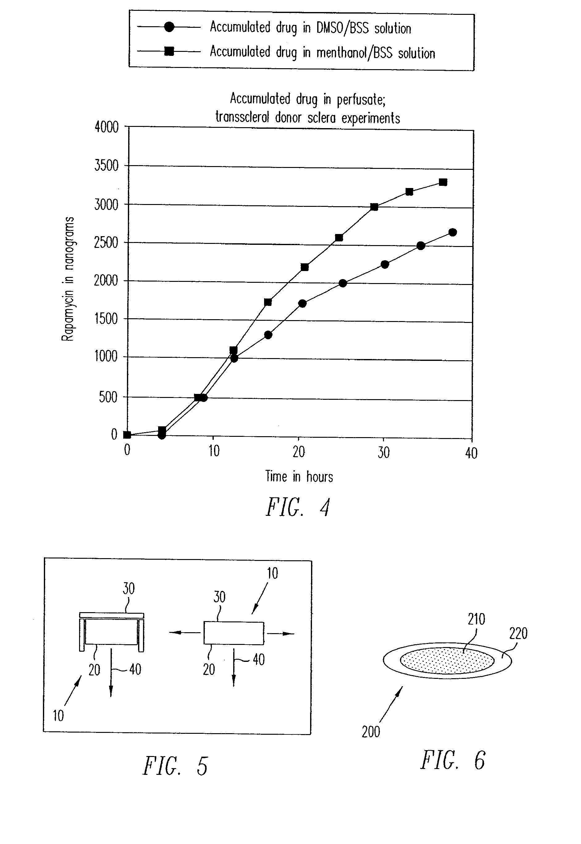
Reversal of Laser Induced CNV by Rapamycin

[0200]Fifteen mice had Bruch's membrane ruptured with laser photocoagulation at 3 locations in each eye. After one week, 5 mice were perfused with fluorescein-labeled dextran and the baseline area of CNV was measured in each eye. At that point, the remaining mice were started on daily periocular injections of 5 μl of corn oil containing 5 μg of rapamycin in one eye and corn oil alone in the fellow eye. After one more week, the mice were perfused with fluorescein-labeled dextran and CNV areas were measured in each eye on choroidal flat mounts. The corn oil solution used is approximately a saturated solution. It is expected that lower doses will be used to deliver therapeutically effective amounts of rapamycin.

[0201]As depicted in FIG. 2, rapamycin treated eyes showed a substantial reduction in CNV area. Baseline lesion CNV area was 0.0105 mm2 with a standard deviation of 0.0037. Untreated eyes had a CNV area of 0.0093 mm2 with a standard devi...

example 3

In-vitro Determination of Flux and Scleral Permeability for Transscleral Rapamycin Delivery

[0203]A two chamber Ussing type permeability apparatus was used to demonstrate delivery of rapamycin across a human sclera. Prior to testing of sclera, loss of therapeutic agent to the glass walls of the experimental apparatus was evaluated. For the evaluation of loss of drug to the experimental apparatus, the uveal and orbital chambers were exposed to a 2 μg / mL rapamycin solution in balanced salt solution. There was small but significant loss of rapamycin to the apparatus when it was exposed to the 2 μg / mL solution of rapamycin in balanced salt solution.

[0204]Transscleral Rapamycin Delivery—DMSO Solution

[0205]A 7 mm disk of fresh donor human sclera was used to separate the two chambers of the Ussing permeability apparatus. The two sides of the sclera were denoted “orbital” to represent the outer surface and “uveal” to represent the internal surface. DMSO and methanol dissolves rapamycin readi...


PUM

PropertyMeasurementUnit
diameteraaaaaaaaaa
diameteraaaaaaaaaa
diameteraaaaaaaaaa

Abstract

Diseases associated with the tissues in the posterior segment of the eye can be effectively treated by administering therapeutic agents transsclerally to those tissues. Compositions, devices, and methods for delivering therapeutic agents so that they cross the sclera and reach these tissues include injecting solutions or suspensions adjacent to or within the sclera and implanting solid structures containing the therapeutic agent adjacent to or within the sclera. These methods may be used for administering rapamycin or related compounds to treat choroidal neovascularization associated with age-related macular degeneration.

Description

FIELD[0001]Described herein are methods, compositions, and devices for the treatment of ocular diseases by the transscleral delivery of therapeutic agents, particularly the treatment of wet AMD by transscleral delivery of rapamycin.BACKGROUND[0002]The retina of the eye contains the cones and rods that detect light. In the center of the retina is the macula lutea, which is about ⅓ to ½ cm in diameter. The macula provides detailed vision, particularly in the center (the fovea), because the cones are higher in density. Blood vessels, ganglion cells, inner nuclear layer and cells, and the plexiform layers are all displaced to one side (rather than resting above the cones), thereby allowing light a more direct path to the cones.[0003]Under the retina are the choroid, comprising a collection of blood vessels embedded within a fibrous tissue, and the deeply pigmented epithelium, which overlays the choroid layer. The choroidal blood vessels provide nutrition to the retina (particularly its ...

Claims


Application Information

Patent Timeline
Patent Type & Authority Applications(United States)
IPC IPC(8): A61K9/14A61P27/02A61K31/439A61K9/00A61K31/436
CPCA61K9/0019A61K31/436A61K9/0048A61P27/02A61P7/10A61P9/10A61K9/10A61K31/4353
Inventor COOPER, EUGENE R.KLEINMAN, DAVID M.NIVAGGIOLI, THIERRYDOR, PHILIPPE JMMUDUMBA, SREENIVASU
Owner SANTEN PHARMA CO LTD
Why Eureka
Try Eureka