
Subconjunctival conformer device and uses thereof

a conformer device and subconjunctival technology, applied in intravenous devices, eye surgery, eye treatment, etc., can solve the imbalance between intraocular fluid production and outflow, blindness worldwide, and irreversible vision loss, so as to enhance and/or direct intraocular fluid flow, minimize scarring, fibrosis, inflammation, etc., to achieve the effect of enhancing and/or directing intraocular fluid flow, minimizing inflammation and scarring

Active Publication Date: 2012-04-12
CUNNINGHAM JR EMMETT T
View PDF6 Cites 62 Cited by
  • Summary
  • Abstract
  • Description
  • Claims
  • Application Information

AI Technical Summary

Benefits of technology

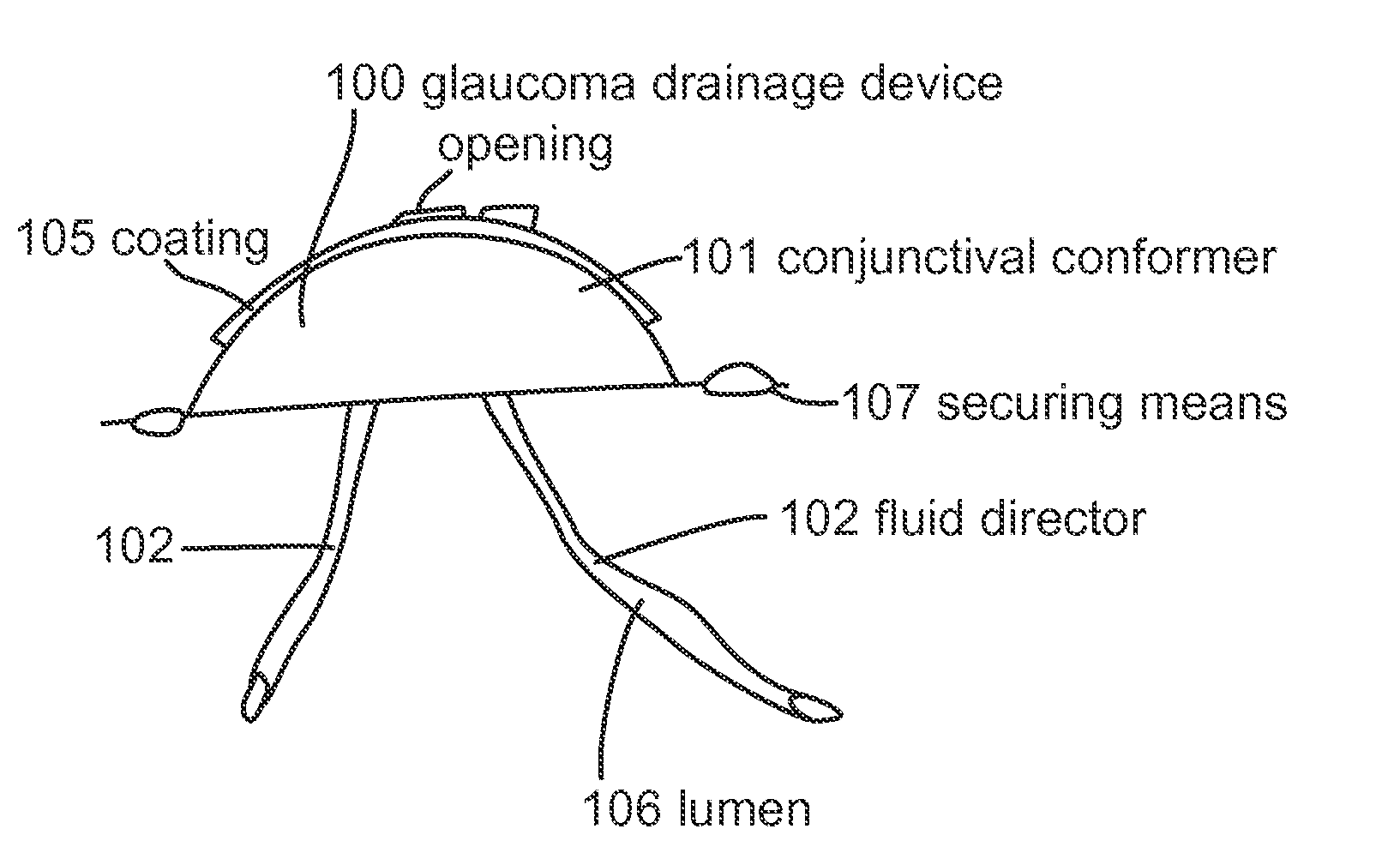
[0011]In some embodiments, the subject device further comprises one or more drug-eluting coatings. The coating can minimize inflammation, scarring, fibrosis, and / or infection. In some embodiments, the drug is an anti-inflammatory agent, anti-proliferative agent, anti-scarring agent, anti-microbial agent, antiseptics, intraocular pressure lowing agent, or vitreolytic agent. In some embodiments, the anti-inflammatory agent is selected from the group consisting of corticosteroids, immunosuppressive agents, non-steroidal anti-inflammatory drugs, anti-inflammatory proteins, peptides, or nucleic acids, and combinations thereof In some embodiments, the anti-proliferative agent is selected from the group consisting of a corticosteroid, an immunosuppressive agent, mitomycin, 5-fluorouracil, heparin, anti-proliferative proteins, peptides, or nucleic acids and combinations thereof. In some embodiments, the anti-scarring agent is mitomycin or 5-flurouracil. In some embodiments, the anti-microbial agent is selected from the group consisting of antibiotics, antivirals, antifungals, and antiparasites. In some embodiments, the intraocular pressure lowing agent is selected from the group consisting of an adrenergic agonist, cholinergic agonist, beta-blocker, carbonic anhydrase inhibitor, prostaglandin analogue, and alpha-adrenergic agonist. In some embodiments, the vitreolytic agent is selected from the group consisting of a dispase, urea, collagenase, hyaluronidase, plasmin, microplasmin, chondroitinase, a protease, and combinations thereof.
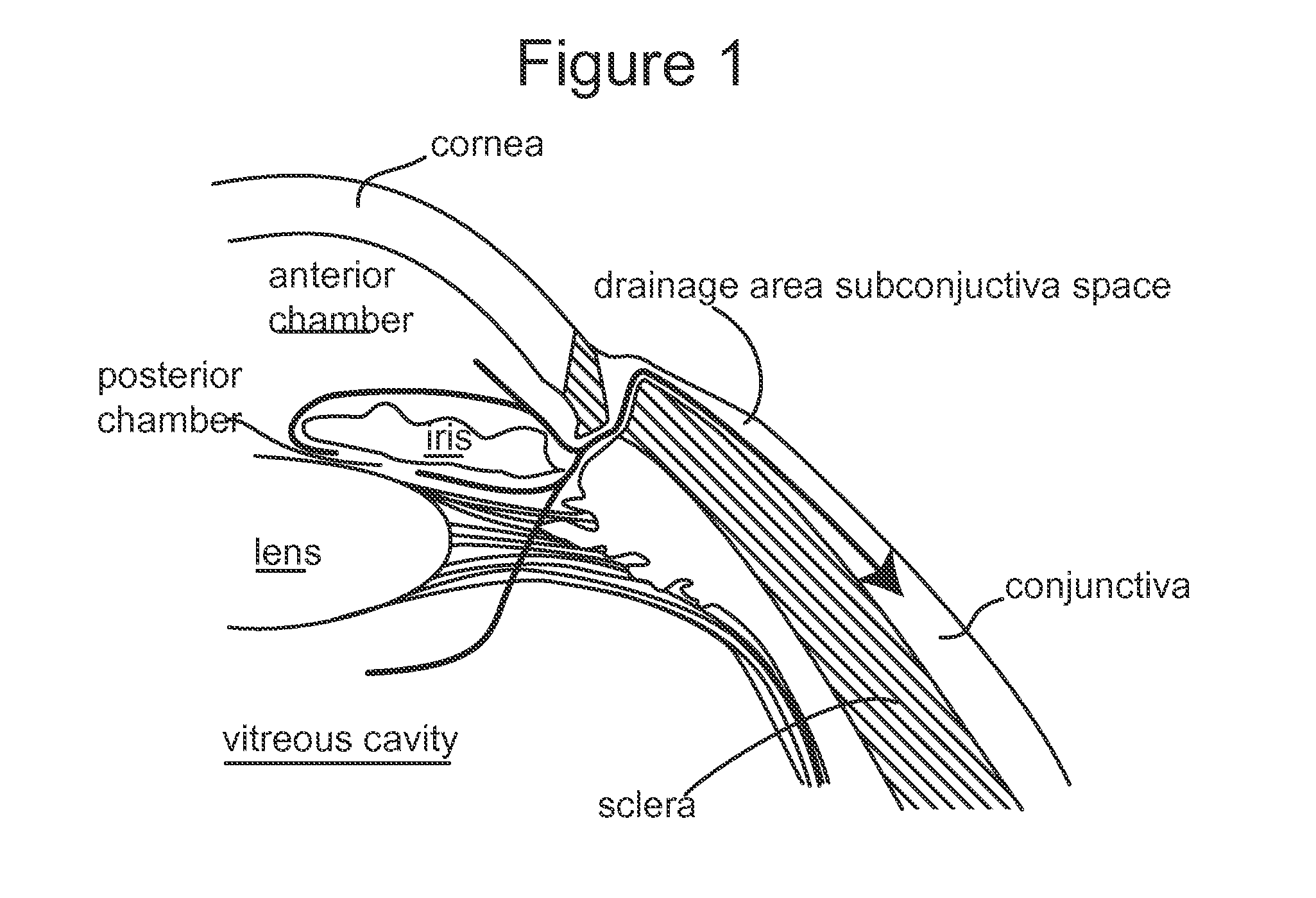
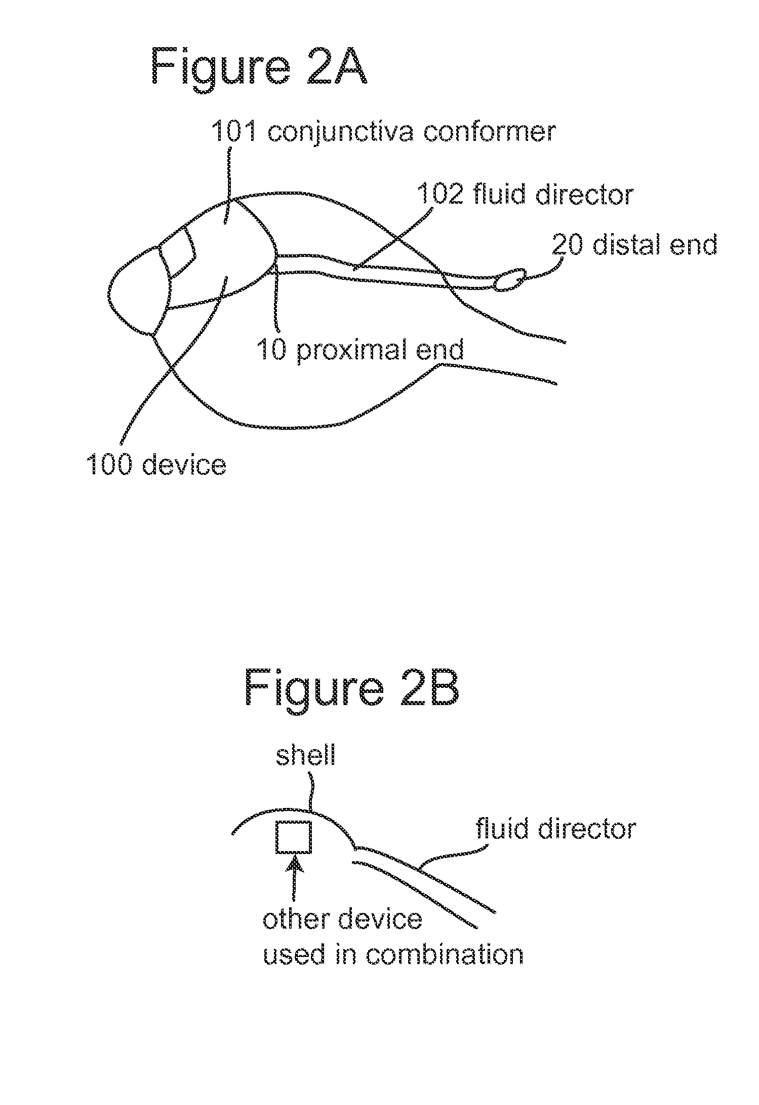
[0012]In some embodiments, the device of the present invention further comprises a securing structure adapted and configured for suturing and fixating the device to the eye wall. The securing structure can be a ring or a flange-like structure. The flange-like structure can be sized and shaped to fit on the surface of the sclera at the pars plana so as to prevent the device from dislocating or migrating. In some embodiments, the subject device is used in combination with eye surgery, for example, trabeculectomy. In some embodiments, the subject device is adapted and configured to be placed in connection with one or more filtering or shunting devices to enhance and / or direct intraocular fluid flow from anterior or posterior chambers or vitreous cavity to subconjunctival space or retrobulbar space. Examples of shunt devices include but are not limited to Ahmed valve, Molteno valve, and Krupin slit valve. In some embodiments, the device is adapted and configured to be placed over, below, around, or adjacent to the filtering or shunting device to minimize scarring, fibrosis, inflammation, and / or infection, and facilitate the egress of intraocular fluid from the filtering or shunting device. In some embodiments, the device is used in combination with another device for monitoring intraocular pressure over a period of time. In some embodiments, intraocular fluid is drained to the subconjunctival space or retrobulbar space at a sufficient flow rate to reduce intraocular pressure. In one example, the subject device is used for treating glaucoma.

Problems solved by technology

Glaucoma is a major cause of blindness worldwide.
If the pressure remains high enough for a long enough period of time, total, irreversible vision loss occurs.
High intraocular pressure results from an imbalance in intraocular fluid production versus outflow.
Trabeculectomy is associated with many problems.
The use of these agents has increased the success rate of trabeculectomy, but also has increased the prevalence of overfiltration and hypotony, or excessively low intraocular pressure (usually less than 6.0 mmHg).
Prolonged hypotony is associated with retinal distortion, loss of vision, and ultimately, loss of structural integrity of the eye,
The conjunctiva over the sclerectomy / trabeculectomy site, know as a “bleb,” can tear, producing excessive filtration and profound hypotony.
In addition, large anteriorly located blebs can be irritating, can disrupt the normal tear film, leading to blurred vision, and can make it difficult for patients to wear contact lenses.
These known devices suffer from certain shortcomings.
One problem with known glaucoma drainage devices is that the location of their insertion can cause damage to the anterior chamber structures, such as the cornea, the iris, or the intraocular lens.
Another problem with such devices is related to complications caused by their occlusion by fibrosis and / or infection.
Another problem with the know devices is their associated difficulty is establishing and regulating a sufficient amount of flow that would allow intraocular pressure to be titrated following the procedure.
Another known problem of such devices is the problems associated with double vision, limitation of eye movement and so on that can be encountered with shunting devices with external reservoirs.

Method used


Image

  • Subconjunctival conformer device and uses thereof
  • Subconjunctival conformer device and uses thereof
  • Subconjunctival conformer device and uses thereof

Examples


example

Results of Preclinical Study in Animal Eyes

Study Device

[0044]A study in 16 swine is performed using a device of the present invention. The device is implanted in one eye of each animal and the non-implanted eye serves as a control.

[0045]Surgical Procedure

[0046]For each animal, the periocular area of the study eye is prepped (eyelash trimming and betadine). The animal is then anesthesized using isofluorane. Under sterile conditions, a lid speculum is used to open the eyelids. An operating microscope is swung into place. A peripheral corneal bridle suture is placed to rotate the eye and expose the superior nasal limbus. A fornix-based conjunctival incision is made in the sclera and hemostasis ensured with bipolar cautery. A partial-thickness triangular scleral flap that measured 4×4 mm is made at the limbus and dissected anteriorly into clear cornea. A second, deeper flap is created at the base of the first flap, and dissected anteriorly to unroof the porcine equivalent of Schlemm's c...


PUM


Abstract

The present invention provides a device for use in an eye with elevated intraocular pressure or glaucoma, the device comprising a subconjunctival conformer shaped to conform to the eye wall and a fluid director that directs or facilitates the flow of intraocular fluid out of the eye and into the subconjunctival or retrobulbar space. The present invention also provides a method of lowering intraocular pressure using the device of the present invention.

Description

CROSS REFERENCE[0001]This application claims priority to U.S. Provisional Application 61 / 392,316, filed Oct. 12, 2010, the contents of which are incorporated herein in their entirety.BACKGROUND OF THE INVENTION[0002]Glaucoma is a major cause of blindness worldwide. The blindness that results from glaucoma can involve both central and peripheral vision and can have a major impact on an individual's ability to lead an independent and productive life. Pathophysiologically, glaucoma is an optic neuropathy (a disorder of the optic nerve) observed most typically in the setting of an elevated intraocular pressure. Dramatic and / or prolonged increases in intraocular pressure cause changes in the appearance (“cupping” or “excavation”) and function (“scotomas” or “blind spots” in the visual field) of the optic nerve. If the pressure remains high enough for a long enough period of time, total, irreversible vision loss occurs. High intraocular pressure results from an imbalance in intraocular fl...

Claims


Application Information

Patent Timeline
IPC IPC(8): A61F9/00
CPCA61F9/00781
Inventor CUNNINGHAM, JR., EMMETT T.
Owner CUNNINGHAM JR EMMETT T
Learn More