Image guided radiation therapy system and shielded radio frequency detector coil for use therein

a radiation therapy system and detector coil technology, applied in the field of radiation therapy, can solve the problems of fiducial markers, increased risk of complications, ptv is typically irradiated, etc., and achieve the effect of reducing the amount of interference and reducing the radiation induced curren

Inactive Publication Date: 2012-06-14
ALBERTA HEALTH SERVICES
View PDF8 Cites 10 Cited by
  • Summary
  • Abstract
  • Description
  • Claims
  • Application Information

AI Technical Summary

Benefits of technology

[0020]Shielding the at least one radiofrequency detector coil from the beam of radiation with an electrically grounded dielectric material significantly reduces the radi

Problems solved by technology

Because of the uncertainty in identifying this volume at the time of treatment, and due to unavoidable patient and tumour motion, an enlarged PTV is typically irradiated.
Because a volume that is larger than the biological extent of the disease and therefore healthy tissue is typically irradiated, there is an increased risk of complications.
However, fiducial markers must be placed using an invasive technique, and are thus less desirable.
IGRT techniques based on x-rays or ultrasound are not ideally suited to IGRT.
For example, x-rays suffer from low soft tissue contrast and are not ideally suited to imaging tumours.
Furthermore, x-ray based techniques use ionizing radiation and result in a supplemental dose deposit to the patient.
Ultrasound cannot be utilized in all locations of the body.
Finally, both x-ray and ultrasound based IGRT techniques are difficult to integrate into a linear accelerator such that they can provide images in any imaging plane in real time at the same moment as the treatment occurs.
There are several significant technological challenges associated with the integration of a linear accelerator with an MRI device.
However, while the documents referred to above provide various advancements, there are technological challenges that are yet

Method used


Image

  • Image guided radiation therapy system and shielded radio frequency detector coil for use therein
  • Image guided radiation therapy system and shielded radio frequency detector coil for use therein
  • Image guided radiation therapy system and shielded radio frequency detector coil for use therein

Examples


Embodiment Construction

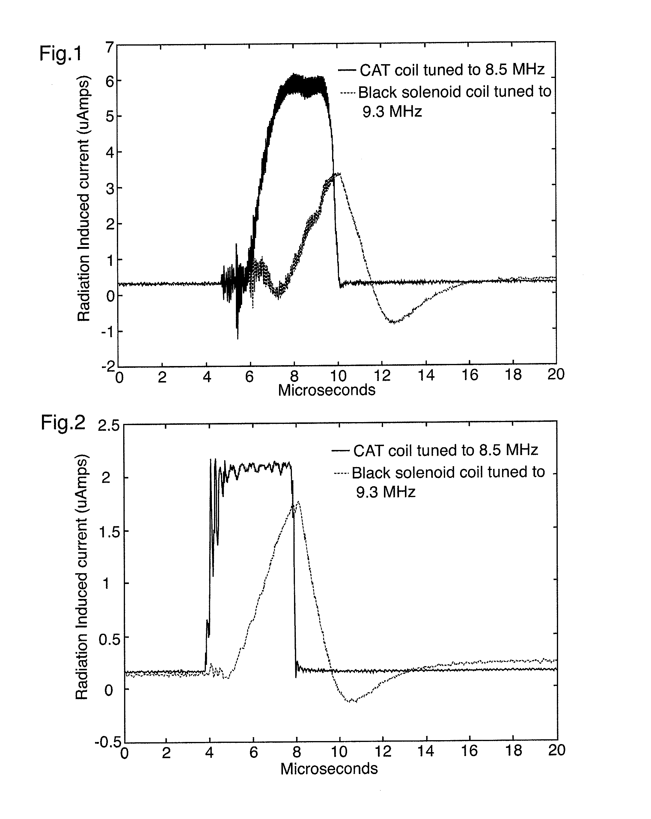
[0041]An investigation of radiation induced current in MRI RF coils was reported in “Radiation Induced Currents in MRI RF Coils: Application to Linac / MRI Integration” (B Burke, B G Fallone, S Rathee; 2010 Institute of Physics and Engineering in Medicine; Phys Med. Biol. 55 (2010) 735-746, which is incorporated entirely herein by reference. This work showed that RIC, or Compton current, is present in MRI RF coils when exposed to the pulsed radiation of a linear accelerator beam. FIGS. 1 and 2 are reproduced from that work, and show the Compton current induced in two MRI RF coils on a Varian600 C linear accelerator, and a Varian Clinac 23iX linear accelerator, respectively.

[0042]It has been found that shielding the radiofrequency detector coils of the MRI imaging system with a grounded dielectric material can significantly reduce or eliminate the net loss of electrons from the coil material when the treatment beam is incident directly on the detector coils. This shielding in turn sign...


PUM


Abstract

A radiation therapy system includes a radiation source capable of generating a beam of radiation; a magnetic resonance imaging (MRI) apparatus comprising at least one radiofrequency detector coil; and an electrically grounded dielectric material between the radiation source and the radiofrequency detector coil for shielding the at least one radiofrequency detector coil from the beam of radiation. Also disclosed is a radiofrequency detector coil for a magnetic resonance imaging (MRI) apparatus sheathed at least in part by a dielectric material that is adapted to be electrically grounded.

Description

CROSS-REFERENCE TO RELATED APPLICATIONS[0001]The present application claims priority under 35 U.S.C. 119(e) from U.S. Provisional Patent Application Ser. No. 61 / 390,172 filed on Oct. 5, 2010, and from U.S. Provisional Patent Application Ser. No. 61 / 489,550 filed on May 24, 2011.FIELD OF THE INVENTION[0002]The present application relates generally to radiation therapy and in particular to an image guided radiation therapy system and shielded MRI radiofrequency detector coil for use therein.BACKGROUND OF THE INVENTION[0003]Image guidance for radiation therapy is an active area of investigation and technology development. Current radiotherapy practice utilizes highly conformal radiation portals that are directed at a precisely defined target region. This target region consists of the Gross Tumour Volume (GTV), the Clinical Target Volume (CTV) and the Planning Target Volume (PTV). The GTV and CTV consist of gross tumour disease and the subclinical microscopic extension of the gross dise...

Claims


Application Information

Patent Timeline
IPC IPC(8): A61B5/055A61N5/00
CPCA61B5/055A61B2562/182A61N2005/1094G01R33/4808A61N2005/1055G01R33/341A61N5/10
Inventor RATHEE, SATYAPALBURKE, BENJAMINGHILA, ANDREIFALLONE, B. GINO
Owner ALBERTA HEALTH SERVICES
Why Eureka
Try Eureka