Inverse Imaging of Electrical Activity of a Heart Muscle

a heart muscle and electrical activity technology, applied in electrocardiography, medical science, sensors, etc., can solve the problems of insufficient information to obtain an accurate estimate of the sequence of electrical activity of the heart muscle, the difficulty of relating the ecg signal directly to the actual electrical activation and recovery process of the heart, and the cumbersome work

Inactive Publication Date: 2012-06-21
CORTIUS HLDG
View PDF8 Cites 53 Cited by
  • Summary
  • Abstract
  • Description
  • Claims
  • Application Information

AI Technical Summary

Benefits of technology

[0095]A first improvement in the initial estimation procedure was the incorporation of global anisotropic propagation in the simulation of ventricular activation. When using an uniform velocity the estimated activation wave revealed earlier epicardial activation for subject NH in the anterior part of the left ventricle [104]. Although no data on individual fiber orientation was available, the global handling of trans-mural anisotropy, estimated using common accepted insights [18,105,106], improved the overall performance.

Problems solved by technology

However, in clinical practice it is difficult to relate the ECG signals directly to the actual electric activation and recovery processes in the heart.
The standard 12-lead electrocardiogram (ECG), commonly used in diagnostic procedures, provides insufficient information for obtaining an accurate estimate of these sequences in all types of abnormality or disease.
This often leads to arrhythmia.
An ion-kinetic model may be suitable, but is more cumbersome to work with.

Method used


Image

  • Inverse Imaging of Electrical Activity of a Heart Muscle
  • Inverse Imaging of Electrical Activity of a Heart Muscle
  • Inverse Imaging of Electrical Activity of a Heart Muscle

Examples


Embodiment Construction

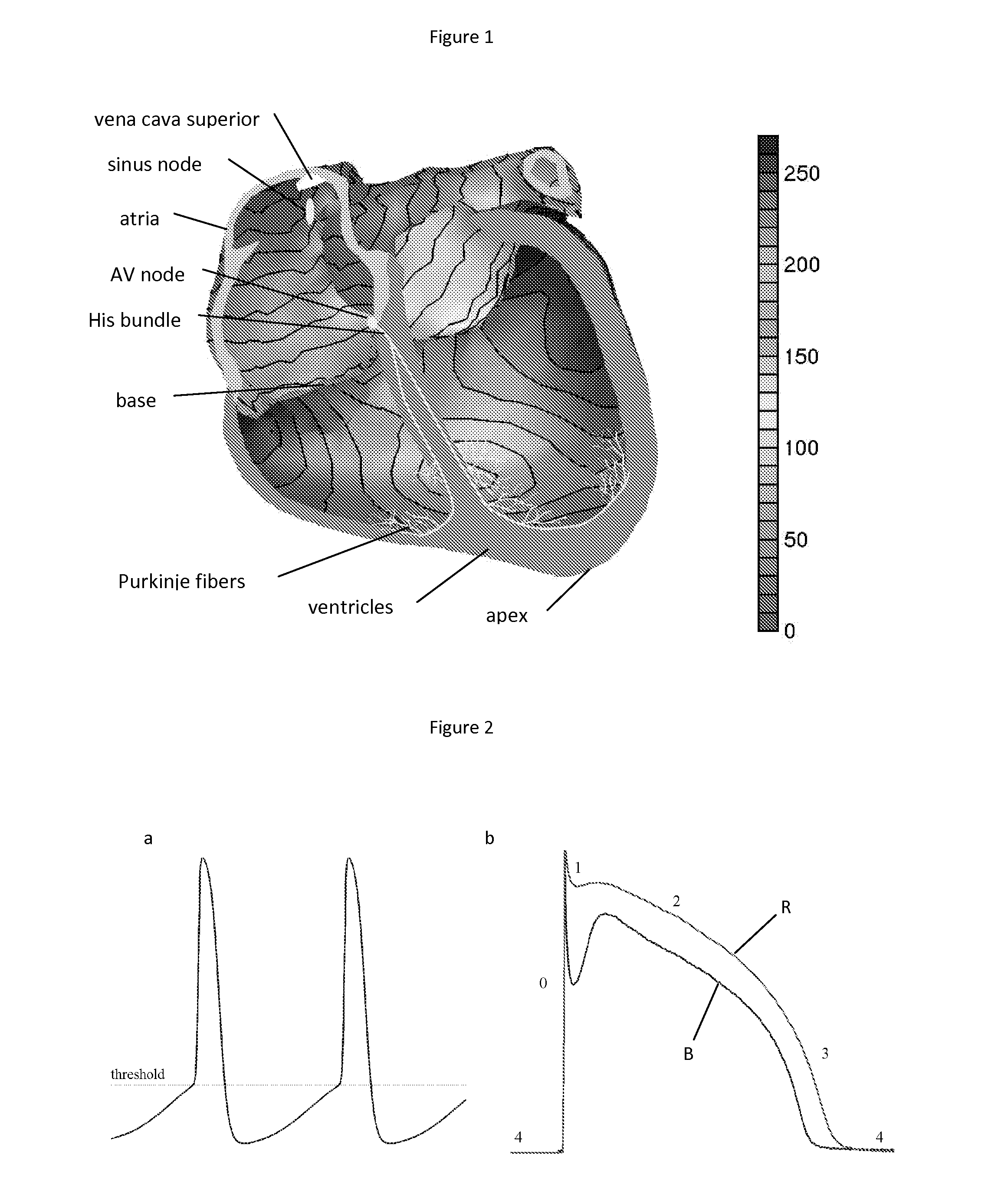
[0027]Since the inception of electrocardiography [33,34] several methods have been developed aimed at providing more information about cardiac electric activity on the basis of potentials observed on the body surface. The differences between these methods relate to the implied physical description of the equivalent generator representing the observed potential field. The earliest of these are the electric current dipole, a key element in vectorcardiography [35,36] and the multipole expansion [37]. Neither of these source models offer a direct view on the timing of myocardial activation and recovery, or other electro-physiologically-tinted features.

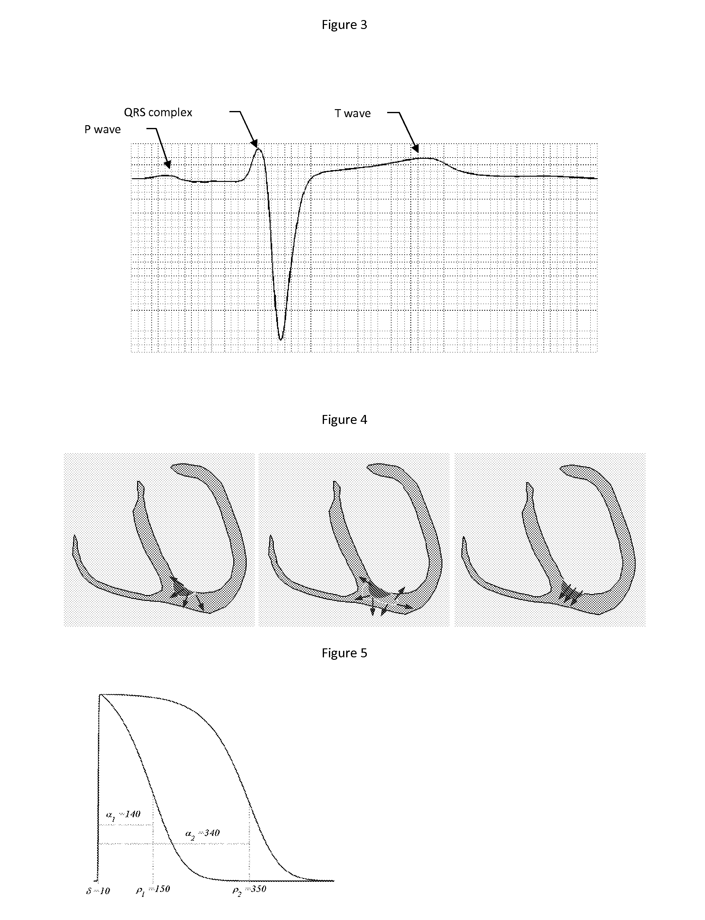
[0028]From the 1970s onwards, the potential of two other types of source descriptions have been explored [38,39]. This development stemmed from increased insight in cardiac electrophysiology and advances in numerical methods and their implementation in ever more powerful computer systems. The results of both methods are scalar functions on...


PUM


Abstract

A method for providing a representation of the distribution, fluctuation and / or movement of electrical activity through heart tissue, said method comprising: obtaining an ECG of the heart comprising said tissue; obtaining a model of the heart geometry; obtaining a model of the torso geometry; relating the measurements per electrode of the ECG to the heart and torso geometry and estimating the distribution, fluctuation and / or movement of electrical activity through heart tissue based upon a fastest route algorithm, shortest path algorithm and / or fast marching algorithm.

Description

[0001]The invention relates to methods for providing images of heart tissue, in particular of heart muscles, in particular images that can provide information on different areas of a heart muscle. In particular the invention relates to non-invasive methods of providing information on heart muscles. Furthermore the invention relates to methods for providing such images based upon ECG's and heart torso geometry measurements and / or models.INTRODUCTION[0002]The biophysical modelling approach used in this application relates the activation and recovery of the heart directly to the electrical processes within the myocardial cells. A proper understanding of the underlying processes within the myocardial cells and the electrical functioning of the whole heart is important for linking the observations on the body surface to the actual activation and recovery of the heart.[0003]At rest, ion pumps in the membrane of the cardiac cell maintain a potential difference over the membrane: the cell i...

Claims


Application Information

Patent Timeline
Patent Type & Authority Applications(United States)
IPC IPC(8): A61B5/0205
CPCA61B2018/00839A61B5/0402A61B5/318
Inventor VAN DAM, PETER MICHAELVAN OOSTEROM, ADRIAAN
Owner CORTIUS HLDG
Why Eureka
Try Eureka