
Mastopexy and Breast Reconstruction Prostheses and Method

a breast reconstruction and breast technology, applied in the field of tissue engineering, can solve the problems of cosmetic improvement at the cost of scars, present specific risks and complications, and drawbacks of techniques, and achieve the effects of improving the performance of mastopexy and breast reconstruction devices, preserving strength, and being convenient to handl

Inactive Publication Date: 2012-06-21
CODORI HURFF JEANNE +1
View PDF11 Cites 58 Cited by
  • Summary
  • Abstract
  • Description
  • Claims
  • Application Information

AI Technical Summary

Benefits of technology

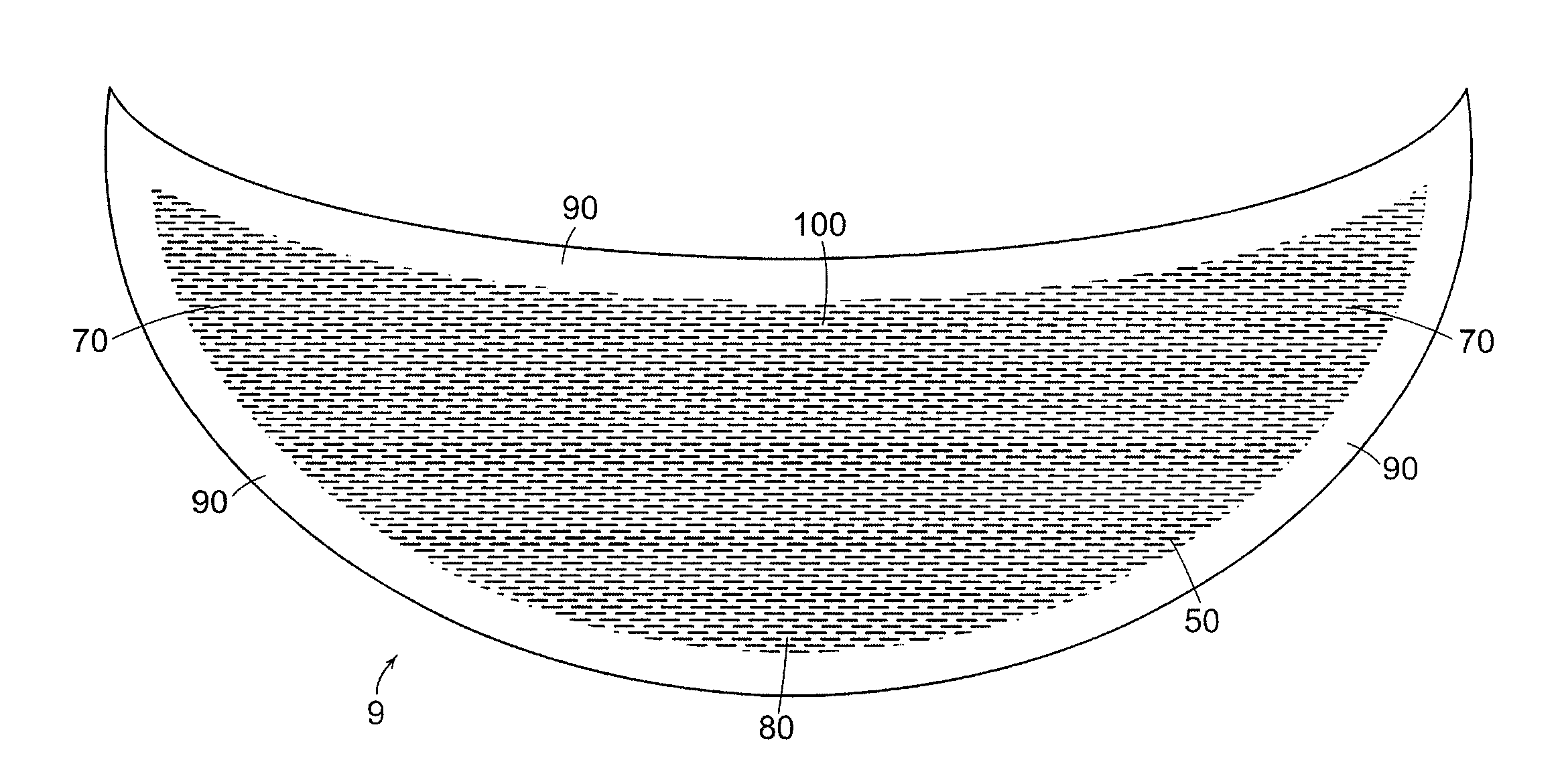
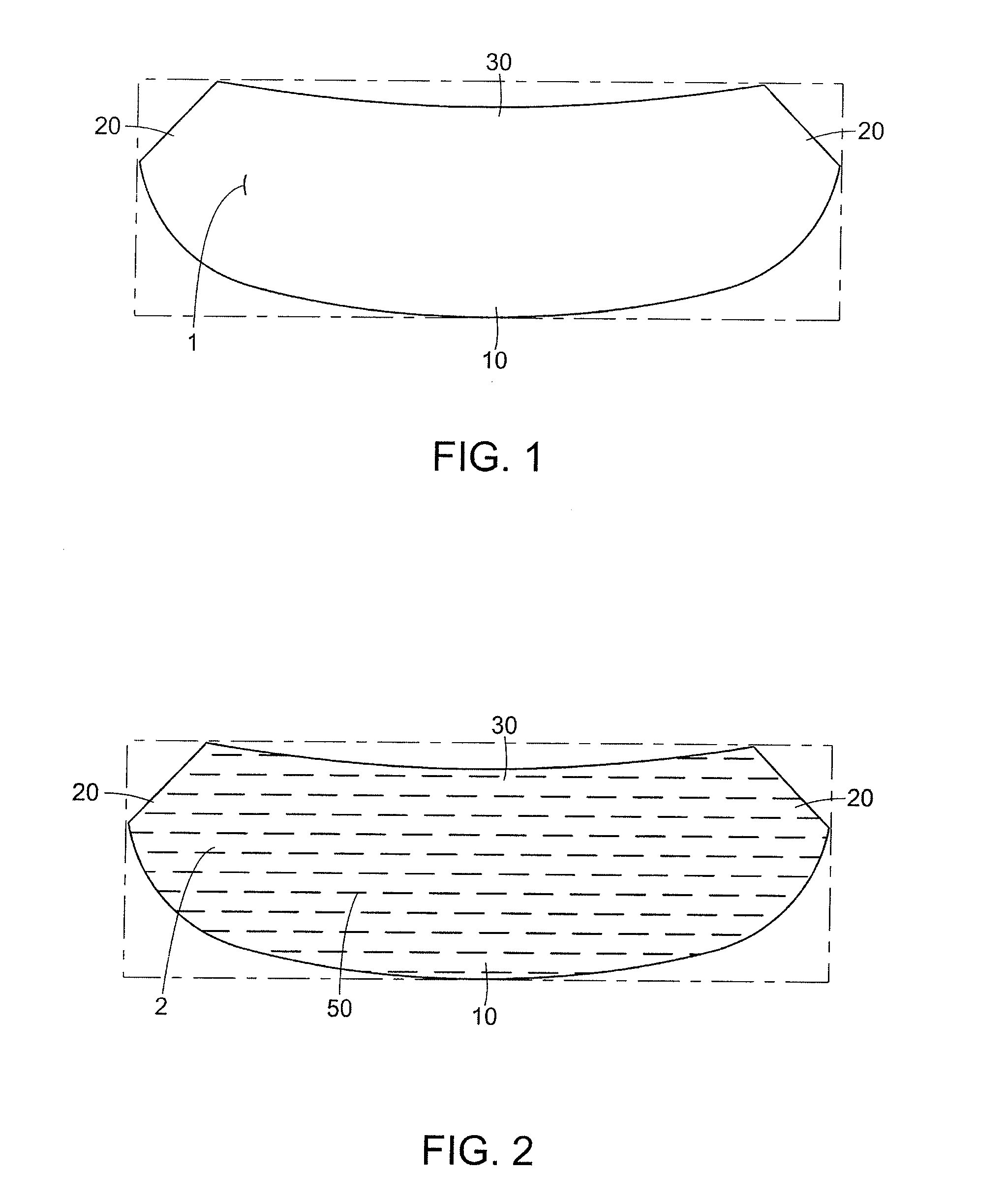
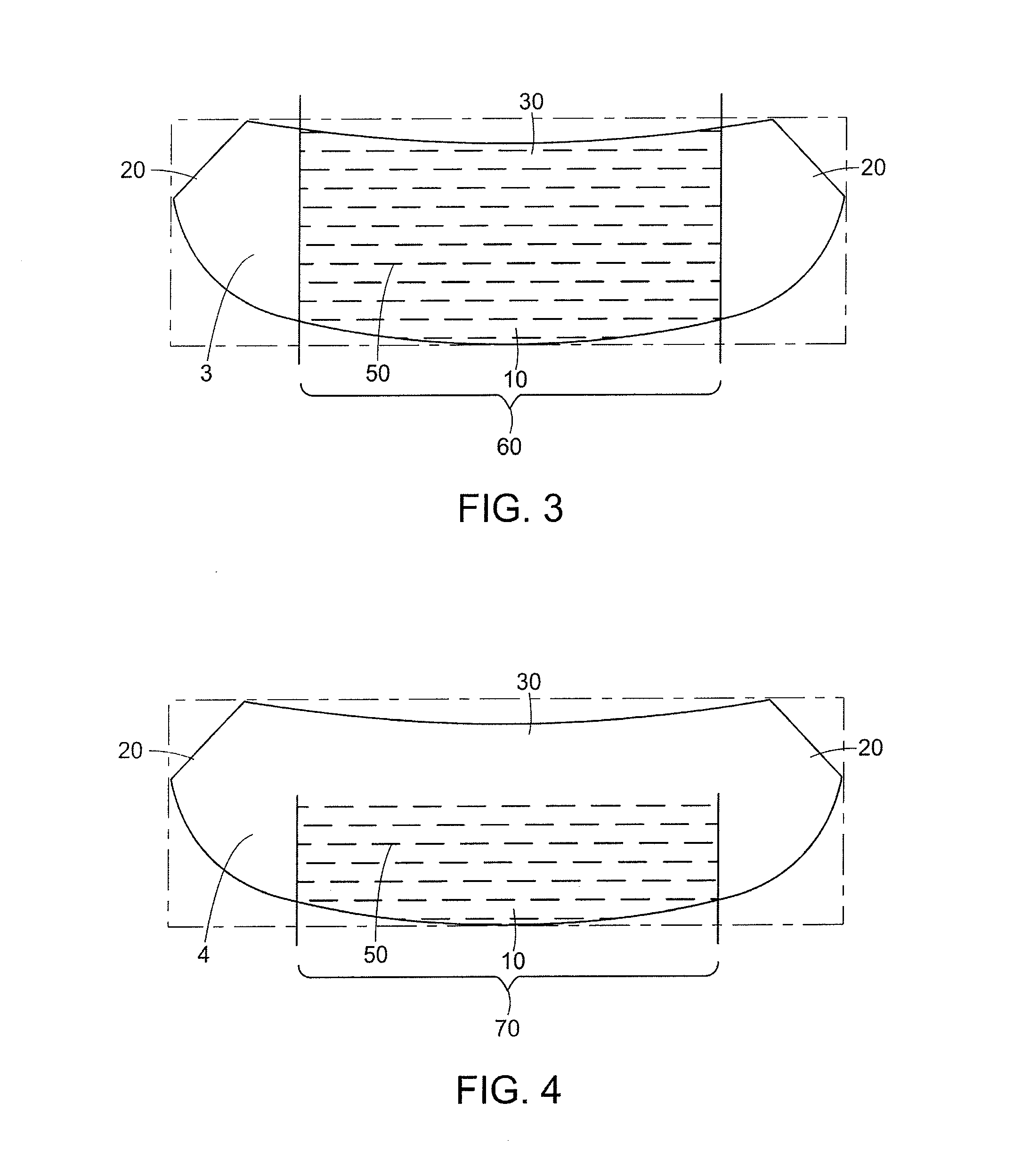
"The patent describes a method for creating a prosthesis for breast reconstruction and mastopexy procedures using a biologically-derived collagen material. The collagen is processed from the intestine or dermis to remove cells and become infiltrated with host cells. The prosthesis is designed to be remodeled by the patient's living cells and does not interfere with radiographic imaging. The prosthesis can be made with a single layer of collagen or multiple layers, and can be shaped like a crescent or arcuate. The purified collagen material is thin, strong, and easily handleable for preparing multi-layered prostheses. The prosthesis can be stretched in one direction to open the slits of the mesh but not in the other direction to preserve strength. Overall, this patent provides a better method for creating prosthesis for breast reconstruction and mastopexy procedures."

Problems solved by technology

Mastopexy presents one of the greatest challenges to the breast surgeon but previous techniques have drawbacks.
The aesthetic goals of these techniques are to obtain a more youthful appearance, improved projection, and reduced ptosis but aesthetic improvement comes at the cost of scars.
In addition, although breast implants can provide the upper pole projection patients often desire, they present specific risks and complications.
Another indication is following implant removal, which can result in breast ptosis and lax skin.
Mastectomy is the most common treatment of localized breast cancer but may negatively impact the patient emotionally, leaving her feeling deformed and mutilated, leading to anger, depression, and anxiety.
Autograft tissue bears a risk of tissue morbidity and total coverage and support of the implant or the expander with the muscle tissue in the mastectomy pocket is a challenge.
Without appropriate coverage, the implant can become exposed and reduce cosmetic outcome.
Heretofore, mastopexy and breast reconstruction materials and prostheses fabricated from biosynthetic materials and methods for their implantation have drawbacks in that they interfere with mammographical imaging that is necessary for detecting breast tissue abnormalities, including cancerous tumors.
Because these mastopexy devices cup a significant portion of the round of the breast, previous implant methods excessively disrupt the breast tissue by separating the tissue layers and cause a slow healing response and create a potential risk for tissue morbidity.
Materials derived from human cadaver tissue, usually from skin, also offer drawbacks in that their supply is limited and reports have demonstrated that their sourcing has been met with ethical challenges and safety concerns.

Method used


Image

  • Mastopexy and Breast Reconstruction Prostheses and Method
  • Mastopexy and Breast Reconstruction Prostheses and Method
  • Mastopexy and Breast Reconstruction Prostheses and Method

Examples


example 1

Chemical Cleaning of Mechanically Cleaned Porcine Small Intestine

[0140]The small intestine of a pig was harvested and mechanically stripped, using a Bitterling gut cleaning machine (Nottingham, UK) which forcibly removes the fat, muscle and mucosal layers from the tunica submucosa using a combination of mechanical action and washing using water. The mechanical action can be described as a series of rollers that compress and strip away the successive layers from the tunica submucosa when the intact intestine is run between them. The tunica submucosa of the small intestine is comparatively harder and stiffer than the surrounding tissue, and the rollers squeeze the softer components from the submucosa. The result of the machine cleaning was such that the submucosal layer of the intestine solely remained.

[0141]The remainder of the procedure, chemical cleaning according to International PCT Application No. WO 98 / 49969 to Abraham, et al., was performed under aseptic conditions and at room...

example 2

Method for Fabricating a Multilayer ICL Construct

[0149]ICL processed according to the method of Example 1 was used to form a multilayer construct having 2 layers of ICL. A sterile sheet of porous polycarbonate (pore size, manufacturer) was laid down in the sterile field of a laminar flow cabinet. ICL was blotted with sterile TEXWIPES (LYM-TECH Scientific, Chicopee, Mass.) to absorb excess water from the material. ICL material was trimmed of its lymphatic tags from the abluminal side and then into pieces about 6 inches in length (approx. 15.2 cm). A first sheet of trimmed ICL was laid on the polycarbonate sheet, mucosal side down, manually removing any air bubbles, folds, and creases. A second sheet of trimmed ICL was laid on the top facing, or abluminal side, of the first sheet with the abluminal side of the second sheet contacting the abluminal side of the first sheet, again manually removing any air bubbles, folds, and creases. The polycarbonate sheet with the ICL layers was angle...

example 3

Implant Studies Using Multilayer ICL Constructs

[0153]New Zealand white rabbits were used for in vivo analysis and all procedures were performed in compliance with Animal Care and Use Committee (ACUC) guidelines. A full thickness defect of approximately two inches was created through the rectus abdominis muscle in each animal and then was repaired with a 6 layer patch prosthesis. Patches were removed at 30, 66, 99 and 180 days post-implant. Three rabbits were sacrificed at each time point and examined for any evidence of herniation, swelling, infection or adhesions. Explanted patches were fixed in formalin and stained with hematoxylin and eosin or alizarin red for histologic evaluation of cell infiltration, inflammatory response and calcification. In some cases, unfixed patches were evaluated to determine the effect of implantation on the mechanical characteristics using uniaxial MTS analysis.

[0154]All animals underwent an uneventful post-operative course with no swelling, herniation...


PUM

PropertyMeasurementUnit
pore sizeaaaaaaaaaa
pore sizeaaaaaaaaaa
pore sizeaaaaaaaaaa

Abstract

Mastopexy and breast reconstruction prostheses and implantation method that allow for radiographic imaging of the breast tissue. The prostheses are arcuate and elongate optionally meshed to conform with breast tissue when implanted. Prostheses are made from naturally occurring extracellular matrix, primarily collagen, that, allows for mammographic imaging without interference as is expected from synthetic materials.

Description

FIELD OF THE INVENTION[0001]This invention is in the field of tissue engineering. The invention is directed to bioengineered graft prostheses prepared from cleaned tissue material derived from animal sources. The bioengineered graft prostheses of the invention are prepared using methods that preserve biocompatibility, cell compatibility, strength, and bioremodelability of the processed tissue matrix. The bioengineered graft prostheses are used for implantation, repair, or for use in a mammalian host.BRIEF DESCRIPTION OF THE BACKGROUND OF THE INVENTION[0002]The field of tissue engineering combines the methods of engineering with the principles of life science to understand the structural and functional relationships in normal and pathological mammalian tissues. The goal of tissue engineering is the development and ultimate application of biological substitutes to restore, maintain, and improve tissue functions.[0003]Collagen is the principal structural protein in the body and constit...

Claims


Application Information

Patent Timeline
Patent Type & Authority Applications(United States)
IPC IPC(8): A61F2/12
CPCA61F2210/0004A61F2/12A61F2/0063
Inventor CODORI-HURFF, JEANNEHAMMOND, DENNIS C.
Owner CODORI HURFF JEANNE
Learn More