Method for Selectively Damaging and Killing Tumor Cells and Apparatus Therefor

a tumor cell and selective damage technology, applied in the field of selective damage and tumor cell killing methods and apparatuses, can solve the problems of difficult operation, do damage to normal tissues in addition to tumor cells, and the applicability of pulse light using xenon flash lamps

Inactive Publication Date: 2012-07-05
TOKAI UNIV +1
View PDF2 Cites 21 Cited by
  • Summary
  • Abstract
  • Description
  • Claims
  • Application Information

AI Technical Summary

Benefits of technology

[0012]With the method and the apparatus of the present invention, it is possible to eliminate tumor cells alone without causing cell death to non-tumor cells, with ease for a short period of time. By the present invention, the application to pinpoint treatment of tumor cancer tissues present in a living body is expected.

Problems solved by technology

These methods, however, have problems that it is difficult to perform pinpoint treatment at tumor cancer tissues present deep in a living body, and these methods do damage to normal tissues in addition to tumor cells.
However, the applicability of the pulse light employing the xenon flash lamp or the like to the selective damaging and killing of the tumor cells has not been reported.

Method used


Image

  • Method for Selectively Damaging and Killing Tumor Cells and Apparatus Therefor
  • Method for Selectively Damaging and Killing Tumor Cells and Apparatus Therefor
  • Method for Selectively Damaging and Killing Tumor Cells and Apparatus Therefor

Examples


example 1

Material / Method

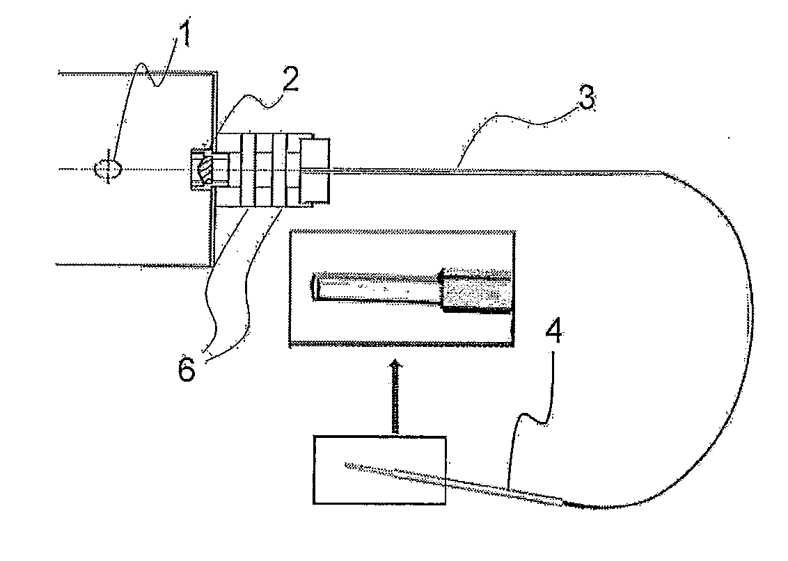
[0047]UV pulse flash light source (using a xenon flash lamp): BHX-200 (COMET Corp.)

Confocal laser scan microscope: LSM510-META (Carl Zeiss Microlmaging, Jena Germany)

Tumor cells:

[0048]1. MCF-7 (cell line originated from human breast cancer)

[0049]2. BT474 (cell line originated from human breast cancer)

[0050]3. Hella (cell line originated from human cervical cancer)

Non-Tumor Cells:

[0051]1. Cos 7 (cell line originated from African green monkey kidney)

[0052]2. MDCK (cell line originated from canine kidney uriniferous tubule epithelial cell)

[0053]At first, utilizing cell intrinsic fluorescence of each cell, morphology information of each cell was obtained. Then, to each cell, MBL Azami-Green (phmAG1-MC1) DNA was transfected using Lipfectamine-2000 (Invitrogen), and the fluorescence was observed. Under the environment of 5% CO2 and 37° C., three-dimensional images were obtained using LSM510-META. Thereafter, BHX-200 was set at a position of 8 cm immediately above the cells,...

example 2

Tumor Cell Lines

[0059]4. Human leukemia cell line MOLT / S

[0060]5. Human leukemia cell line MOLT / TMQ 200 (line 200-time resistant to trimetrexate (TMQ) anticarcinogenic agents)

[0061]6. Human leukemia cell line K562 / S1

[0062]7. Human leukemia cell line K562 / S2

[0063]8. Human leukemia cell line K562 / ARA-C (line resistant to Cytarabine anticarcinogenic agent)

Method

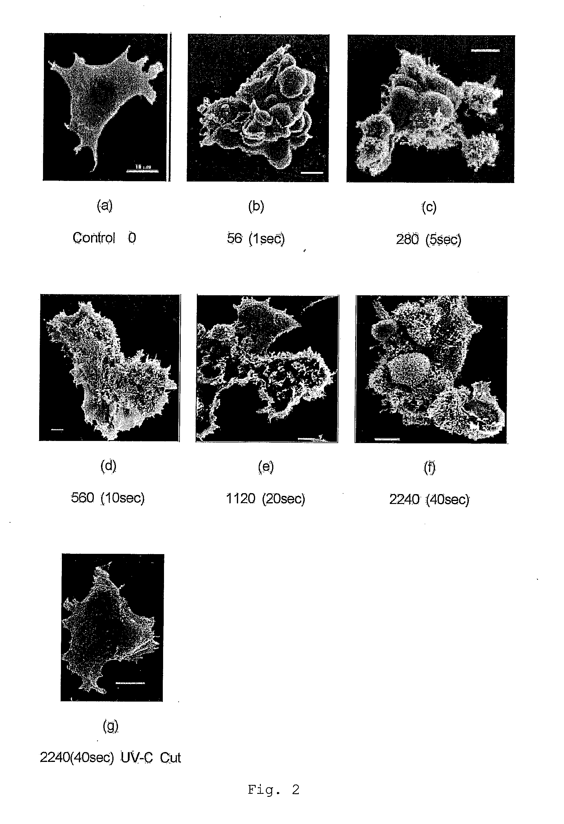
[0064]To each cell described above, propidium iodide (PI) was added. At first, utilizing intrinsic fluorescence of each cell, morphology information of each cell was obtained, similarly to Example 1. Then, under the environment of 5% CO2 and 37° C., three-dimensional images were obtained using LSM510-META. Thereafter, BHX-200 was set at a position of 8 cm immediately above the cells, and the cells were irradiated with a UV pulse flash (the frequency of irradiation: 0, 14, 56, 560, 2240 times). Thereafter, three-dimensional images were obtained again. The life and death of the cells was observed based on PI reaction. After the irr...

example 3

Tumor Cell Lines

[0067]9. Human fibrosarcoma cell line HT-1080

[0068]10. Human prostate cancer cell line DU145

[0069]11. Human prostate cancer cell line PC3

[0070]12. Human malignant chorioepithelioma cell line BeWo

Method

[0071]To each cell described above, propidium iodide (PI) was added. At first, utilizing intrinsic fluorescence of each cell, morphology information of each cell was obtained, similarly to Example 1. Then, under the environment of 5% CO2 and 37° C., three-dimensional images were obtained using LSM510-META. Thereafter, BHX-200 was set at a position of 8 cm immediately above the cells, and the cells were irradiated with a UV pulse flash (the frequency of irradiation: 0, 14, 56, 560, 2240 times). Thereafter, three-dimensional images were obtained again. The life and death of the cells was observed based on PI reaction. After the irradiation with the UV pulse flash, the cells were cultured continuously for 24 hours. The cells were observed again to determine a viability. T...


PUM

PropertyMeasurementUnit
distanceaaaaaaaaaa
timeaaaaaaaaaa
thicknessaaaaaaaaaa

Abstract

A method and an apparatus capable of selectively damaging and killing tumor cells includes a step of irradiating tumor cells with a UV pulse flash having continuous emission spectra ranging at least from 230 to 270 nm, outside a living body of a human or a living body of a non-human animal or in a living body of a non-human animal. The UV pulse flash may have an accumulated irradiation amount per unit area that is achieved at a distance of 8 cm from a light source having an integrated output of, for example, 90, 180-7100 or 14200 J.

Description

TECHNICAL FIELD[0001]The present invention relates to a method and an apparatus for selectively damaging and killing tumor cells using a specific UV pulse flash.BACKGROUND ART[0002]Conventional known methods for disrupting cancer cells include radiation methods using radiation, gamma ray or baryon beam. These methods, however, have problems that it is difficult to perform pinpoint treatment at tumor cancer tissues present deep in a living body, and these methods do damage to normal tissues in addition to tumor cells.[0003]It is well known that ultraviolet ray has a sterilizing effect, and a low-pressure mercury lamp (UV lamp) has been long used as a sterilizing lamp. In recent years, a “light pulse sterilization” using a xenon flash lamp has come to be used (for example, see Patent Document 1). The xenon flash lamp can emit a light having a wavelength spectra ranging from 200 to 300 nm, at which range the sterilizing effect is supposedly strong, instantaneously at a microsecond orde...

Claims


Application Information

Patent Timeline
Patent Type & Authority Applications(United States)
IPC IPC(8): A61N5/06C12N13/00C12M1/42A61N5/067
CPCA61B18/18A61B2018/1807A61N2005/0661A61N5/0613A61N2005/0654A61N5/0601
Inventor ITOH, JOHBU
Owner TOKAI UNIV
Why Eureka
Try Eureka