Microvesicles derived from nucleated, mammalian cells and use thereof

a technology of microvesicles and mammalian cells, applied in the field of microvesicles derived from nucleated mammalian cells, can solve the problems that the stealth liposome itself cannot deliver drugs to target cells or tissues, cannot pass clinical tests and successfully commercialized, and have not yet passed clinical trials and commercialization, etc., to achieve the effect of enhancing therapeutic efficacy, reducing agony and inconvenience of cancer patients, and enhancing therapeutic efficacy

Inactive Publication Date: 2012-07-12
AEON MEDIX
View PDF1 Cites 36 Cited by
  • Summary
  • Abstract
  • Description
  • Claims
  • Application Information

AI Technical Summary

Benefits of technology

[0033]The nucleated mammalian cell-derived microvesicles or shedding microvesicles with therapeutic and/or diagnostic substances thereto in accordance with the present invention can deliver the substances to target cells or tissues selectively and effectively, whereby the possible adverse effects which might occur upon the delivery of the therapeutic substances to non-target can be eliminated, reducing the agony and inconvenience of cancer patients during treatment. In addition, the specific delivery of diagnostic substances to target cells or tissues by the microvesicles enhances therapeutic efficacy. Particularly, metastasized cancer to which conventional chemotherapy is difficult to be applied can be effectively treated with the microvesicles of the present invention, w

Problems solved by technology

However, liposomes and stealth liposomes themselves cannot deliver drugs to target cells or tissues because they lack the ability to recognize the target cells or tissues.
To allow liposomes to bind to a specific target, studies have recently been directed toward the impartment of targeting ligands, such as monoclonal antibodies, to liposomes, but none of them have yet passed clinical tests and been successfully commercialized.
When vesicles, usually comprised of bacterial cell membranes, are derived fro

Method used


Image

  • Microvesicles derived from nucleated, mammalian cells and use thereof
  • Microvesicles derived from nucleated, mammalian cells and use thereof
  • Microvesicles derived from nucleated, mammalian cells and use thereof

Examples


example 1

Preparation of Microvesicles by Extrusion

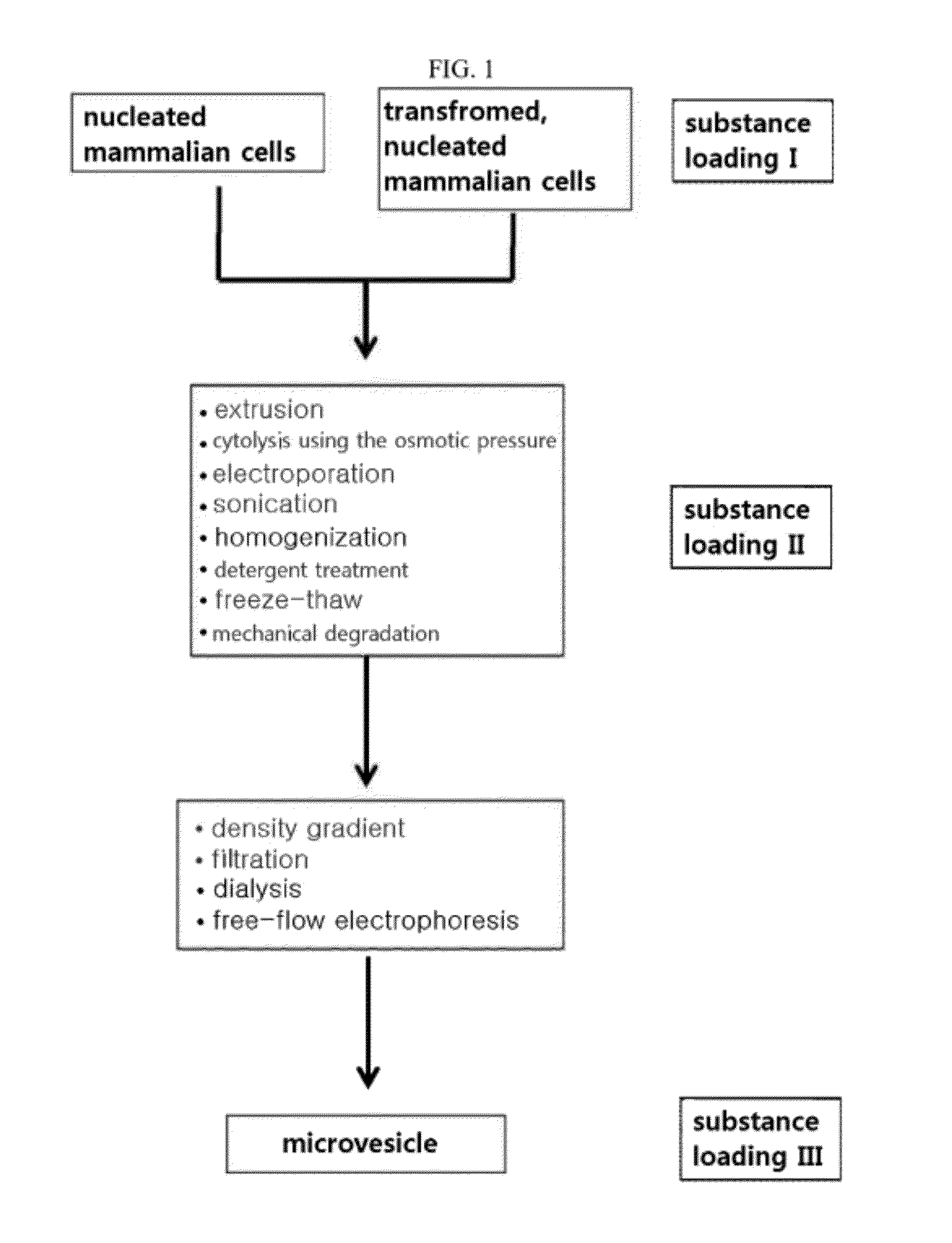
[0186]FIG. 1 is a scheme showing a process of preparing microvesicles loaded with various substances including targeting materials, therapeutic materials and diagnostic materials from nucleated mammalian cells, whether transformed or not.

[0187]According to the procedure illustrated in the scheme of FIG. 1, microvesicles were prepared from monocytes or macrophages. From among those suggested in FIG. 1, extrusion and a density gradient were selected.

[0188]The monocyte U937 (ATCC No. CRL-1593.2) or the macrophage Raw264.7 (ATCC No. TIB-71) was resuspended at a density of 5×10⁶ cells / ml in 3 mL of PBS (phosphate buffered saline). The cell suspension was passed three times through each of the membrane filters with a pore size of 10 μm, 5 μm and 1 μm, in that order. In a 5 mL untracentrifuge tube were sequentially placed 1 mL of 50% OptiPrep, 1 mL of 5% OptiPrep and 3 mL of the cell suspension effluent from the membrane filters. Ultracentrifugation...

example 2

Preparation of Microvesicles by Sonication

[0189]According to the procedure illustrated in the scheme of FIG. 1, microvesicles were prepared from monocytes or macrophages. From among those suggested in FIG. 1, sonication and a density gradient were selected.

[0190]Monocytes or macrophages were suspended at a density of 2×10⁷ cells / ml in 3 mL of PBS, followed by 30 cycles of sonication with the sonicator (UP 400S, Hielscher) at amplitude 50%, and cycle 0.5 and then with a water bath sonicator for 30 min. In a 5 mL untracentrifuge tube were sequentially placed 1 mL of 50% OptiPrep, 1 mL of 5% OptiPrep and 3 mL of the sonicated cell suspension. Ultracentrifugation at 100,000×g for 2 hours formed a layer of microvesicles between 50% OptiPrep and 5% OptiPrep.

example 3

Analysis of Property of Monocyte-Derived Microvesicles

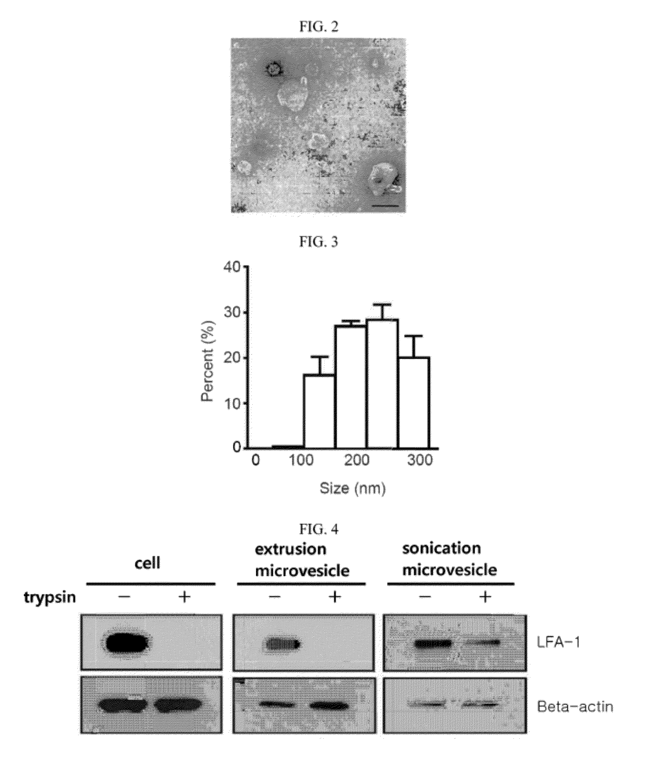
[0191]The microvesicles generated from monocytes in Example 1 were adsorbed for 3 min to a glow-discharged carbon-coated copper grid. The grid was washed with distilled water and stained for 1 min with uranylacetate before observation under a JEM101 electron microscope (Jeol, Japan). The electron microscope image is shown in FIG. 2.

[0192]As can be seen in the Transmission electron microscope (TEM) image of FIG. 2, the microvesicles constructed from monocytes by extrusion consisted of a lipid bilayer and is generally spherical with a size of 100˜200 nm.

[0193]The microvesicles generated from monocytes in Example 1 were diluted to a concentration of 5 μg / ml in 1 mL of PBS which was then placed in a cuvette and analyzed for particle sizes using a dynamic light scattering (DLS) particle size analyzer. The results are given in FIG. 3. As can be seen, the microvesicles ranged in size from 200 to 300 nm with a mean size of 250 nm.

[0194]W...


PUM


Abstract

The present invention relates to a microvesicle that is derived from nucleated mammalian cells, which are smaller than the nucleated cells. The microvesicles of the present invention can be used in the delivery of a therapeutic or diagnostic substance to specific tissues or cells, and more particularly, relates to microvesicles derived from monocytes, macrophages, dendritic cells, stem cells or the like, which can be used to deliver specific therapeutic or diagnostic substances for treating and/or diagnosing tissue associated with cancer, diseased blood vessels, inflammation, or the like.

Description

TECHNICAL FIELD[0001]The present invention relates to microvesicles derived from nucleated mammalian cells and the use thereof in the delivery of therapeutic and / or diagnostic substances.BACKGROUND ART[0002]A drug delivery system (DDS) is intended to aid the delivery of medicine to a target site within the body to bring about a therapeutic effect. For example, if a medicine is excreted too fast from the body due to its low absorption or bioavailability rates, a DDS may be used to modify the drug release profile. Medicines with serious adverse effects need to be delivered to target tissues or cells only. Many currently available anticancer agents, for example, exhibit cytotoxicity on normal cells as well as on cancerous cells. The substantial delivery of anticancer agents to cancerous cells or tissues would reduce the agony and inconvenience of cancer patients during treatment.[0003]Since the first use thereof in the 1960s, liposomes have been widely studied for their use in DDS. Adv...

Claims


Application Information

Patent Timeline
IPC IPC(8): A61K49/00A61K9/00
CPCA61K9/5068A61K45/06A61K35/15A61K35/545A61K9/1278A61K9/127A61K2300/00
Inventor GHO, YONG SONGKIM, YOON KEUNJANG, SU CHULKIM, OH YOUNCHOI, DONG-SICYOON, YAE JIN
Owner AEON MEDIX
Why Eureka
Try Eureka