Method for measuring autophagy

a technology of autophagy and autophagy, which is applied in the field of autophagy measurement, can solve the problems of insufficient research on the subject, inability to detect microautophagy or chaperone-mediated autophagy by the method, and time-consuming techniques, and achieves accurate identification of small differences in activity levels, great changes in fluorescence intensity ratio, and potent resistance to degradation

Active Publication Date: 2012-07-12
JAPAN SCI & TECH CORP
View PDF0 Cites 3 Cited by
  • Summary
  • Abstract
  • Description
  • Claims
  • Application Information

AI Technical Summary

Benefits of technology

[0011]The present inventors have now found a method that enables more accurate measurement of the activity of the autophagy that is the system in cells for degrading cytoplasmic materials such as proteins or organelles. More specifically, the present inventors have now found a novel method for measuring autophagy in cells, characterized by the use of a single fluorescent protein, which is resistant to degrading activities and which changes spectra depending on pH changes, as a probe, while utilizing the fact that autophagy is a reaction that the cytoplasmic materials are incorporated into the lysosome or vacuole having an acidic pH from the cytoplasm having a neutral pH. Specifically, the activity of autophagy in cells can be more accurately measured by using, as a probe reagent, a single fluorescent protein having spectra that change the fluorescence intensities in opposite directions between an acidic condition and a neutral condition.
[0023]According to this invention, autophagy activity can be detected as greater changes in the fluorescence intensity ratio using, as a probe reagent, a single fluorescent protein having potent resistance to degradation and changing the fluorescence intensities in opposite directions between the acidic and neutral conditions, compared with the use of two conventional fluorescent proteins in combination. This enables more accurate identification of small differences in activity levels. Since measurement problems caused by different properties of two fluorescent proteins, such as folding speeds or quenching speeds, are dissolved, more quantitative measurement of autophagy becomes possible.

Problems solved by technology

However, such techniques required skills and times due to insufficient specificity for autophagy and complicated procedures.
However, because the formation of autophagosome is a phenominon observed only in the case of macroautophagy, it is impossible to detect microautophagy or chaperone-mediated autophagy by the method as mentioned above.
At present, however, research thereon has not advanced as that of macroautophagy, and there are no effective methods for measurement.
It is thus impossible to determine the total amount of all types of autophagy occurring in cells.
Because Rosado et al employ two fluorescent proteins, there is a problem that their technique makes the accurate measurement of autophagy activities difficult for the following reasons: that because the size of a label becomes large, the activity or localization of the proteins may probably be inhibited due to steric hindrance when the fluorescent protein is fused to a target protein; that two fluorescent proteins may probably generate improper signals when they are cleaved with protease in cells; that because pH-independent fluorescence properties of the two fluorescent proteins differ from each other in terms of quenching properties caused by the folding speed or photobleaching in cells, the value of the ratio may probably vary depending on experimental conditions; that because changes in the ratio depend only on fluorescence changes of the super ecliptic pHluorin, significant changes cannot be observed; and the like.

Method used


Image

  • Method for measuring autophagy
  • Method for measuring autophagy
  • Method for measuring autophagy

Examples


example 1

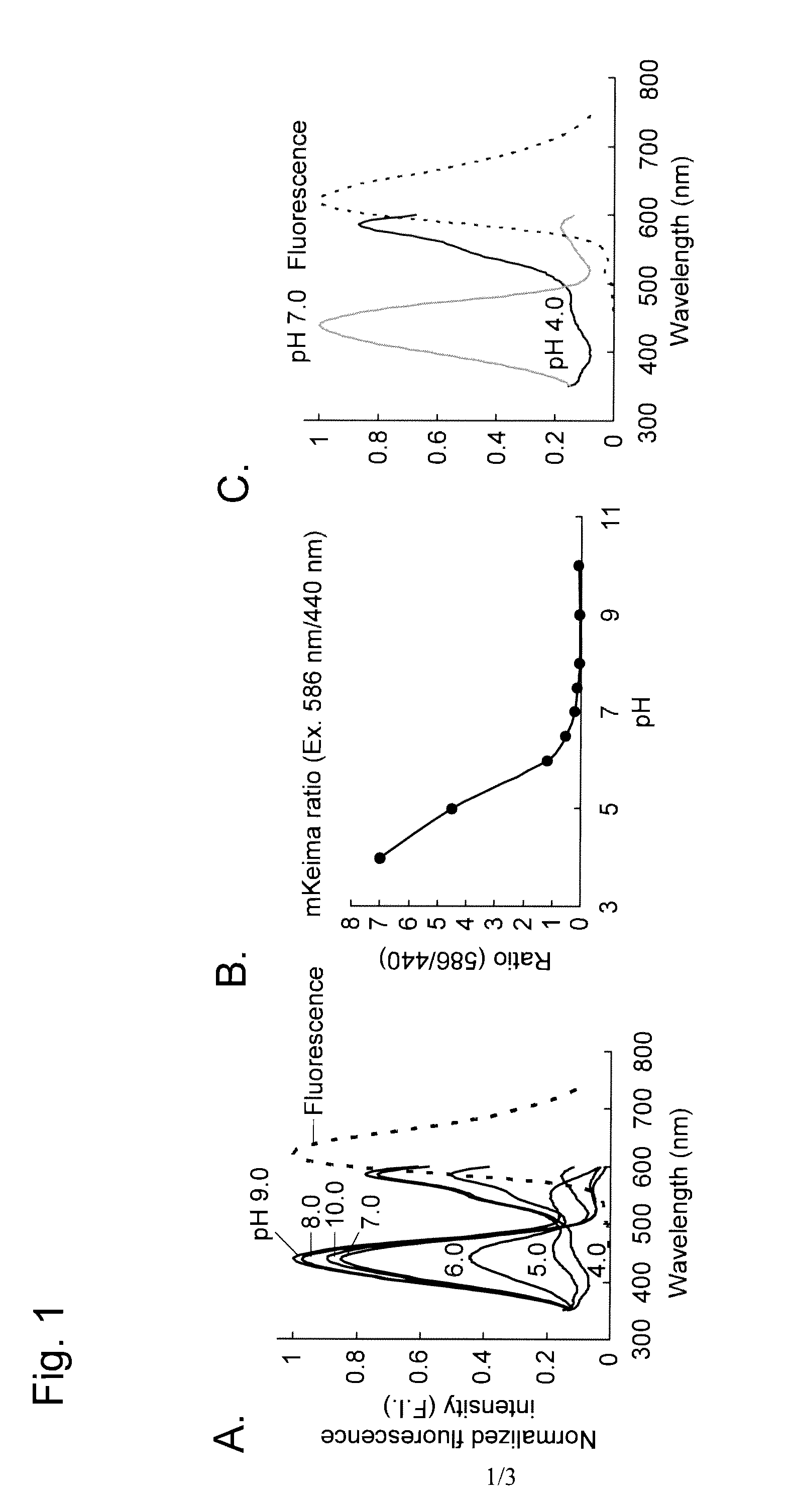
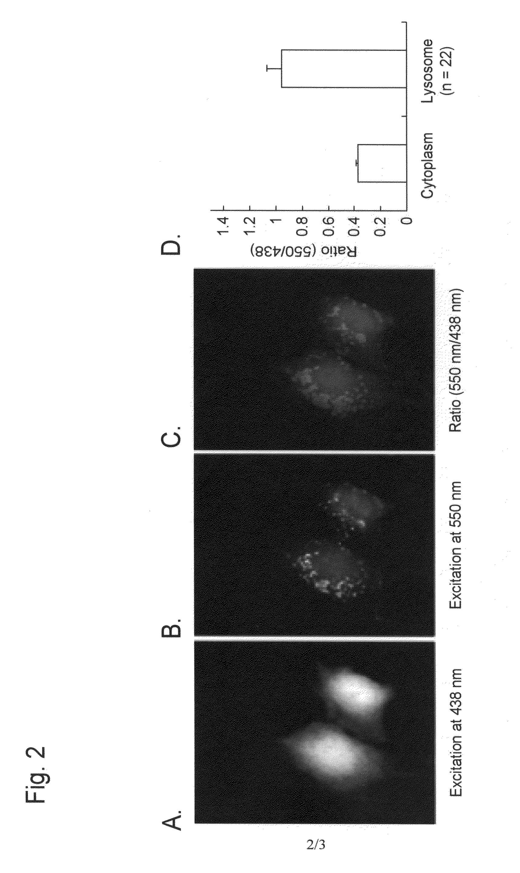
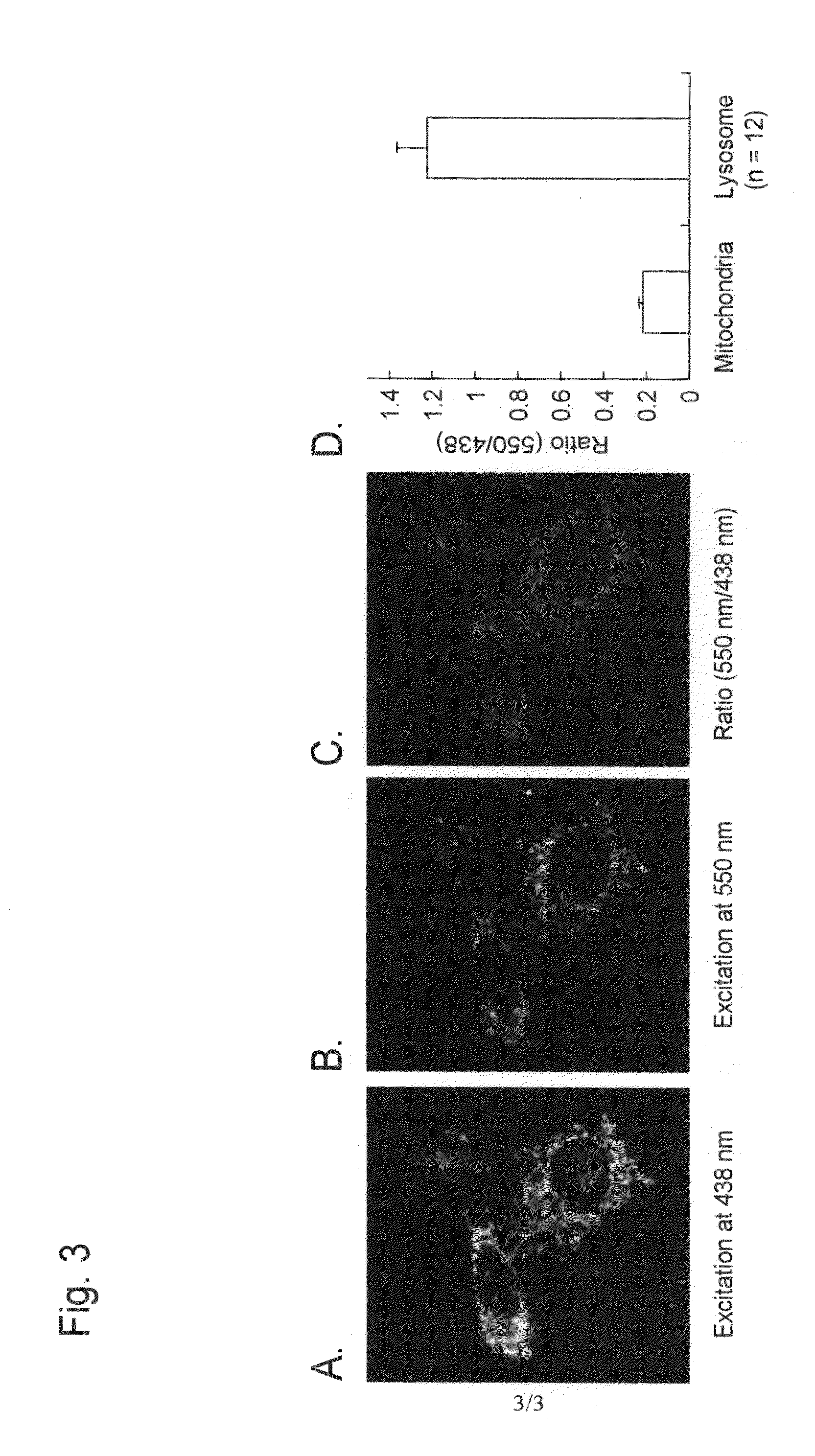
[0094]An example of autophagy measurement with the use of a Montipora sp-derived fluorescent protein (a monomeric protein, mKeima) as a fluorescent probe reagent is described below.

[0095]

[0096]mKeima was purified in the following manner, and pH-dependent spectral properties were studied.

[0097]pRSETB (Invitrogen) into which mKeima cDNA had been inserted (note: cDNA cloning is described in Kogure, T. et al., 2006, Nat. Biotechnol., 24: 577-581) was introduced into the JM109 (DE3) competent cells. The competent cells were applied to an LA plate and cultured at 37° C. overnight. The resulting colonies were transferred to 100 ml LA medium and then subjected to shake culture at 18° C. for 72 hours. The colonies were lysed by means of freezing and thawing, and then the mKeima-containing supernatant obtained after centrifugation was applied to a nickel column (Qiagen) and subjected to elution through the column. In order to remove imidazole used during the above procedure, mKeima was finall...


PUM

PropertyMeasurementUnit
diameteraaaaaaaaaa
pHaaaaaaaaaa
fluorescence wavelengthaaaaaaaaaa

Abstract

This invention relates to a method for measuring autophagy in cells, comprising using, as a probe reagent, a single fluorescent protein, to measure a change in fluorescence properties of the fluorescent probe reagent depending on pH changes associated with autophagy, thereby determining the presence or activity of autophagy, wherein the single fluorescent protein is resistant to degrading enzyme activity in the lysosome or vacuole of the cell, it is not denatured or inactivated under acidic to neutral pH environment, and it is capable of changing excitation spectra or fluorescence spectra when located under the environments of acidic region and neutral region.

Description

TECHNICAL FIELD[0001]This invention relates to a method for measuring autophagy.[0002]This invention also relates to a method of screening for therapeutic agents for a disease associated with autophagy anomalies using the method for measuring autophagy.BACKGROUND ART[0003]Autophagy is a pathway of degrading cytoplasmic materials ubiquitously observed in eucaryocytes. Autophagy is classified into three types based on its mechanisms; i.e., macroautophagy, microautophagy, and chaperone-mediated autophagy. In any pathway, cytoplasmic materials are ultimately translocated into lysosomes (vacuoles in the case of yeast or plants) and degraded by degrading enzymes existing therein. Examples of the cytoplasmic materials to be degraded include not only protein molecules but also organelles such as mitochondria and endoplasmic reticulum. Since autophagy is induced by nutrient starvation, supply of nutrient components to cells via recycling of the degraded cytoplasmic materials had been conside...

Claims


Application Information

Patent Timeline
Patent Type & Authority Applications(United States)
IPC IPC(8): C12Q1/02
CPCG01N21/6428G01N21/80G01N2021/6421G01N33/5035G01N2021/6419G01N33/5005
Inventor MIYAWAKI, ATSUSHIKATAYAMA, HIROYUKI
Owner JAPAN SCI & TECH CORP
Why Eureka
Try Eureka