Interconnected porous non-degradable poly(VINYL) alcohol implant and method of manufacture

a non-degradable polyvinyl alcohol and interconnected technology, applied in the field of manufacturing porous non-degradable polyvinyl alcohol implants, can solve the problems of inability to perform joint replacement in younger patient populations, and inability to prevent the progression of osteoarthritis

Active Publication Date: 2012-07-12
NEW YORK SOC FOR THE RUPTURED & CRIPPLED MAINTAINING THE HOSPITAL FOR SPECIAL SURGERY
View PDF1 Cites 14 Cited by
  • Summary
  • Abstract
  • Description
  • Claims
  • Application Information

AI Technical Summary

Benefits of technology

[0008]The present invention overcomes the problems in the art by providing a method that allows for precise control over the morphology of the interconnected porous network and the mechanical properties of an interconnected porous non-degradable polymer implant during manufacture. The resulting interconnected porous non-biodegradable polymer implant is suitable for implantation into a mammal, to treat, repair or replace defects and / or injury to musculoskeletal tissue including bone, tendon, ligaments, cartilage and the discs of the spine. In a preferred embodiment the implant is used to stabilize a chrondral defect, which is a defect in the articular cartilage at the end of the bones. In a most preferred embodiment, the chondral defect is found in the knee. The implant would not only repair the defect but protect the surrounding tissue from degradation.

Problems solved by technology

However, all of these techniques have their limitations, and do not prevent the progression of the osteoarthritis, which often propagates from the focal defect.
Performance of joint replacements in younger patient populations is less satisfactory than for older patients, oftentimes leading to multiple revision surgeries, each with successively diminishing longevity.
(1990)), many have failed due to the lack of a porous periphery which would be required to facilitate cell migration and integration (Maher et al.
Additionally, the mechanical properties of the scaffolds developed thus far are unable to carry joint loads of native tissue at the time of implantation (Maher et al.
Moreover, the techniques developed thus far for creating porous PVA scaffolds either do not enable control over the mechanical properties of the end-product or do not enable the formation of a truly interconnected porous network.
However, this method of manufacture did not enable control over the mechanical properties, nor control over porosity (Dai et al.
Moreover, control over the mechanical properties and the porosity is not possible using the aforementioned techniques.

Method used


Image

  • Interconnected porous non-degradable poly(VINYL) alcohol implant and method of manufacture
  • Interconnected porous non-degradable poly(VINYL) alcohol implant and method of manufacture
  • Interconnected porous non-degradable poly(VINYL) alcohol implant and method of manufacture

Examples


example 1

Manufacture of the Interconnected PVA Implant

[0137]Materials and Methods

[0138]All handling and fabrication techniques were performed aseptically to minimize contamination with bacteria and other infectious agents. The method of manufacture involves four steps:

[0139]a. Hydration of gelatin sponges,

[0140]b. Replacement of water with poly(vinyl) alcohol solutions,

[0141]c. Freeze-thawing of construct, and

[0142]d. Removal of gelatin sponge to form macroporous network.

[0143]Hydration of gelatin sponges: Gelatin (denatured collagen) sponges (Ethicon-Johnson & Johnson, Somerville, N.J.) were first soaked in deionized water until the entire sponge was saturated with water via capillary action and under applied vacuum (30 minutes on / off for 6 hours) for 1 day. The sponges were transferred to 50 mL conical tubes and repeatedly centrifuged at 3000 g for 1 hour at a time, with gentle agitation of the tube between centrifugations to restore its original shape, until all remaining air bubbles had ...

example 2

Control of the Mechanical Properties of the Implant for a Given Morphology

[0151]Materials and Methods

[0152]10% and 20% PVA scaffolds were manufactured using the materials and methods described in Example 1.

[0153]Cylindrical scaffolds (Ø5×2-3.5 mm) were cored out for mechanical testing (n=5 per group). Samples were first tested in unconfined compression in a phosphate buffered saline bath. Initial contact with the specimen surface was determined when an instantaneous applied load reading of 0.2-0.5 g was measured and the initial specimen height was determined at this point. Then applied strain (displacement / original height) compressions were applied in 5% increments to a maximum of 25% strain, with the sample being allowed to equilibrate after each step for 10-15 minutes. After each test, the samples were unloaded and allowed to recover to their original height in a saline bath. Samples were then retested in confined compression loading configuration.

[0154]The loads were converted to...

example 3

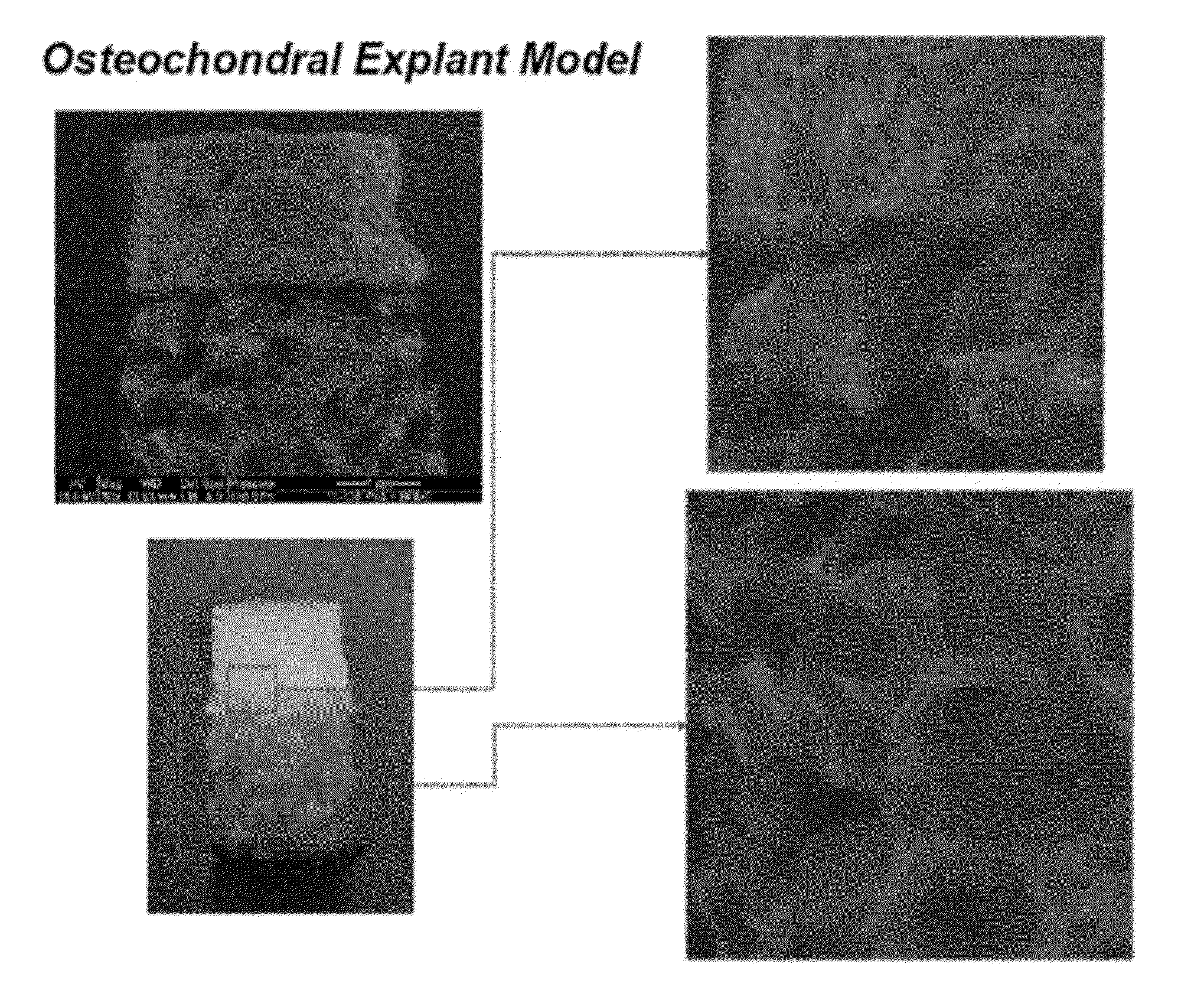
Implants can Support Cell Survival and Proliferation

[0158]Materials and Methods

[0159]PVA scaffolds (Ø5.0×2.5 mm) prepared as described in Example 1 were rehydrated in 400 μL of Dulbecco's Modified Eagle Medium (DMEM) containing 20×106 chondrocytes / mL in a 24-well plate. After this initial rehydration, scaffolds were covered with an addition 2 mL of cell suspension and incubated at 37° C. with 5% CO2 for 30 minutes. Afterwards, the media was changed with fresh DMEM with 5% fetal bovine serum and antibiotics. After 48 hours, cell-seeded scaffolds were placed in a custom loading bioreactor and loaded in unconfined compression from 0→1 N at 0.5 Hz for 5 minutes at 25° C. Free-swelling (unloaded) controls were maintained adjacent to the bioreactor during loading. After loading, all samples were cultured for an additional 48 hours in DMEM with 10 μM bromodeoxyuridine (BrdU) and then washed for 2 hours in fresh media, fixed in 10% formalin, embedded in paraffin wax, sectioned, and then ass...


PUM

PropertyMeasurementUnit
pore diameteraaaaaaaaaa

Abstract

The present invention relates to a method of manufacture of an interconnected porous non-biodegradable polymer implant suitable for implantation into a mammal for the treatment, repair or replacement of defects or injury in musculoskeletal tissue, wherein the mechanical properties of the implant can be controlled by varying the concentration of the non-biodegradable polymer and/or varying the duration and number of freeze-thaw cycles and the interconnected porous non-biodegradable polymer implant has sufficient percent porosity and pore diameter to facilitate integration of cells and attachment within the mammal via ingrowth of surrounding tissue. The present invention also relates to an implant manufactured by the method.

Description

CROSS REFERENCE TO RELATED APPLICATION[0001]The present application claims priority to U.S. patent application Ser. No. 61 / 432,098, filed Jan. 12, 2011, which is hereby incorporated by reference in its entirety.FIELD OF INVENTION[0002]The present invention is in the field of manufacturing porous non-degradable poly(vinyl) alcohol implants suitable for implantation into a mammal, to treat, repair or replace defects and / or injury to musculoskeletal tissue.BACKGROUND OF THE INVENTION[0003]Articular cartilage is a hydrated and lubricated joint tissue that allows for the relative movement of opposing joint surfaces under high loads (Scholten et al. (2011): Buckwalter et al. (1998); Mow et al. (1993)). Mature articular cartilage does not possess an intrinsic ability to heal, since it is avascular and lacks a source of mesenchymal cells (Scholten et al. (2010); Buckwalter et al. (1998)).[0004]Current operative procedures for the treatment of articular cartilage damage generally fall into f...

Claims


Application Information

Patent Timeline
Patent Type & Authority Applications(United States)
IPC IPC(8): C08F116/06C08J9/26
CPCC08J3/005C08J9/26C08J2201/046C08J2329/04C08J2400/16C08L29/00A61L27/58A61L2430/06A61L2430/24A61L27/16C08L29/04
Inventor MAHER, SUZANNE A.NG, KENNETHCHEN, TONYWANIVENHAUS, FLORIAN
Owner NEW YORK SOC FOR THE RUPTURED & CRIPPLED MAINTAINING THE HOSPITAL FOR SPECIAL SURGERY
Why Eureka
Try Eureka