

Blood vessel segmentation with three dimensional spectral domain optical coherence tomography
a three-dimensional spectral domain, optical coherence tomography technology, applied in image enhancement, instruments, angiography, etc., can solve the problems of limited approach, reduced vessel pattern visibility, and difficulty in segmentation on oct imag
Active Publication Date: 2012-08-23
UNIVERSITY OF PITTSBURGH
View PDF9 Cites 63 Cited by
- Summary
- Abstract
- Description
- Claims
- Application Information
AI Technical Summary
Benefits of technology
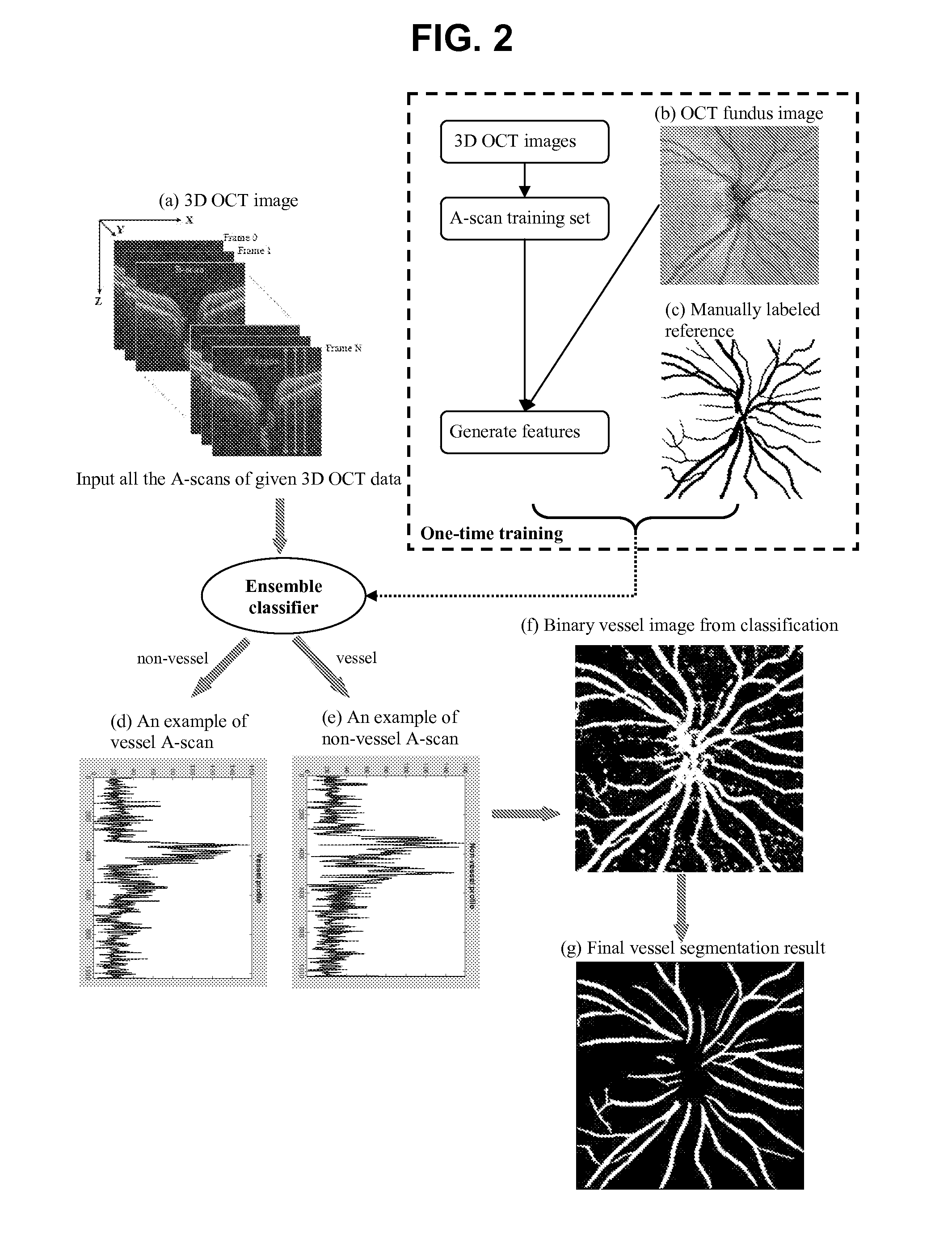
[0008]It is an object of the present invention to provide a three-dimensional boosting learning algorithm trained to automatically identify and / or segment retinal blood vessels in a 3D OCT image without replying pre-segmentation. This allows the clinicians to obtain accurate pattern of the blood vessels for clinical analysis, for retinal image registration, and for early diagnosis and monitoring of the progression of glaucoma and other retinal diseases.
Problems solved by technology
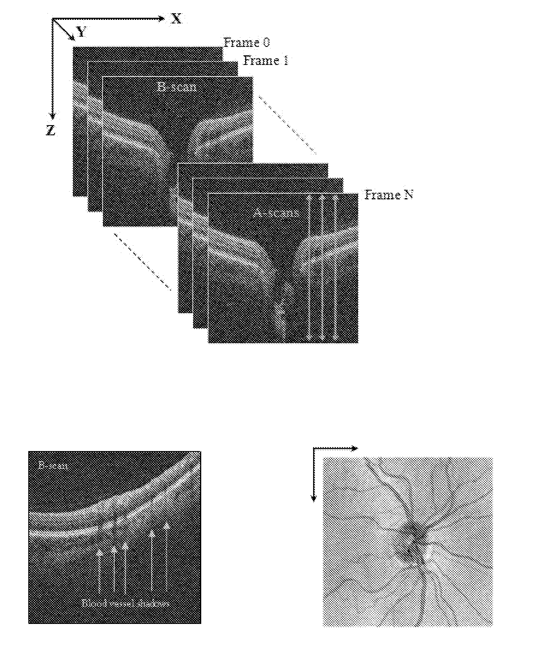
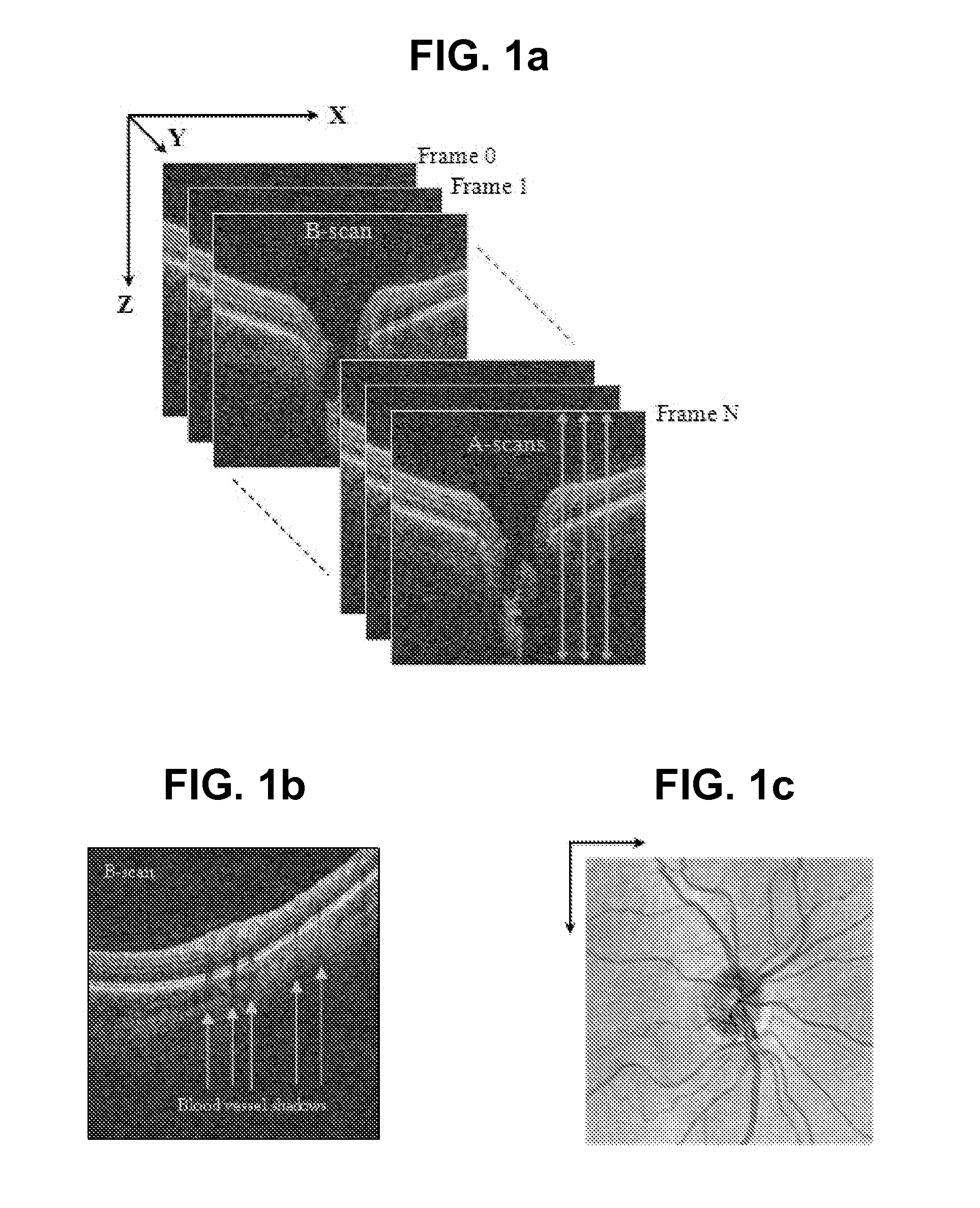
When an OCT fundus image is generated (FIG. 1(c)) by averaging each A-scan, the visibility of vessel pattern may decrease due to the noise and high reflection on the top layers of the retina.
This makes vessel segmentation on OCT image a challenge, compared to a conventional 2D fundus image.
This approach is limited in that its performance highly depends on retinal layer segmentation.
Method used
View moreImage
Smart ImageViewing Examples

Examples
working examples
Non-Limiting Working Examples
[0055]The present invention is further illustrated by reference to the following description of methodology within the invention and corresponding results. See [12].
Login to View More PUM
 Abstract
In the context of the early detection and monitoring of eye diseases, such as glaucoma and diabetic retinopathy, the use of optical coherence tomography presents the difficulty, with respect to blood vessel segmentation, of weak visibility of vessel pattern in the OCT fundus image. To address this problem, a boosting learning approach uses three-dimensional (3D) information to effect automated segmentation of retinal blood vessels. The automated blood vessel segmentation technique described herein is based on 3D spectral domain OCT and provides accurate vessel pattern for clinical analysis, for retinal image registration, and for early diagnosis and monitoring of the progression of glaucoma and other retinal diseases. The technique employs a machine learning algorithm to identify blood vessel automatically in 3D OCT image, in a manner that does not rely on retinal layer segmentation.
Description
CROSS-REFERENCE TO RELATED PATENT APPLICATIONS[0001]This application claims priority from U.S. Provisional Application Ser. No. 61 / 182,495, filed May 29, 2009, which is incorporated herein by reference in its entirety.STATEMENT REGARDING FEDERALLY SPONSORED RESEARCH[0002]This invention was made with government support under RO1-EY013178-8, awarded by the National Institutes of Health. The United States government has certain rights in this invention.BACKGROUND OF THE INVENTION[0003]Spectral domain optical coherence tomography (SD-OCT) is a new high resolution imaging technique, capable of achieving micrometer resolution in depth. It allows detailed imaging of the eye structures. Three-dimensional (3D) OCT, which can yield 3D (cube) images of the retina, is a promising technique for automated analysis, early detection, and monitoring the progression of eye diseases, such as glaucoma, diabetic retinopathy and others.[0004]A blood vessel on a retinal image is not only an indicator of v...
Claims
Login to View More Application Information
Patent Timeline
 Patent Type & AuthorityApplications(United States)
IPC IPC(8): G06K9/62G06K9/00
CPCA61B5/0073G01B9/02083G01N21/4795G06K9/0061G06K9/4609G06K2009/00932G06T7/0081G06T7/0087G06T19/00G06T2207/10101G06T2207/30101G06T2210/41G01B9/02044G01B9/02091A61B5/02007A61B5/0066G06T7/11G06T7/143G06V40/193G06V40/14G06V10/443
InventorXU, JUANTOLLIVER, DAVIDISHIKAWA, HIROSHIWOLLSTEIN, CHAIM GADSCHUMAN, JOEL S.
OwnerUNIVERSITY OF PITTSBURGH