Rnascope® HPV assay for determining HPV status in head and neck cancers and cervical lesions

a technology of hpv and assay, which is applied in the direction of biomass after treatment, biochemical apparatus and processes, specific use bioreactors/fermenters, etc., can solve the problems of cancer death for women, limitations of cervical cancer screening programs, and inability to biopsy

Inactive Publication Date: 2012-08-23
ADVANCED CELL DIAGNOSTICS INC
View PDF5 Cites 29 Cited by
  • Summary
  • Abstract
  • Description
  • Claims
  • Application Information

AI Technical Summary

Problems solved by technology

Cervical cancer is one of the most common malignancies affecting women worldwide and a major cause of cancer death for women globally.
Cervical cancer screening programs are effective in preventing cancer and reducing mortality (16); however, there are limitations.
The major limitations of cervical cancer screening programs arise from the fact that Papanicolaou (Pap) smears and even biopsy cannot distinguish benign transient HPV infections from those HPV lesions that will progress (19, 22, 23).
The current cervical cancer screening methods have several limitations.
For example, the current clinically approved methods are unable to distinguish transient HPV infections from true premalignant cervical lesions.
The current clinically approved methods also cannot predict the likelihood that a cervical lesion in a patient will progress to a high grade lesion or cancer.
However, it remains impractical in the clinical setting to quantify E6 / E7 mRNA using reverse transcription real time PCR (RT-PCR) due to the cumbersome procedures of RNA extraction from formalin-fixed paraffin-embedded (FFPE) tissue and RT-PCR.
However, existing RNA in situ hybridization (ISH) methods lack sufficient sensitivity and specificity for reliable HPV E6 / E7 mRNA detection in FFPE tissues.
However, DNA ISH has low sensitivity and p16 expression may be unspecific to HPV, leading some workers to propose a combined algorithm (8, 9).
Although RNAscope® assay is designed to detect mRNA in situ, applying this technology platform to detect HPV subtypes in cancers such as HNC and cervical cancer is still challenging in several aspects.
Second, the multiple oligo target probes required for detecting high-risk HPV subtypes make it difficult to design an RNAscope® assay.
The behavior of such a large number of oligos in a single in situ hybridization experiment has never been tested and its effect is hard to predict.

Method used


Image

  • Rnascope® HPV assay for determining HPV status in head and neck cancers and cervical lesions
  • Rnascope® HPV assay for determining HPV status in head and neck cancers and cervical lesions
  • Rnascope® HPV assay for determining HPV status in head and neck cancers and cervical lesions

Examples


example 1

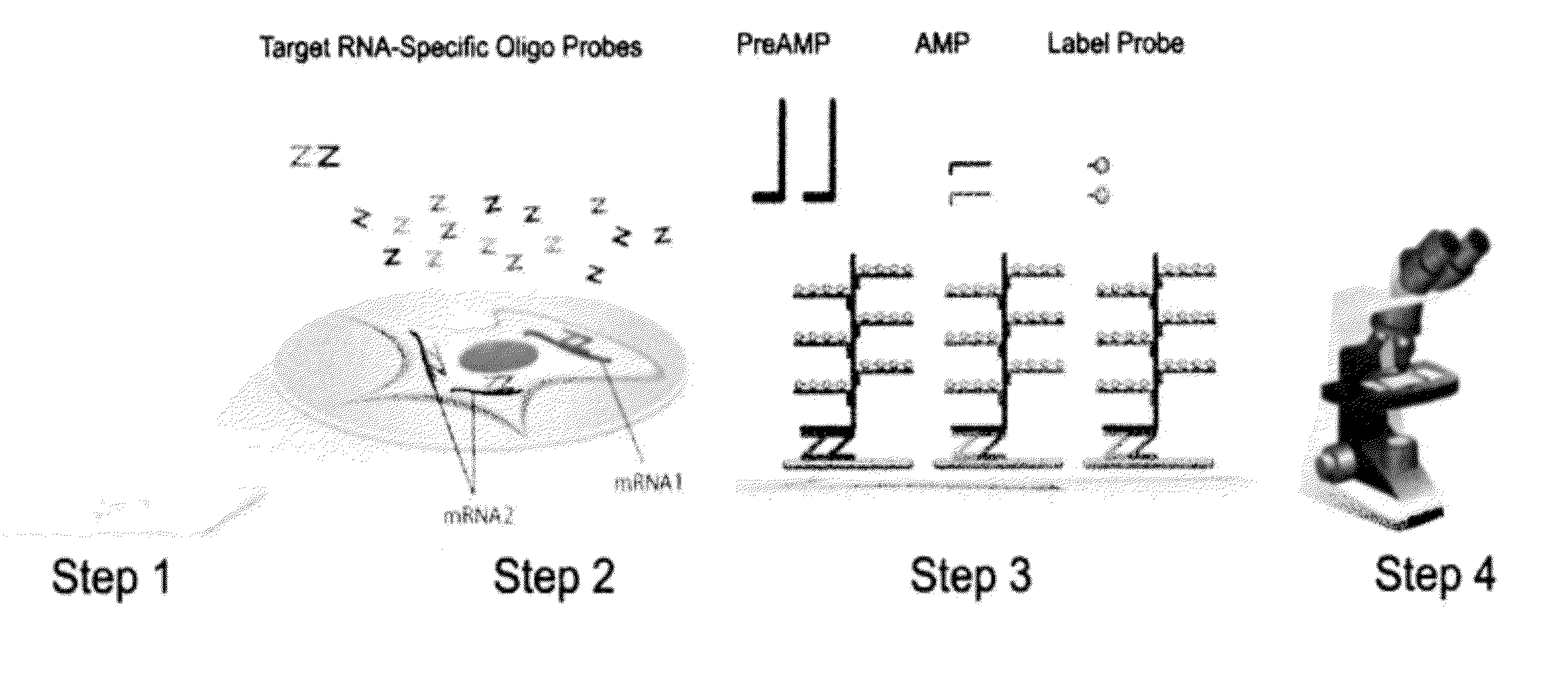
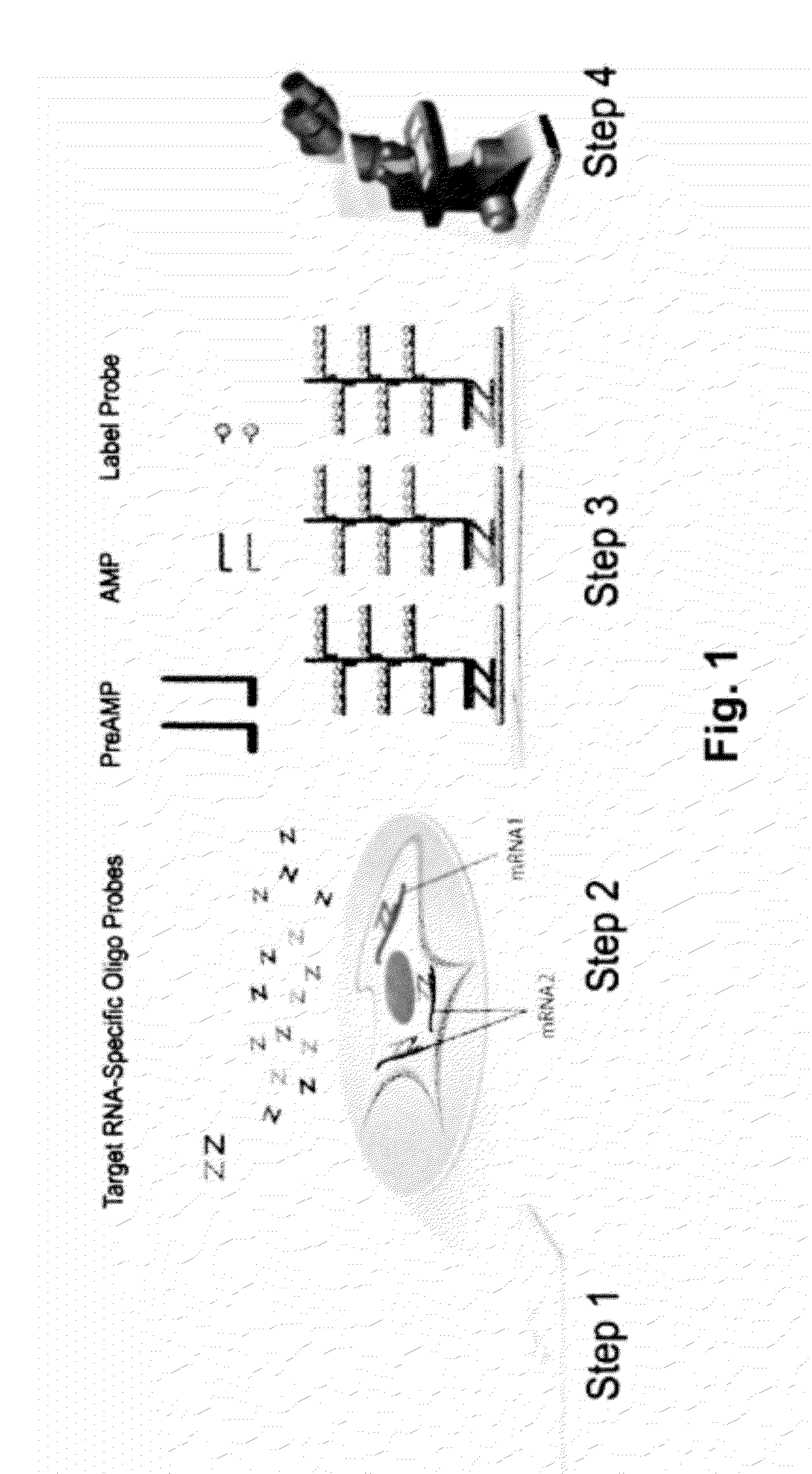
Protocol for the RNAscope® HPV Assay

[0074]For each tumor sample, 5-micron tissue sections were cut and mounted on glass slides. The following steps were performed for each tumor sample:

[0075]1. Obtaining a sample from a subject. The sample can be a tissue sample obtained from a patient without preservation treatment or in a formalin fixed, paraffin embedded tissue section.

[0076]2. Optionally, if the sample is in a FFPE tissue section, the sample will be treated, first by heating the FFPE tissue section in a citrate buffer, and followed by digestion with a protease.

[0077]3. Performing an RNAscope® HPV assay on the sample, which includes the following tests:[0078]i) performing negative control using target probes against the bacterial gene dapB;[0079]ii) performing positive control using target probes against human gene UBC;[0080]iii) performing a test using HPV target probe sets containing either a pool of high-risk HPV subtypes or individual target probe set specific to one of these...

example 2

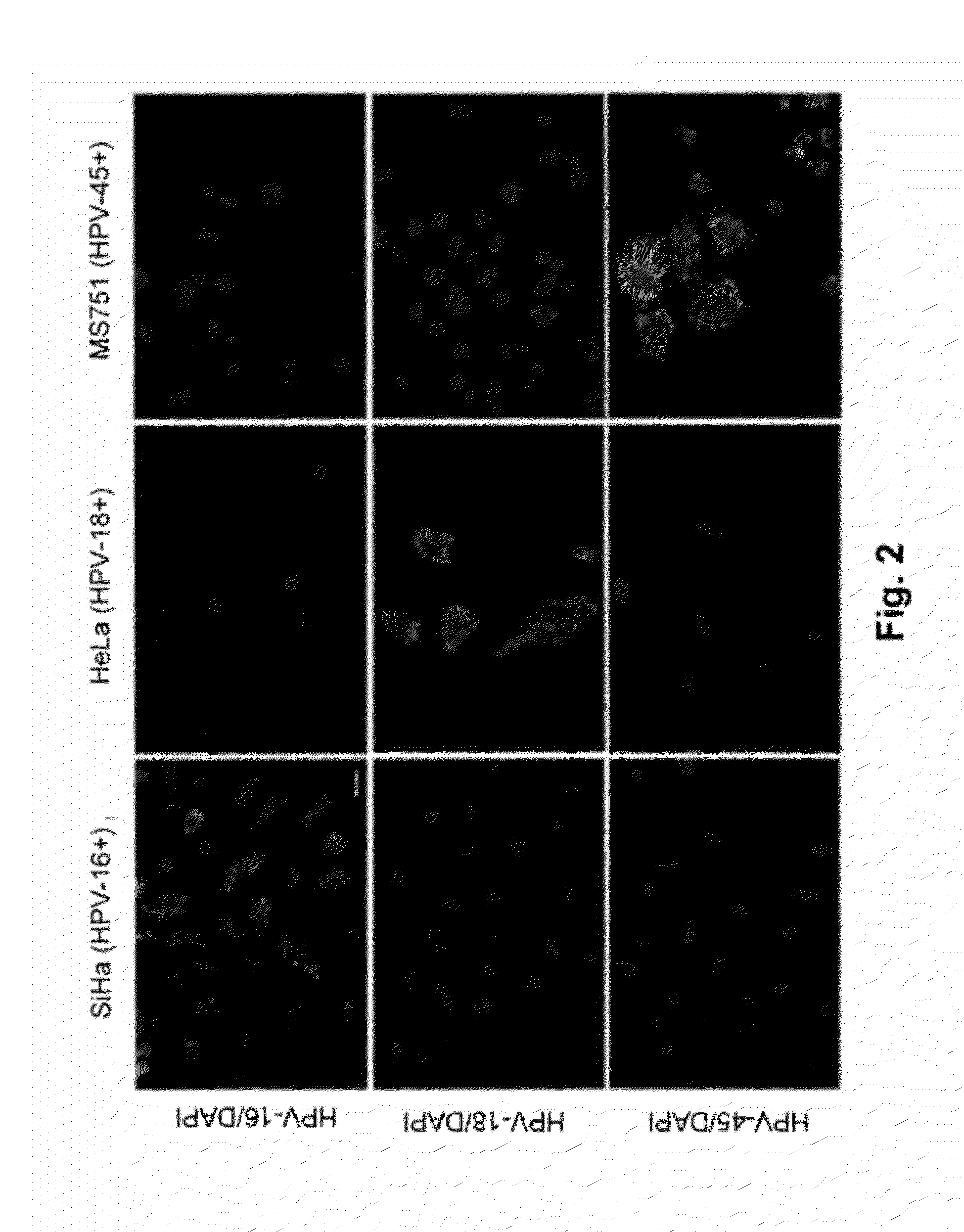
RNAscope® Detection of E6 / E7 mRNA of HPV Subtypes in Subtype-Specific Cell Lines

[0082]In order to demonstrate the feasibility of the RNAscope® HPV assay to detect and discriminate among different HPV subtypes, an experiment was conducted to detect HPV subtypes in cell lines which are known to contain only specific HPV subtypes. The protocol for the RNAscope® HPV assay described in Example 1 was used. The target probe sets are designed to be specific for E6 / E7 mRNA of HPV-16, HPV-18, and HPV-45, respectively. Hybridization of the probe sets to their target was detected using an alkaline phosphatase conjugated signal amplification system followed by development with Fast Red, which results in a red, fluorescent precipitate. As shown in FIG. 2, the HPV-16 target probe set produced a positive signal only in SiHa and CaSki cells, both of which harbor only the HPV-16 subtype. The HPV-18 target probe set produced a positive signal only in Hela cell lines which harbor only the HPV-18 subtyp...

example 3

RNAscope® Detection of HPV Subtypes in Head and Neck Cancer Tissue Samples

[0083]The following experiment was conducted to detect E6 / E7 mRNA of HPV high-risk subtypes in FFPE tissue samples obtained from HNC patients. In the experiment, the protocol for the RNAscope® HPV assay described in Example 1 was used. The two groups of target probe sets were created. In one group, the target probe sets contain only target probes that only bind to HPV-16. In the other group, the target probe sets contain target probes that bind to HPV-18, 31, 33, 35, 52, and 58 (called “HR-HPV probe set”). Multiple HNC patient samples who are diagnosed with cervical lesions were tested using the above groups of target probe sets. An exemplary result is shown in FIG. 3. The result shows detection of positive E6 / E7 mRNA of HPV high-risk subgroup in both patients. Both patients are known to have HPV-related HNC. Thus, the test proves that the RNAscope® HPV assay with the specifically designed HPV target probe set...


PUM

PropertyMeasurementUnit
reverse transcription real time PCRaaaaaaaaaa
epitluorescent microscopeaaaaaaaaaa
prognosis poweraaaaaaaaaa

Abstract

The present invention provides a method and a kit for determining whether a head and neck cancer is HPV-related. In one embodiment, an RNAscope® HPV assay was designed to detect the presence of E6 / E7 mRNA of certain high-risk HPV subtypes related to head and neck cancer. The present invention also provides a method and a kit for determining whether a cervical lesion is a benign lesion or a cervical intraepithethial neoplasm lesion. The present invention further provides a method for determining the progression of cervical intraepithethial neoplasm based on the spatial pattern and levels of the E6 / E7 mRNA of certain high-risk HPV subtypes. The present invention also provides a method for determining the risk of developing cervical cancer in a human diagnosed with cervical intraepithethial neoplasm based on presence and absence of the certain subgroups of high-risk HPV subtypes.

Description

CROSS-REFERENCE TO RELATED APPLICATIONS[0001]This application claims priority to and benefit of U.S. Provisional Application No. 61 / 437,337, filed Jan. 28, 2011, entitled “RNAscope™ HPV for Determining HPV Status in Head and Neck Cancers”.[0002]The entire contents and disclosures of the aforementioned applications is incorporated herein by reference into this application.FIELD OF INVENTION[0003]Provided herein are methods and kits for determining whether a head and neck cancer is related to human papillomavirus (HPV) based on the presence or the absence of E6 / E7 mRNA of high-risk HPV subtypes. Also provided herein are methods and kits for diagnosing whether or not a cervical lesion in a subject is benign based on the presence or absence of E6 / E7 mRNA of high-risk HPV subtypes. Further provided herein are methods for determining the progression of cervical intraepithelial neoplasm (CIN) based on the spatial pattern and expression level of E6 / E7 mRNA of high-risk HPV subtypes. Also pr...

Claims


Application Information

Patent Timeline
Patent Type & Authority Applications(United States)
IPC IPC(8): C12Q1/70C12M1/34
CPCC12Q1/6841C12Q1/6886C12Q2600/118C12Q2600/112C12Q1/708
Inventor MA, XIAO-JUNFLANAGAN, JOHNLUO, YULING
Owner ADVANCED CELL DIAGNOSTICS INC
Why Eureka
Try Eureka