
Measurement of PKA for Cancer Detection

Active Publication Date: 2012-12-20
TRAXXSSON
View PDF0 Cites 2 Cited by
  • Summary
  • Abstract
  • Description
  • Claims
  • Application Information

AI Technical Summary

Benefits of technology

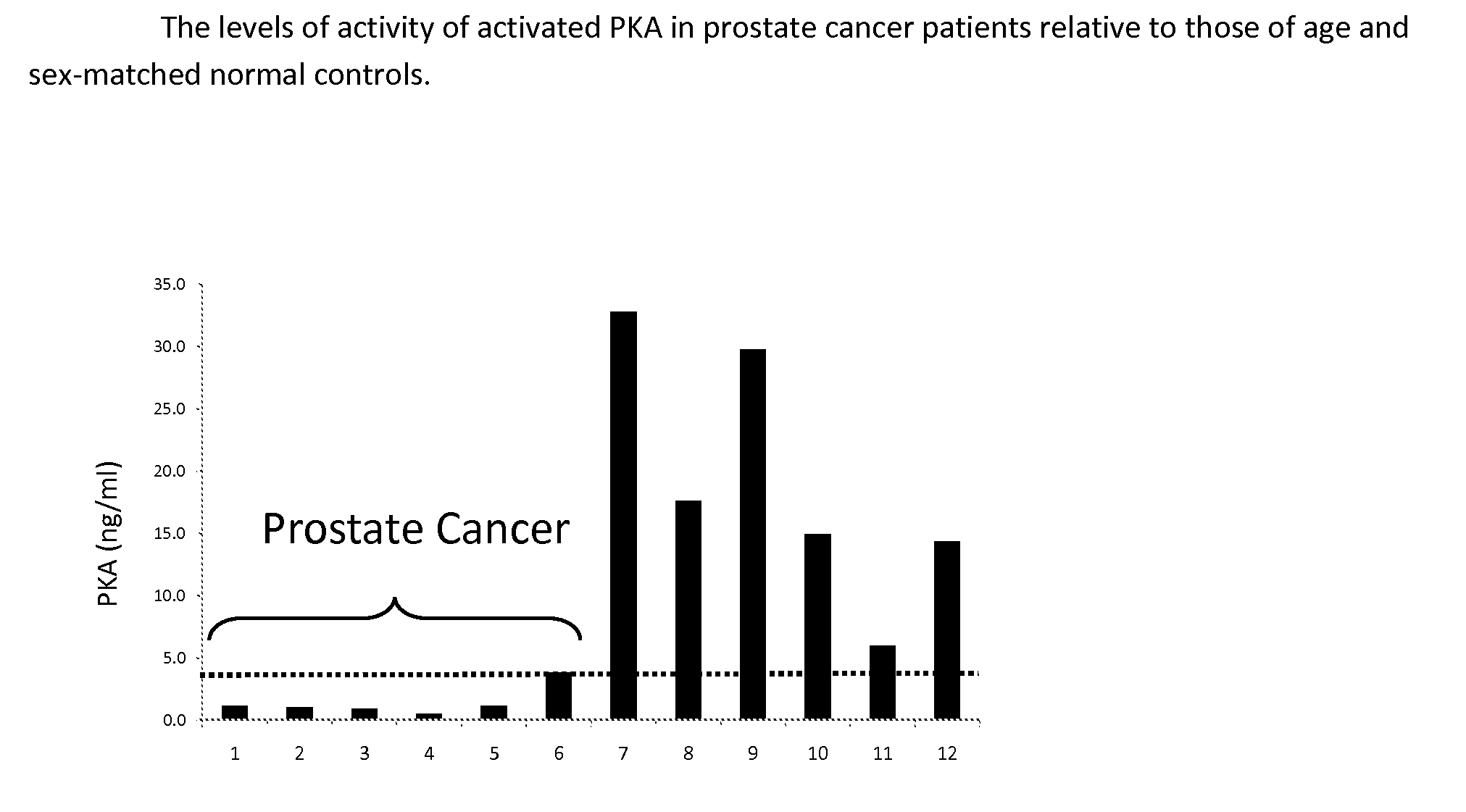
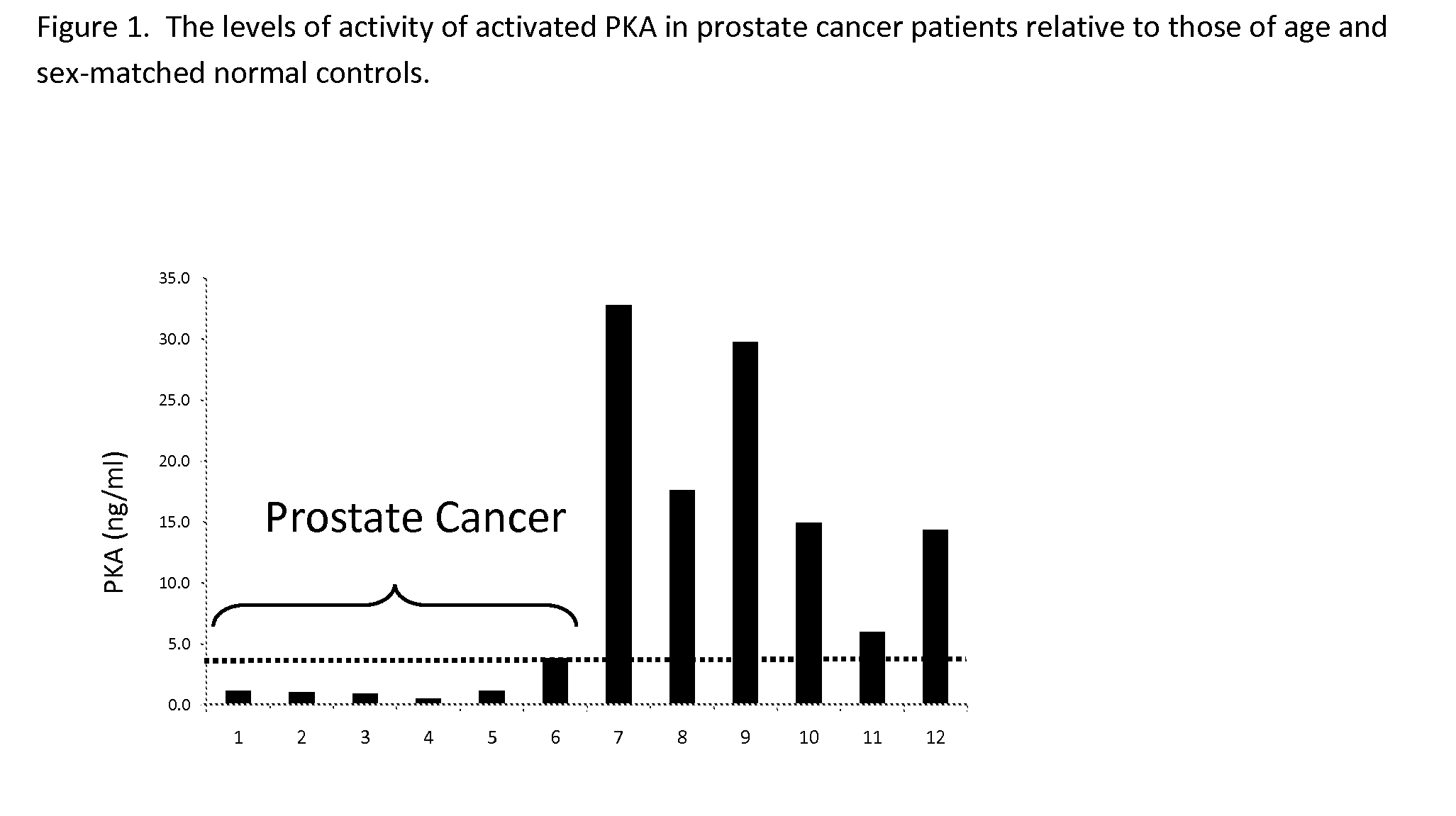
[0019]Extremely low levels of PKA activity are detectable in non-reduced blood samples making accurate measurement of enzyme activity very difficult. The addition of an antioxidant to the PKA assay activates the enzyme making it much easier to measure the PKA activity and to measure differences, especially decreases, in enzyme activity. Assaying activated PKA activity in blood samples provides identification of individuals who have cancer by virtue of their low levels of activated PKA activity. The detection of elevated levels of anti-PKA antibodies in addition to measurement of low activated PKA activity provides additional evidence that individuals have cancer.

Problems solved by technology

Mammograms have a poorer record than PSA tests.
What makes breast cancer screening costly is that an estimated 95% of the positive mammograms are false positives—the patient does not have cancer [13].
This brings the national cost for the estimated 2 million unnecessary breast biopsies to more than $5.1 billion annually.
Many of these tests are not specific for cancer or specific for a particular type of cancer, rendering them useless as cancer screening tests.

Method used


Image

  • Measurement of PKA for Cancer Detection
  • Measurement of PKA for Cancer Detection
  • Measurement of PKA for Cancer Detection

Examples


embodiment 1

[0024]Serum samples from patients with early and late-stage cancer were obtained from ProMedDx, LLC. In one embodiment blood samples from prostate cancer patients and normal controls presumably without cancer were assayed for activated PKA activity. In this assay activated PKA in samples was mixed with a defined peptide used as a substrate. Phosphorylation of the peptide was detected using biotinylated phosphoserine antibody, which was in turn was detected in an ELISA format using peroxidase-conjugated to streptavidin. Detection of the bound peroxidase was established using a color-producing peroxidase substrate included in the assay kit. Bovine PKA catalytic unit was used at varying concentrations to develop a standard activity curve. The detail of the assay protocol is described below.

[0025]Modified MESACUP Protein Kinase A Activity Assay[0026]1. Reference: Kit Instructions[0027]2. Materials[0028]a. MESACUP Protein Kinase Assay Kit (MBL Code No. 5230)[0029]b. ATP: 10 mM in water[0...

embodiment 2

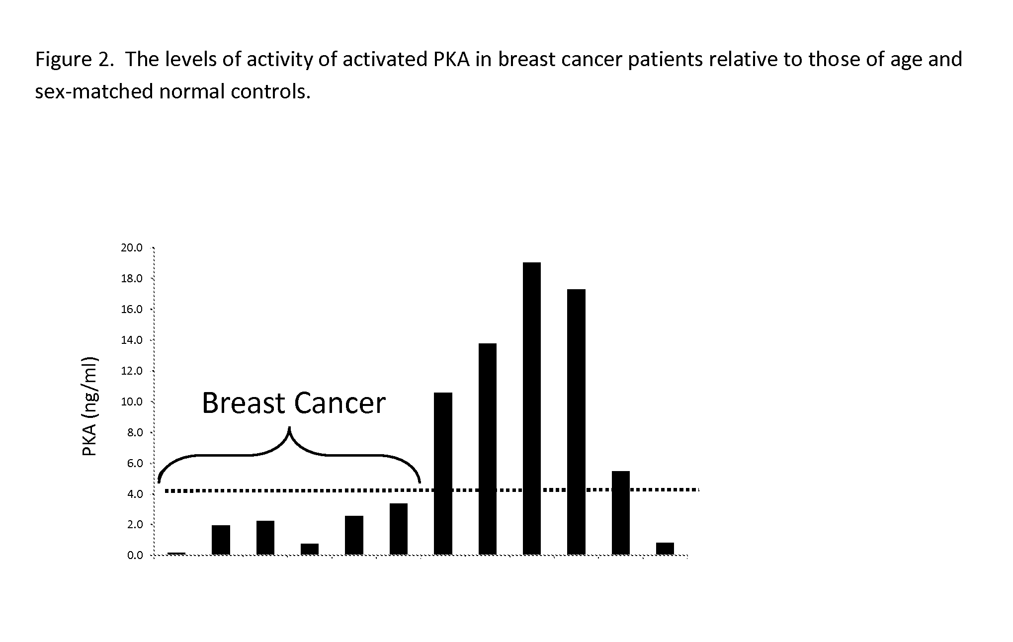
[0082]Blood samples from breast cancer patients and from age and sex-matched controls presumably without cancer were analyzed for activated PKA activity using the same protocol as used in Embodiment 1. The activity levels of activated PKA in blood from prostate cancer patients were lower (below 4 ng / ml) than those for samples from normal controls (FIG. 2).

embodiment 3

[0083]The same prostate cancer patient samples and related control samples used in Embodiment 1 were tested for anti-PKA antibodies as described below.

[0084]PKA Autoantibody ELISA[0085]1. Materials[0086]a. MaxiSorp 96 well polystyrene plates (NUNC Prod. No. 439454)[0087]b. PKA diluent: 25 mM KH2PO4, 5 mM EDTA, 150 mM NaCl, 50% (w / v) glycerol, 1 mg / ml BSA, 5 mM -mercaptoethanol, pH 6.5[0088]c. Protein Kinase A (Sigma Prod. No. P2645)[0089]i. Dissolve in PKA diluent and dilute to 50 g / ml[0090]ii. Store at −20° C.[0091]d. Coating buffer: 10 mM sodium phosphate, 150 mM sodium chloride, pH 7.4[0092]e. Coating wash buffer: 20 mM HEPES, 150 mM sodium chloride, 30 mM sucrose, pH 7.0 with 0.1% BSA[0093]f. Blocker Casein (Pierce Prod. No. 37532)[0094]g. Assay buffer: 10 mM sodium phosphate, 150 mM sodium chloride, pH 7.4 with 0.25% BSA and 0.1% Tween 20[0095]h. Assay wash buffer: 10 mM citrate, 150 mM sodium chloride, pH 5.1 with 0.1% Tween 20[0096]i. Detection antibody: peroxidase-conjugated...


PUM

PropertyMeasurementUnit
Molar densityaaaaaaaaaa

Abstract

The present invention relates to a method of detecting the presence of cancer by measuring the level of enzyme activity and autoantibodies in the blood of an individual. In particular the present invention relates to methods for measurement of activated cAMP-dependent protein kinase A (PKA) activity and antibodies to PKA, a kit for activated PKA activity measurement, and the use of the measured levels of these analytes for determining the presence of cancer.

Description

[0001]This application claims priority of U.S. provisional patent 61 / 251,769 filed on Oct. 15, 2009.TECHNICAL FIELD[0002]The present invention is in the field of diagnostics and relates to methods for determining the presence, activity, and concentrations of certain protein biomarkers and their use in determining the presence of cancer.BACKGROUND ART[0003]The cAMP-dependent protein kinase (PKA) is activated by the binding of cAMP to the regulatory subunit (R), of the molecule and results in the release of the active catalytic kinase subunit (C). Most of the effects of cAMP in eukaryotic systems are the result of phosphorylation of proteins at serine or threonine residues by PKA. There are several isoforms of both subunits of PKA. PKA is localized subcellularly by binding to multidomain scaffolding proteins known as AKAPs which bind to the R subunits of the holoenzyme [1]. More than 50 AKAPs are known which localize PKA in various cell types. The PKA-specific inhibitor (PKI) acts by ...

Claims


Application Information

Patent Timeline
IPC IPC(8): G01N33/573
CPCG01N33/57434G01N2333/91205
Inventor PUSKAS, ROBERTHELD, DOUGLAS
Owner TRAXXSSON
Learn More