

Apparatus and method for obtaining and providing imaging information associated with at least one portion of a sample, and effecting such portion(s)
a technology of imaging information and apparatus, applied in the field of apparatus and methods for obtaining and providing imaging information associated with at least one portion of a sample, and affecting such portion(s), can solve the problems of poor image quality of current devices, hampered wide adoption of microendoscopy, and good image quality with small diameter bundles, etc., to overcome limitations and deficiencies, improve image quality, and safe and effective treatment
- Summary
- Abstract
- Description
- Claims
- Application Information
AI Technical Summary
Benefits of technology
Problems solved by technology
Method used
Image

Examples
Embodiment Construction
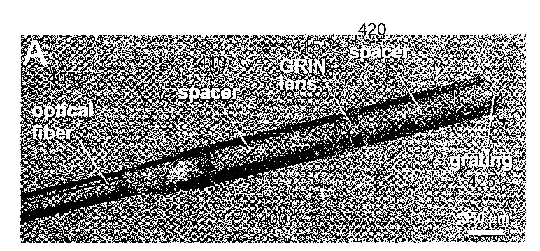
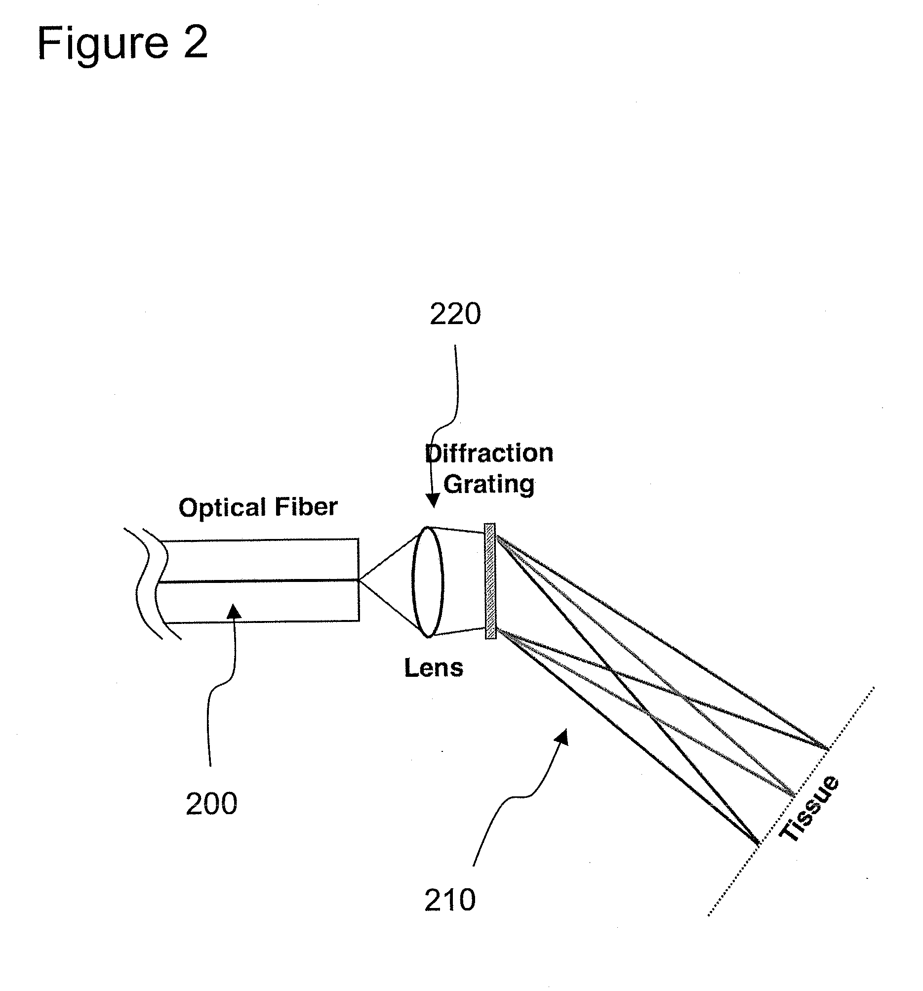
[0018]One of the objects of the present invention is to provide an ultraminiature (e.g., 350 μm diameter) endoscopic imaging apparatus / device / arrangement / system with integrated laser therapy capabilities for microsurgical applications inside the body, e.g., for safe and effective treatment of twin-twin transfusion syndrome (TTTS). It is possible to overcome the limitations and deficiencies of the current TTTS therapy devices by providing a much smaller microsurgical endoscope that provides more informative images with a better image quality. This exemplary enhancement includes spectrally encoded endoscopy (SEE) concepts that use wavelength division multiplexing to obtain high-resolution images through a single optical fiber. The exemplary SEE system and probe can be provided for color differentiation of arterial and venous placental vessels and can add Doppler imaging to quantify blood flow. Additionally, it is possible to incorporate therapeutic laser delivery through the same prob...
PUM
 Abstract
Description
Claims
Application Information