UV imaging for intraoperative tumor delineation

a tumor and intraoperative technology, applied in the field of cameras, can solve the problems of long time to acquire tissue data, ultrasound is limited by signal artifacts, and the scan cannot be used intraoperatively

Inactive Publication Date: 2013-05-02
CALIFORNIA INST OF TECH
View PDF8 Cites 4 Cited by
  • Summary
  • Abstract
  • Description
  • Claims
  • Application Information

AI Technical Summary

Benefits of technology

[0010]According to one aspect, the invention features a medical imaging system. The medical imaging system comprises a camera having a back illuminated silicon imaging detector that is sensitive in the visible and in the UV portions of the electromagnetic spectrum, the camera having a filter with a pass band in the UV, the filter being controllable to allow the camera to receive UV without visible illumination when the filter is engaged, and being controllable to allow the camera to receive visible illumination when the filter is removed from an optical path, the camera having at least one control port and having at least one output port; as required if not already present in a location of use, a UV illumination source configured to illuminate the field ...

Problems solved by technology

On the other hand, PET scans cannot be used intraoperatively.
Ultrasound is limited by signal artifacts caused by blood and surgical trauma at the resection margin once the resection starts.
Unfortunately, in the case of photosensitizers the photosignaling is dependent on the injection of specific light sensitive compounds.
In the case of time resolved laser induced fluorescenc...

Method used


Image

  • UV imaging for intraoperative tumor delineation
  • UV imaging for intraoperative tumor delineation
  • UV imaging for intraoperative tumor delineation

Examples


Embodiment Construction

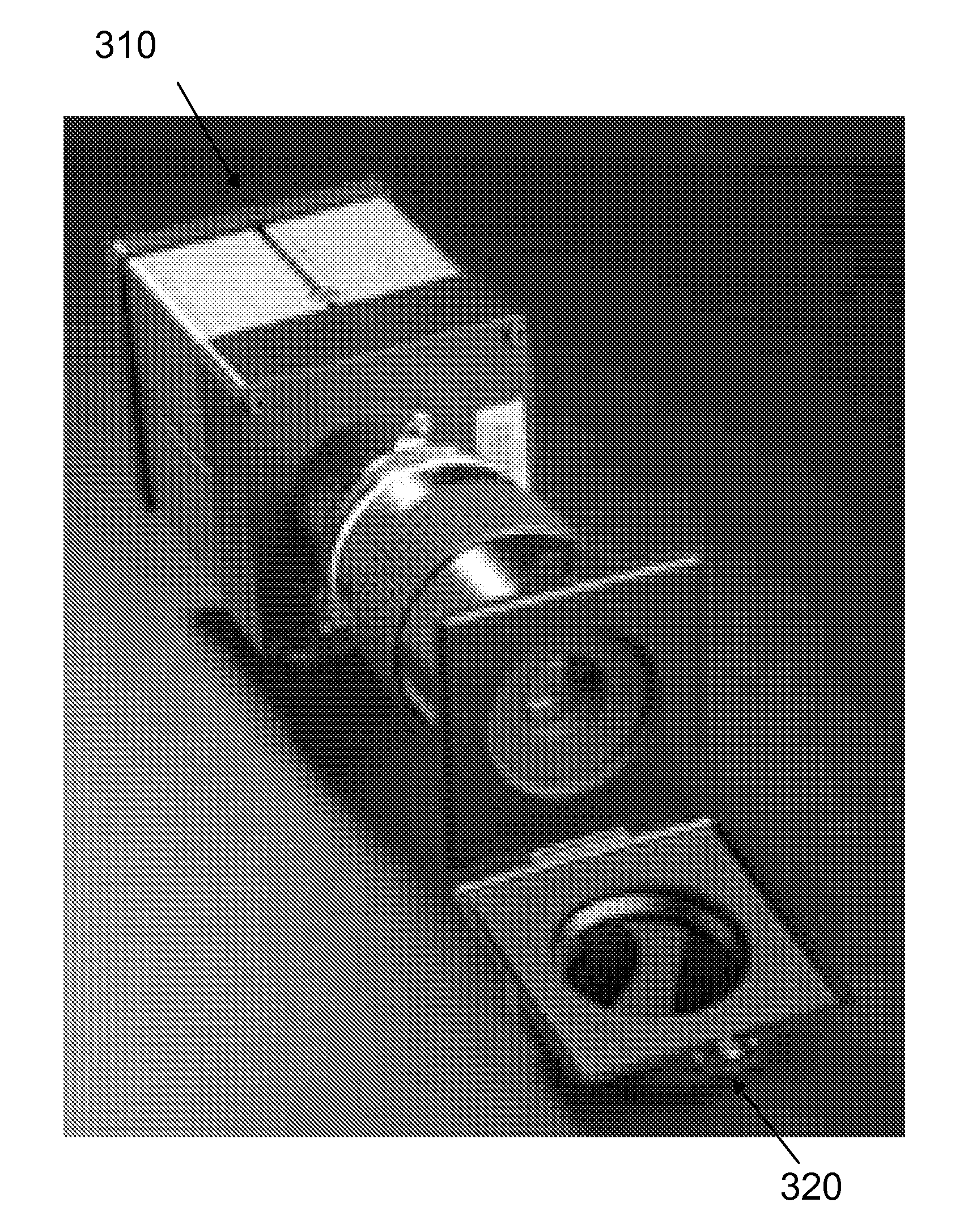
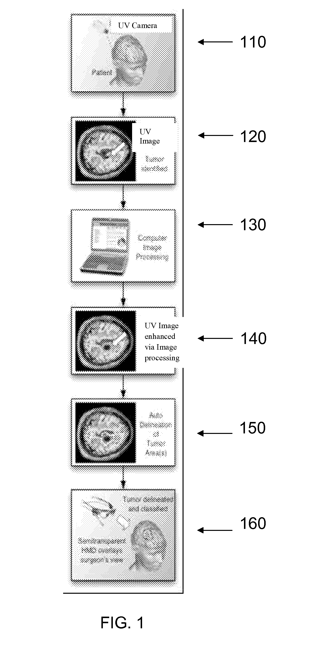
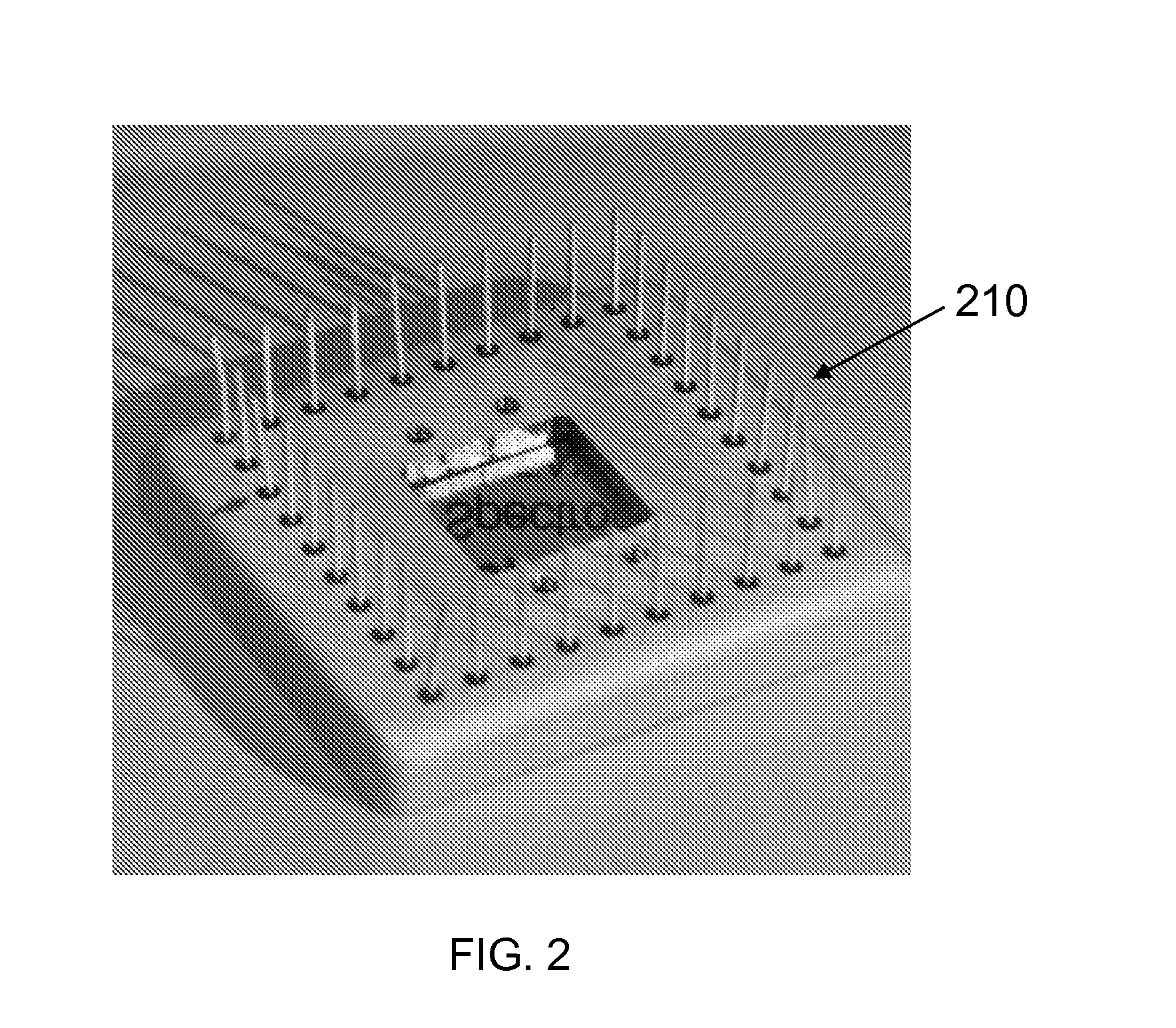
[0044]Ultraviolet imaging (UVI) of brain tissue is expected to be a useful tool for intraoperative delineation of tumor resection margins. One of the significant variables affecting the survival and quality of life of patients with gliomas is the completeness of tumor resection. Although recent advances in neuroimaging have opened a new window of opportunity for neurosurgeons to obtain more extensive information regarding the location, the invasiveness and metabolic properties of brain tumors, current techniques still cannot provide real-time intraoperative feedback about the completeness of the resection.

[0045]Intraoperative use of a UV camera according to principles of the invention are expected to provide a tool for neurosurgeons to achieve 100% tumor resection. Previous experimental studies have provided significant capability of similar techniques involving UV observations with other instruments in enhancing optical imaging of human skin cancer, as described in the Rehua paper ...


PUM


Abstract

A medical imaging system and method. A UV/visible camera uses a back illuminated silicon imaging detector to observe a surface of a brain of a human subject in vivo during brain surgery for excision of a cancerous tumor. The detector can be a CCD detector or a CMOS detector. Under UV illumination, the camera can record images that can be processed to detect the location and extent of a cancerous tumor because the presence of auto-fluorescent NADH variations can be detected between normal and cancerous cells. The image data is processed in a general purpose programmable computer. In some instances, an image is also taken using visible light, and the identified cancerous region is displayed as an overlay on the visible image.

Description

CROSS-REFERENCE TO RELATED APPLICATIONS[0001]This application claims priority to and the benefit of co-pending U.S. provisional patent application Ser. No. 61 / 554,122 filed Nov. 1, 2011, which application is incorporated herein by reference in its entirety.STATEMENT REGARDING FEDERALLY FUNDED RESEARCH OR DEVELOPMENT[0002]The invention described herein was made in the performance of work under a NASA contract, and is subject to the provisions of Public Law 96-517 (35 USC 202) in which the Contractor has elected to retain title.THE NAMES OF THE PARTIES TO A JOINT RESEARCH AGREEMENT[0003]Caltech—JPL and Cedars Sinai Medical Center located in Los Angeles, Calif. have an agreement under which the camera of the invention is used under operating room conditions.FIELD OF THE INVENTION[0004]The invention relates to cameras in general and particularly to a camera system and methods used to detect cancerous cells.BACKGROUND OF THE INVENTION[0005]Computed tomography (CT), magnetic resonance ima...

Claims


Application Information

Patent Timeline
IPC IPC(8): A61B5/00
CPCA61B5/0077A61B5/0071A61B5/0042A61B5/7425
Inventor NIKZAD, SHOULEHHOENK, MICHAEL E.JONES, TODD J.CHENG, SAMUEL R.
Owner CALIFORNIA INST OF TECH
Why Eureka
Try Eureka