Novel Liposome Nanoparticles for Tumor Magnetic Resonance Imaging

a tumor magnetic resonance imaging and nanoparticle technology, applied in the direction of diagnostic recording/measuring, dispersed delivery, biocide, etc., can solve the problems of low sensitivity, limited mr imaging utility, and difficult formulation of high-concentration liposomes using this lipid

Inactive Publication Date: 2013-05-23
IMPERIAL INNOVATIONS LTD +1
View PDF2 Cites 35 Cited by
  • Summary
  • Abstract
  • Description
  • Claims
  • Application Information

AI Technical Summary

Problems solved by technology

This is due to the fact that MRI suffers from an inherent lack of sensitivity and often in order to diagnose pathology correctly, a paramagnetic contrast agent is injected intravenously into patients to further enhance the magnetic resonance (MR) signal and hence site of disease.
However, as these agents are non-specific and are cleared within a few hours of injection,

Method used


Image

  • Novel Liposome Nanoparticles for Tumor Magnetic Resonance Imaging
  • Novel Liposome Nanoparticles for Tumor Magnetic Resonance Imaging
  • Novel Liposome Nanoparticles for Tumor Magnetic Resonance Imaging

Examples


fifth embodiment

Preferred aspects of this fifth embodiment include:

(31) use according to (30) in the preparation of a magnetic resonance contrast agent for enhancing a magnetic resonance image of a tumour in a mammal.

(32) use according to (30) or (31), wherein the concentration of said liposomes in said magnetic resonance contrast agent is 1-50 mg / mL, more preferably 1-30 mg / mL.

In a sixth aspect of the present invention, there is provided:

(33) a method of magnetic resonance imaging of an organ or organ structure in a mammal, comprising the steps of:

(a) administering the magnetic resonance contrast agent according to (27) or (28) to a patient; and

(b) taking images of the organ of interest in the patient.

sixth embodiment

Preferred aspects of this sixth embodiment include:

(34) a method according to (33), wherein said magnetic resonance contrast agent is used for enhancing a magnetic resonance image of a tumour in a mammal.

(35) a method according to (33) or (34), wherein the concentration of liposomes in said magnetic resonance contrast agent is 1-50 mg / mL, more preferably 1-30 mg / mL.

In a seventh aspect of the present invention, there is provided:

(36) a method of magnetic resonance imaging of an organ or organ structure in a mammal pre-administered with the magnetic contrast agent according to (27) or (28) comprising the step of:

(i) taking images of the organ of interest in the patient.

In an eighth aspect of the present invention, there is provided:

(37) a method of making a liposome according to (1) to (26) comprising mixing a solution of Gd.DOTA.DSA (gadolinium(III)2-14,7-bis-carboxymethyl-10-[(N,N-distearylamidomethyl-N′-amido-methyl]-1,4,7,10-tetra-azacyclododec-1-yl}-acetic acid) and a solution of...

eighth embodiment

A preferred aspect of the eighth embodiment includes:

(38) drying the mixture (e.g. in vacuo) and optionally rehydrating the resulting liposome.

In a ninth aspect of the present invention, there is provided:

(39) a method of making a magnetic contrast agent according to (27) or (28) comprising mixing a liposome of (1) to (26) and a pharmaceutically acceptable carrier.

Preferred aspects of the eighth and ninth embodiments are the same as those listed above in relation to the first, second and third aspects.

DETAILED DESCRIPTION OF THE INVENTION

[0020]We will now discuss the present invention in further detail. The present invention may also be further understood by reference to FIGS. 1 to 29, wherein:

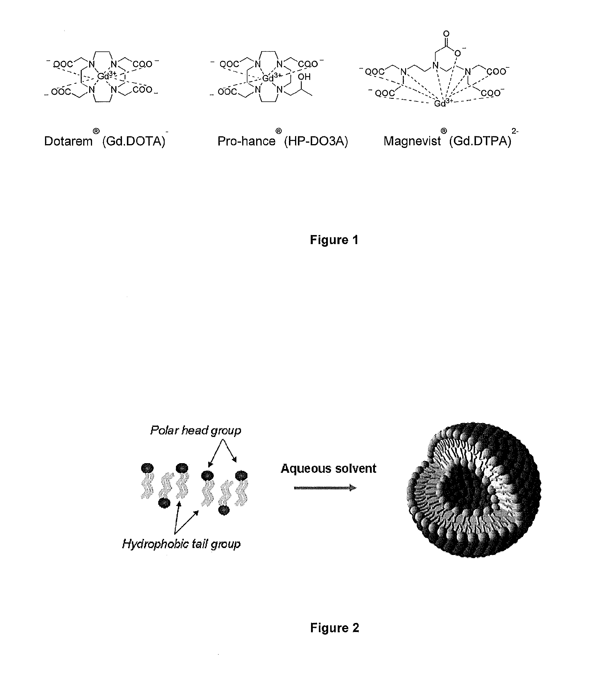
[0021]FIG. 1 shows Gadolinium based clinical contrast agents approved by the FDA;

[0022]FIG. 2 shows liposome formation from amphipathic lipids;

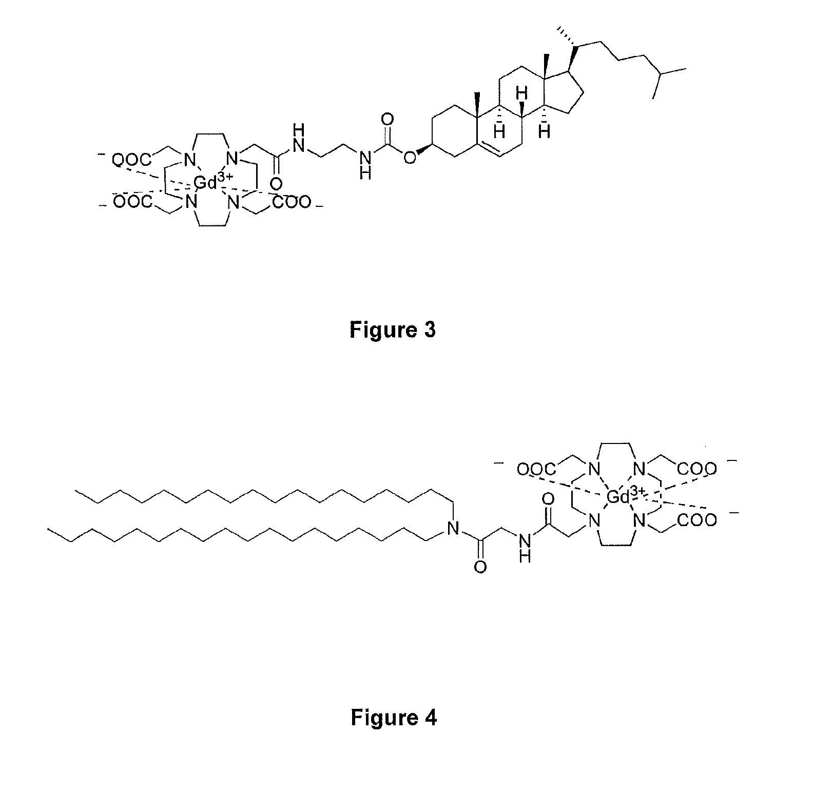
[0023]FIG. 3 shows Gd.DOTA.Chol, a T1 lipidic contrast agent component of MAGfect;

[0024]FIG. 4 shows the paramagnetic gadolinium lipid target, Gd.DOTA....


PUM

PropertyMeasurementUnit
Concentrationaaaaaaaaaa
Particle sizeaaaaaaaaaa

Abstract

The present invention provides novel liposomes comprising Gd.DOTA.DSA (gadolinium(III)2-{4,7-bis-carboxymethyl-10-[(N,N-distearylamidomethyl-N′-amido-methyl]-1,4,7,10-tetra-azacyclododec-1-yl}-acetic acid), characterised in that said liposome further comprises a neutral, fully saturated phospholipid component (e.g. DSPC (1,2-distearoyl-sn-glycero-3-phosphocholine]), which are of particular use in the preparation of magnetic resonance contrast agents for enhancing a magnetic resonance image of tumours in a mammal.

Description

FIELD OF THE INVENTION[0001]The present invention relates to novel liposomes suitable for use in the preparation of a contrast agent for use in enhancing magnetic resonance imaging (MRI), particularly in enhancing magnetic resonance images of tumours.BACKGROUND TO THE INVENTION[0002]Cancer imaging is one of the most important disease areas where molecular imaging is set to play a major role, both in the detection of cancer and subsequent treatment. For effective cancer imaging by magnetic resonance imaging (MRI) there is a clear need to develop effective and biocompatible molecular imaging probes.1,2 In this field, nanotechnology has a lot to offer as nanomedicine is set to make considerable contributions in the important areas of drug delivery, disease detection, and therapy. The application of nanotechnology platforms to cancer imaging has opened up opportunities for the use of multifunctional nanoparticle systems such as liposomes, in the study of cancer detection and therapy.[00...

Claims


Application Information

Patent Timeline
IPC IPC(8): A61K49/18
CPCA61K49/1812Y10S977/773B82Y5/00
Inventor KAMALY, NAZILAKALBER, TAMMY LOUISEKENNY, GAVIN DAVIDTHANOU, MAYAMILLER, ANDREW DAVIDBELL, JIMMY
Owner IMPERIAL INNOVATIONS LTD
Why Eureka
Try Eureka