System and method for ultrasonic examination of the breast

Inactive Publication Date: 2013-10-10
BERMAN MICHAEL
View PDF0 Cites 44 Cited by
  • Summary
  • Abstract
  • Description
  • Claims
  • Application Information

AI Technical Summary

Benefits of technology

[0091]The system of the invention may comprise one or more C-Arm-tomography-sensors and one or more 2D-array-sensors. The sensors may be connected to a step-motor-assembly configured to drive the ultrasound sensors over the scanning device. The step-motor-assembly may include any one or more of a motor, an encoder, a processor, an indexer and a driver. The C-

Problems solved by technology

However, conventional mammography does not detect all breast cancers, including some that are palpable, and as many as three-quarters of all breast lesions biopsied because of a suspicious finding on a mammogram turn out to be benign.
Mammograms are particularly difficult to interpret for women with dense breast tissue, who are at increased risk of breast cancer.
The dense tissue interferes with the identification of abnormalities associated with tumors.
However, the varied, heterogeneous, complex structure of the breast makes good breast ultrasound imaging more difficult than in many other regions of the body.
Conventional ultrasound has a limited field of view, is not reproducible and produces results that are a trade-off between penetration and image resolution.
It is generally perceived that ultrasound is incapable of reliably detecting micro-calcifications, these being early indications of breast cancer.
Conventional two dimensional [2D] ultrasound procedures dynamically distort the breast during the examination, thereby making it difficult to determine the exact location of tumors or other masses.
The 2D nature of conventional ultrasound and the distortion of the breast during the examination, make it difficult to g

Method used


Image

  • System and method for ultrasonic examination of the breast
  • System and method for ultrasonic examination of the breast
  • System and method for ultrasonic examination of the breast

Examples


Embodiment Construction

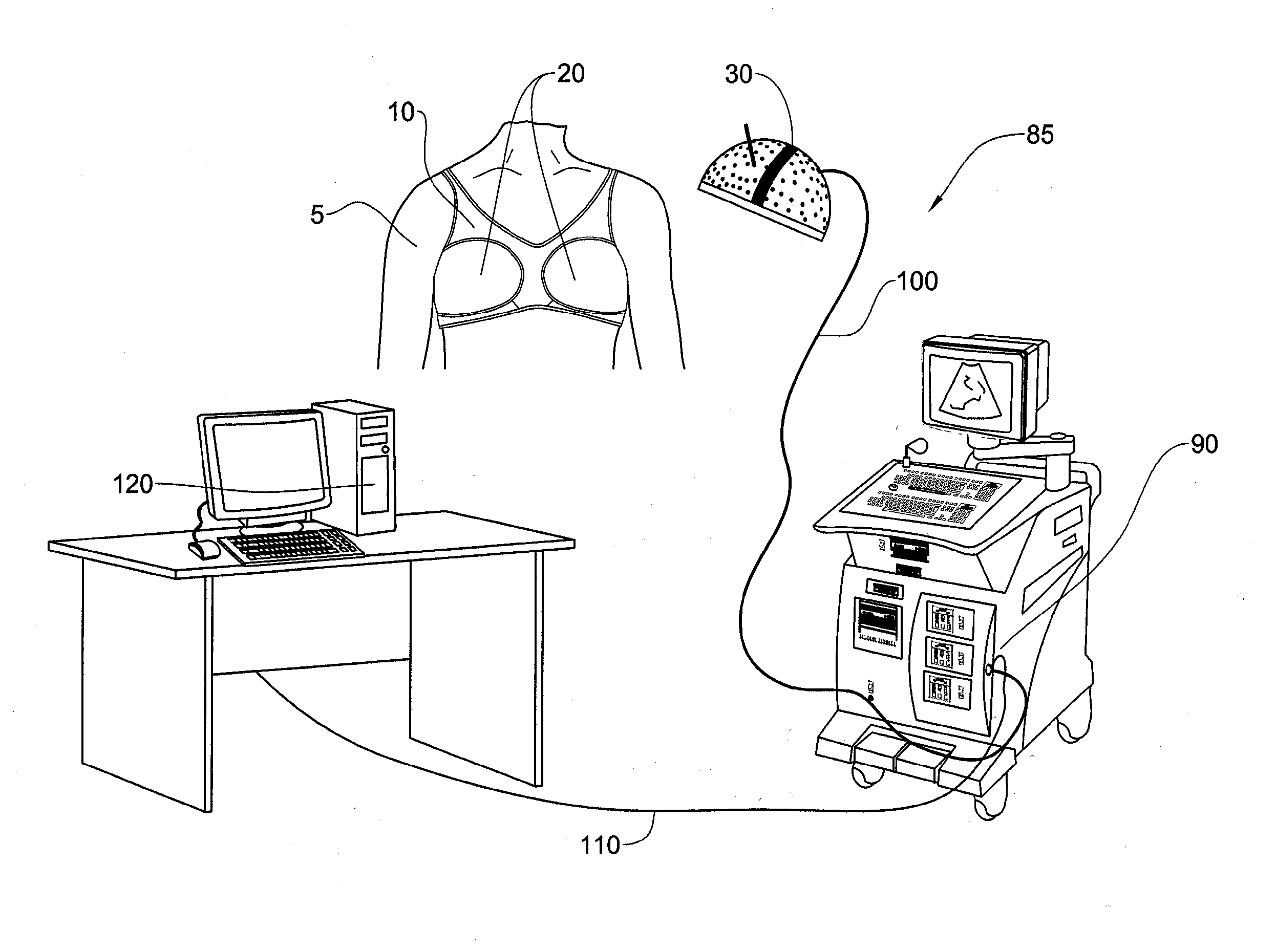
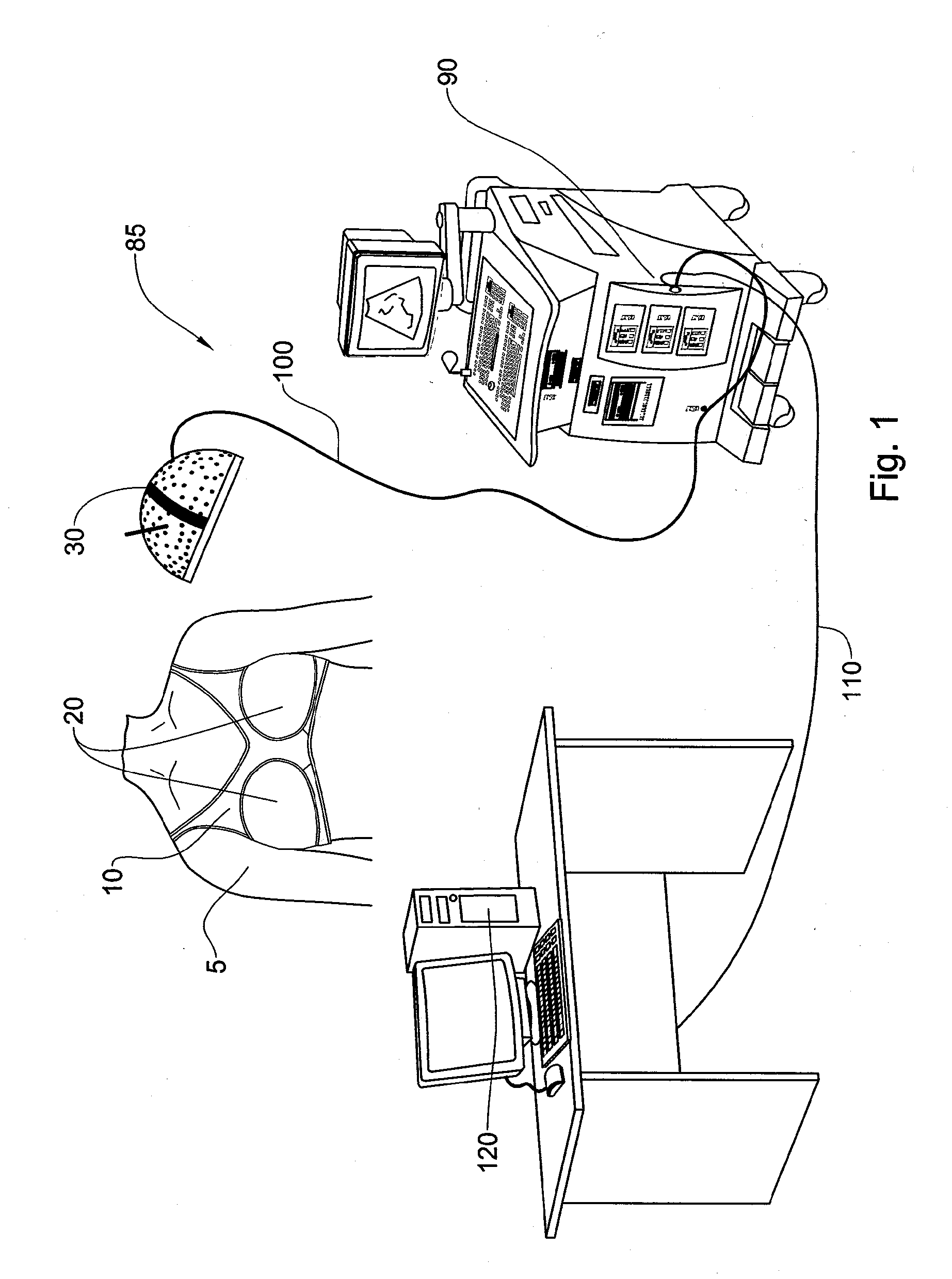
[0122]For the sake of clarity, and ease of description, the present invention will be described in relation to breast imaging, it being evident that the system and method of the invention can be modified to image any desired body part.

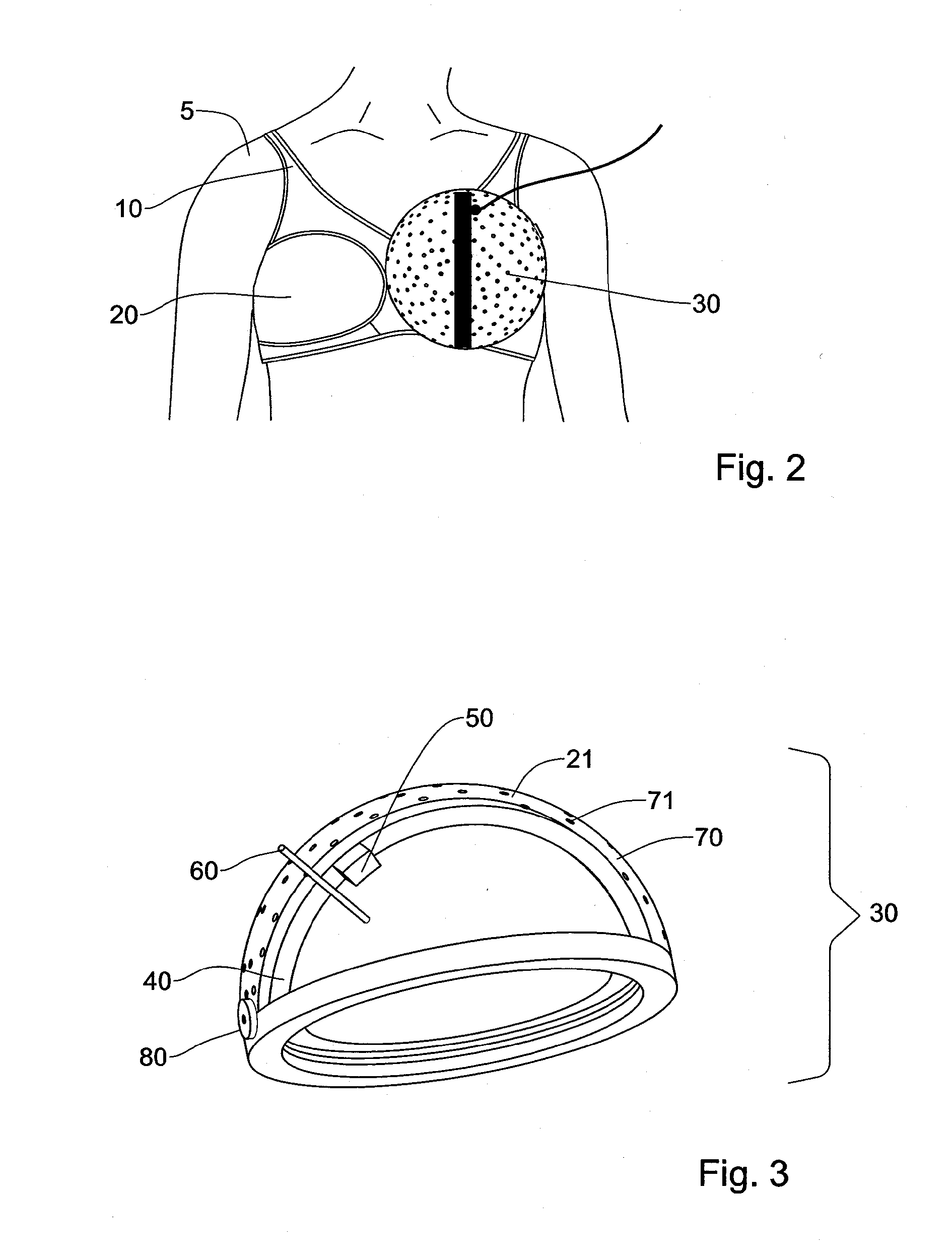
[0123]FIG. 1 shows a system 85 for ultrasound imaging of a breast in accordance with one embodiment of the invention. The system 85 comprises a dome shaped scanning device 30, described in detail below, configured to receive in its interior a breast of an individual 5. The scanning-device 30 is anchored to an ultrasound system 90 over a cable-assembly 100. A control-cable 110 connects the ultrasound system 90 to a workstation 120. The work station 120 may include a CRT screen 123 for displaying images. A user input device, such as a keypad 124 allows a user to input various parameters relating to the examination, such as personal details of the individual being examined, or the parameters of the ultrasound radiation (frequency, intensity, etc.).

[0124]I...


PUM


Abstract

The invention provides a system and method for limited view ultrasound imaging of a 2D section or a 3D volume of a body part. Ultrasound sensors configured are spatially or temporally arrayed in a limited view circular arc or over at least part of a concave surface such as a hemisphere. A processor calculates from detected ultrasound radiation a beam forming (BF) functional and calculates from the free amplitudes a point spread function (PSF). A filter g(k) is calculated from the Fourier transform HBF(k) of the PSF that is used to generate an image of the 2D section or the 3D volume of the body part.

Description

PRIORITY INFORMATION[0001]This National Stage application claims priority to U.S. Provisional Application No. 61 / 420,098 filed on Dec. 6, 2010.FIELD OF THE INVENTION[0002]This invention relates to medical devices and more specifically to such devices for medical imaging by ultrasound.BACKGROUND OF THE INVENTION[0003]The following prior art publications are considered to be relevant for an understanding of the prior art.[0004]1. Report on “Mammography and beyond: developing technologies for the early detection of breast cancer”,[0005]2. U.S.A. Institute of Medicine and the Governing Board of the National Research Council, National Academy Press, 2101 Constitution Avenue, N.W., Box 285, Washington, D.C. 20055.[0006]3. Chandra M. Sehgal et al, Journal of Mammary Gland Biology and Neoplasia, Volume 11, Number 2, April, 2006.[0007]4. W. E. Svensso, “Breast Ultrasound Update”, ULTRASOUND, February 2006, Volume 14, Number 1, 20-33.[0008]5. J. F. Greenleaf et al on quantitative cross-sectio...

Claims


Application Information

Patent Timeline
IPC IPC(8): A61B8/00A61B8/08
CPCA61B8/0825A61B8/406A61B8/461A61B8/4461A61B8/4477A61B8/466
Inventor BERMAN, MICHAEL
Owner BERMAN MICHAEL
Why Eureka
Try Eureka