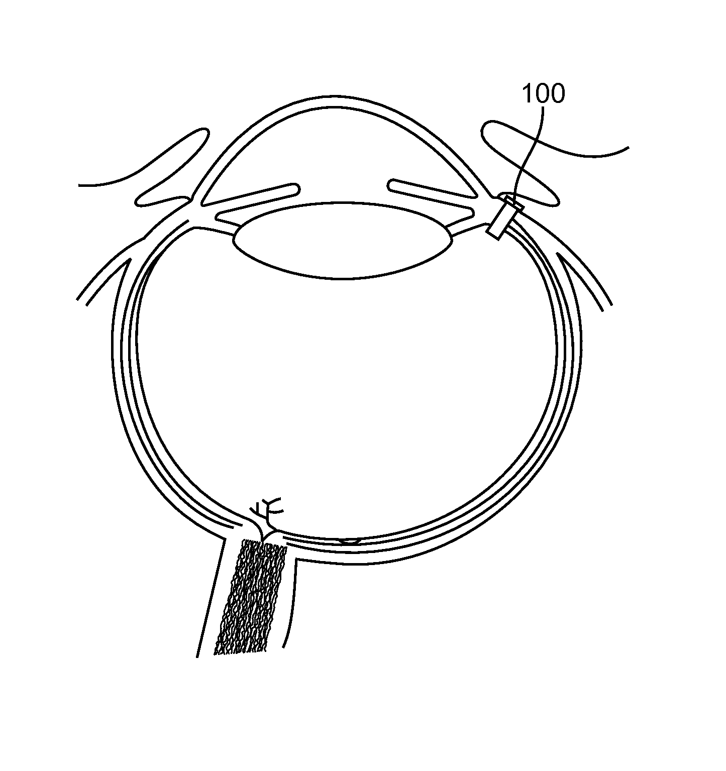
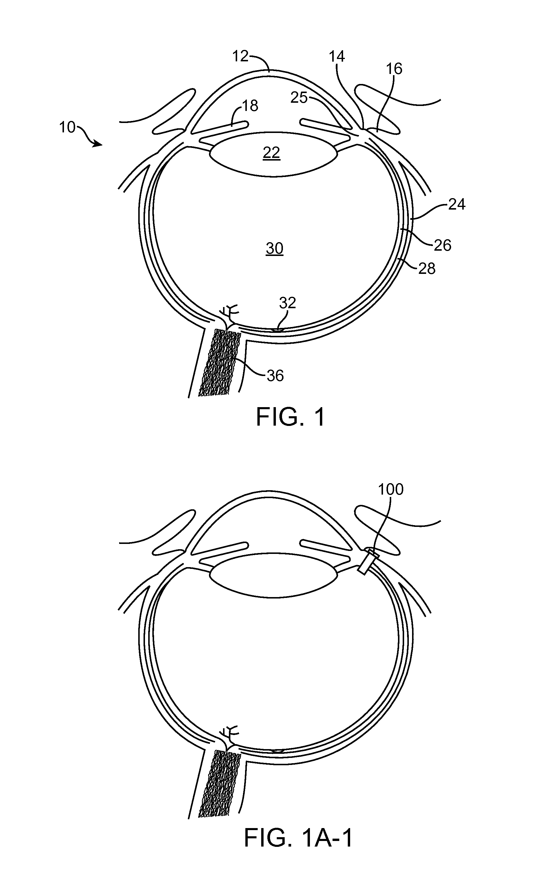
Subconjuctival implant for posterior segment drug delivery
- Summary
- Abstract
- Description
- Claims
- Application Information
AI Technical Summary
Benefits of technology
Problems solved by technology
Method used
Image

Examples
example 5
Release of Protein Through a Cylindrical Sintered Porous Titanium Cylinder
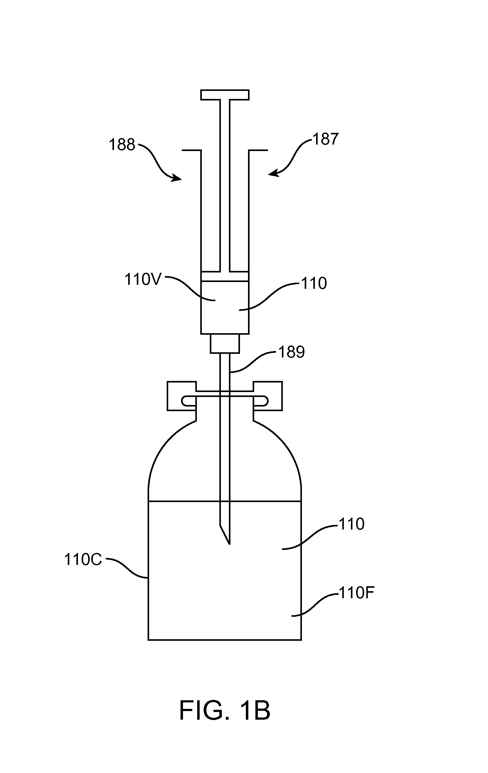
[0344]Reservoirs were fabricated from syringes and sintered porous titanium cylinders (available from Applied Porous Technologies, Inc., Mott Corporation or Chand Eisenmann Metallurgical). These were sintered porous cylinders with a diameter of 0.062 inches and a thickness of 0.039 inches prepared from titanium particles. The porosity is 0.17 with mean pore sizes on the order of 3 to 5 micrometers. The porous cylinder is characterized as 0.2 media grade according to measurements of bubble point. The porous cylinders were press-fit into sleeves machined from Delrin. The sleeves exposed one entire planar face to the solution in the reservoir and the other entire planar face to the receiver solution in the vials, corresponding to an area of 1.9 square millimeters. The tips were cut off of 1 mL polypropylene syringes and machined to accept a polymer sleeve with outer diameter slightly larger than the inner diamete...
example 6
Release of Protein Through Masked Sintered Porous Titanium Cylinders
[0348]Reservoirs were fabricated from syringes and porous sintered titanium cylinders similar to that described in Example 5. The porous sintered titanium cylinders (available from Applied Porous Technologies, Inc., Mott Corporation or Chand Eisenmann Metallurgical) had a diameter of 0.082 inch, a thickness of 0.039 inch, a media grade of 0.2 and were prepared from titanium particles. The porosity is 0.17 with mean pore sizes on the order of 3 to 5 micrometers. The porous cylinder is characterized as 0.2 media grade according to measurements of bubble point. The porous cylinders were press fit into sleeves machined from Delrin. The sleeves exposed one entire planar face to the solution in the reservoir and the other entire planar face to the receiver solution in the vials, corresponding to an area of 3.4 square millimeters. The tips were cut off of 1 mL polycarbonate syringes and machined to accept a polymer sleeve ...
example 7
Release of Protein Through Sintered Porous Stainless Steel Cylinder (Media Grade 0.1)
[0354]Prototype devices were fabricated from tubing and sintered porous stainless steel cylinders (available from Applied Porous Technologies, Inc., Mott Corporation or Chand Eisenmann Metallurgical) which are cylindrical with diameter 0.155 inch and thickness 0.188 inch prepared from 316L stainless steel particles. The porous cylinder is characterized as 0.1 media grade according to measurements of bubble point. This study was performed with these large, off-the-shelf porous cylinders with an area of 12 mm2 in order to characterize the resistive properties of 0.1 media grade stainless steel.
[0355]These devices were prepared using Teflon-FEP heat shrink tubing (Zeus, #37950) and a hot air gun to shrink around the porous cylinders on one end and a custom prepared septum on the other end (Nusil MED1 4013 silicone molded to 0.145 inch diameter). The reservoir volume (46+ / −2 uL) was determined from the ...
PUM
 Abstract
Description
Claims
Application Information
 - Generate Ideas
- Intellectual Property
- Life Sciences
- Materials
- Tech Scout
- Unparalleled Data Quality
- Higher Quality Content
- 60% Fewer Hallucinations
Browse by: Latest US Patents, China's latest patents, Technical Efficacy Thesaurus, Application Domain, Technology Topic, Popular Technical Reports.
© 2025 PatSnap. All rights reserved.Legal|Privacy policy|Modern Slavery Act Transparency Statement|Sitemap|About US| Contact US: help@patsnap.com