Stain-free protein quantification and normalization

a protein and quantification technology, applied in the field of stain-free protein quantification and normalization, can solve the problems of inability to expect, limited recognition of the usefulness of stain-free technique to date, and inability to compare different protein bands in different samples

Active Publication Date: 2013-10-31
BIO RAD LAB INC
View PDF5 Cites 17 Cited by
  • Summary
  • Abstract
  • Description
  • Claims
  • Application Information

AI Technical Summary

Benefits of technology

[0012]Provided herein are methods of using haloalkylated tryptophan fluorescence to quantify and compare the total amount(s) of protein in one or more target protein samples, and normalize the amount(s) of one or more proteins within or among protein samples.

Problems solved by technology

For this reason, recognition of the usefulness of the stain-free technique to date has been limited.
Comparisons between different protein bands in different samples would not be useful, since the different proteins are most likely to have different tryptophan contents and thus cannot be expected to produce the same signal intensities when present in the same amounts.
The tryptophan variances do not necessarily limit the usefulness of the technique to the comparison of individual proteins, however.
Not only is the process time consuming, but inevitably, the stripping process will remove some level of antigen, thereby compromising downstream results.
Unfortunately this method assumes that there is no variability within the process and this is not always the case.
Although this method accounts for variations in the sample preparation and gel loading it does not account for blotting efficiency and it adds time and complexity to the process.
This method accounts for variability in sample preparation and transfer efficiency but it still adds time and complexity to the process.

Method used


Image

  • Stain-free protein quantification and normalization
  • Stain-free protein quantification and normalization
  • Stain-free protein quantification and normalization

Examples


example 1

Correlation of Haloalkylated Tryptophan Fluorescence Intensity with Cell Lysate Total Protein Quantity

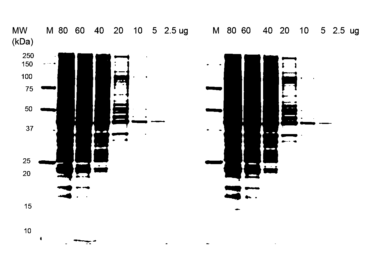
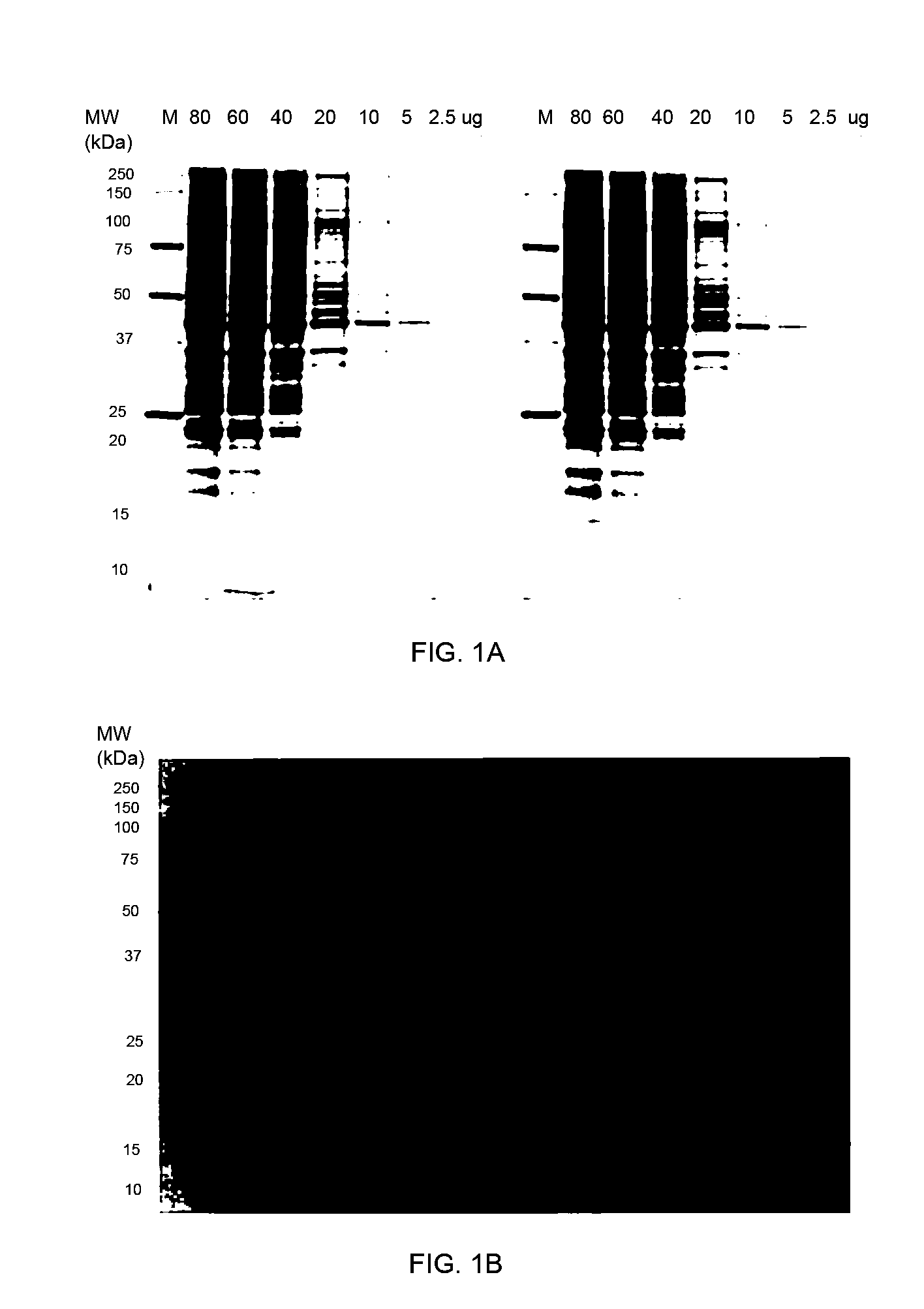
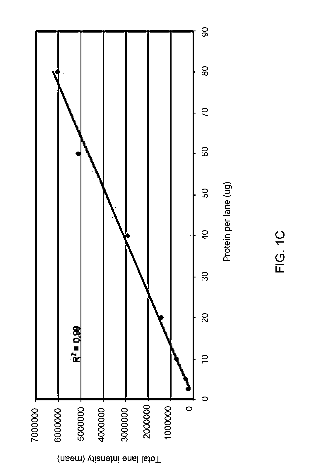
[0084]Stain-free technology enables fluorescent visualization of SDS PAGE gels and corresponding blots. FIG. 1a shows an electropherogram of HeLa cell lysate (2.5-80 μg total protein per lane) separated by TGX “any kD” Criterion Stain-Free 1-D SDS gel (18 slots) and imaged with ChemiDoc MP, where the M lane designates the marker proteins. FIG. 1b shows a Western blot of the same cell lysate separated in the same gels with the same lane loadings, the blot being on a nitrocellulose membrane and imaged with ChemiDoc MP, where the M lane again represents the marker proteins. The quality of SDS-PAGE separations before blotting can be easily monitored and the transfer efficiency of the blotting process can be quickly inspected by imaging both the membrane (see FIG. 1b) and the SDS gel after the blotting process. The relative amount of total protein in each lane on the blot can be calculat...

example 2

Invariance of Total Haloalkylated Tryptophan Fluorescence with Origin and Composition of Protein Extract

[0086]To determine whether the source and composition of a diverse mixture of proteins significantly affects the total amount of haloalkylated tryptophan fluorescence detected from the mixture, protein extracts were obtained from different organisms. The extracts were then diluted to known concentrations to obtain reference protein samples, which were detected on blotting membranes following haloalkylation. The total amount of haloalkylated tryptophan fluorescence detected from each sample was correlated with the total amount of protein in the sample.

[0087]Protein extracts were obtained from rat liver, soybean, and human serum, and the concentration of protein in each extract was determined using established spectrophotometric methods. Reference protein samples prepared from the extracts (60, 40, 20, 10, 5, 2.5, 1.25 μg) were loaded in duplicate onto Criterion TGX 12-2 well “any k...

example 3

Western Blot Analysis with Stain-Free Technology Validates Semi-Quantitative Protein Profiling Studies

[0089]Proteomic technologies like 2-D PAGE are customarily used in semi-quantitative protein profiling studies. After identifying and characterizing proteins that are expressed in different amounts by mass spectrometry, the quantitative data needs to be confirmed by a second, independent method like Western blotting.

[0090]The effect of γ-irradiation on a human LCL cell line (lymphoblastoid cell line) was studied by 2-D PAGE. Among many other proteins that were expressed in different amounts, MCM7, a DNA replication factor, was identified to be down-regulated by about a factor of two in the irradiated LCL sample as compared to a non-irradiated control. To validate this result, a quantitative Western blotting experiment was performed with monoclonal antibodies raised against MCM7. Two data normalization methods, namely data normalization with the GAPDH HKP and the stain-free technolog...


PUM

PropertyMeasurementUnit
wavelengthaaaaaaaaaa
temperatureaaaaaaaaaa

Abstract

Disclosed herein are methods of protein quantification and normalization using haloalkylated tryptophan fluorescence. Complex protein samples, i.e., samples that each contain 1,000 or more distinct proteins, from diverse sources that do not have common protein profiles are treated with a halo-substituted organic compound (i.e. haloalkane) that reacts with tryptophan residues to form fluorescent products. Irradiation of the samples with ultraviolet light and the detection and quantification of the resultant fluorescent emissions from all proteins in each sample are then used to obtain comparative values for total protein content among the various samples. The values thus obtained are found to be valid indications of comparative total protein content, despite the fact that the tryptophan levels vary widely among the various proteins in any single sample and the samples, due to the diversity of their origins, tend to differ among themselves in the identities and relative amounts of the proteins that they contain. Protein samples are also normalized to correct for differences in sample dilution, sample loading, and protein transfer inconsistencies, by using stain-free detection of total protein in each of the samples, or detection of subsamples within each sample.

Description

CROSS-REFERENCES TO RELATED APPLICATIONS[0001]The present application claims priority from U.S. Provisional Application No. 61 / 639,692, entitled “Stain-free total protein quantification of complex samples from diverse sources” and filed Apr. 27, 2012, the entire contents of which are herein incorporated by reference for all purposes. The present application also claims priority from U.S. Provisional Application No. 61 / 639,686, entitled “Stain-free technology for western blot total normalization” and filed Apr. 27, 2012, the entire contents of which are herein incorporated by reference for all purposes.BACKGROUND OF THE INVENTION[0002]Laboratory procedures in biotechnology frequently include the detection, identification, and quantitation of individual proteins. Certain procedures also however require the quantitation of entire mixtures of proteins aside from the individual proteins in each mixture, and with or without separating or differentiating the individual proteins from each o...

Claims


Application Information

Patent Timeline
Patent Type & Authority Applications(United States)
IPC IPC(8): G01N33/53G16B25/10
CPCG01N33/53G16B99/00G01N33/6827G16B25/10G06T7/0012G06T11/60G06T2207/30004
Inventor FREEBY, STEVELIU, NINGMCDONALD, KEVINPAULUS, ARANPOSCH, ANTON
Owner BIO RAD LAB INC
Why Eureka
Try Eureka