Diagnosis of melanoma by nucleic acid analysis

a nucleic acid analysis and melanoma technology, applied in the field of melanoma diagnosis by nucleic acid analysis, can solve the problems of dermatologists' limitations, missed diagnosis of some melanomas, and difficult procedure for diagnosing melanoma, so as to improve the ability of clinicians to identify, high accuracy, and high sensitivity of methods

Inactive Publication Date: 2015-01-01
DERMTECH INT
View PDF1 Cites 9 Cited by
  • Summary
  • Abstract
  • Description
  • Claims
  • Application Information

AI Technical Summary

Benefits of technology

[0009]Described herein, in certain embodiments are pigmented skin lesion assays. In some embodiments, the pigmented skin lesion assay is employed for the diagnoses of melanoma. In some embodiments, the methods provided herein address unmet needs in pigmented lesion diagnosis. For example, the pigmented lesion assays provided herein demonstrate a high accuracy (e.g., at least about 80%, at least about 90%, at least about 93%, at least about 95%, or greater) in differentiating melanoma from non-melanoma using histopathology as a reference standard. In certain instances, the high sensitivity of the methods provided herein will improve the clinician's ability to identify pigmented lesions that are demonstrating malignant changes so that they can be surgically biopsied for histopathologic diagnosis. In certain instances, the high specificity will minimize the number of surgical biopsies required to identify these melanomas. In some embodiments, the negative predictive value of the assays provided exceeds about 98%, about 98.5%, about 99%, or about 99.5%. Thus, in some embodiments, the probability of a false negative test is less than about 2%, less than about 1.5%, less than about 1%, or less than about 0.5%.
[0010]In some embodiments, the assays provided herein analyze the expression of at least 2 genes in tissue samples collected using an adhesive patch. In some embodiments, relative to non-melanoma skin lesions, one of the two genes is down-regulated and the one of the two genes is up-regulated. In some embodiments, analysis of the two genes allows for accurate identification of gene expression consistent with melanoma.
[0011]In some embodiments, the samples for the assays described herein are obtained via non-invasive methods. In some embodiments, the samples are obtained using an adhesive patch applied to the skin. Because the assays provided herein are, at least in certain embodiments, performed on samples collected using non-invasive methods, the pigmented skin lesion assays provided herein provide a valuable non-invasive tool for the clinician to determine the need for one or more surgical biopsies. As demonstrated herein, including but not limited to in the examples provided, the pigmented skin lesion assays described herein have high sensitivity and high specificity. Practice of the assays provided herein, in certain embodiments, substantially reduces the number of unnecessary surgical biopsies performed. In addition, because the assays provided herein demonstrate a high sensitivity, the accuracy of diagnosis in lesions that are sent to pathology is improved which provides greater patient care.

Problems solved by technology

Diagnosis of melanoma has historically remained a difficult procedure.
This results in the need for invasive surgical biopsies of large numbers of pigmented lesions for every possible melanoma that is detected (many samples of which are later determined to not be melanoma at all), and also missed diagnosis some melanomas in their early stages.
The limitations of visual detection are apparent to dermatologists who are in need of ways to better determine whether suspicious lesions are melanoma or not without having to perform a biopsy first.
This still leads to an unacceptable sensitivity in melanoma detection and still results in the need to biopsy large numbers of benign lesions to detect a few melanomas and missed diagnoses.
In addition, optical-based devices, such as MelaFind remain controversial in the field of dermatology due to concerns over false positive rates leading to unnecessary biopsies and false negatives leading to undiagnosed cancers.
Currently, there are no technologies in the U.S. or outside the U.S. that are considered the standard of care for aiding the assessment of pigmented skin lesions to the Applicants' knowledge.
As a result, the performance of the technology (e.g., accuracy, sensitivity and specificity) exceeds that of other technologies.

Method used


Image

  • Diagnosis of melanoma by nucleic acid analysis
  • Diagnosis of melanoma by nucleic acid analysis
  • Diagnosis of melanoma by nucleic acid analysis

Examples


example 1

Tape Stripping to Recover Nucleic Acids from Pigmented Lesions

[0160]The following procedure was used to recover nucleic acids from pigmented lesions and / or skin suspected of melanoma of a subject. In contrast to normal skin, lesional skin should have a preoperative biopsy diameter of greater than or equal to about 6 mm, but less than that of the tape disc. Multiple lesions must be at least about 4 mm apart. The area of tape that touches the lesion should be generously demarcated on the tape with an insoluble ink pen so that this area may be cut away from the surrounding tape at the laboratory as part of the RNA extraction procedure.

[0161]As above, tapes were handled with gloved hands at all times. The site is shaved, if necessary, to remove non-vellus hairs. The site is then cleansed with an alcohol wipe (70% isopropyl alcohol). The site is then air-dried completely before application of the tape. It is recommended to approximately 2 minutes to ensure the site is completely dry befo...

example 2

RNA Quantitation and Profiling

[0165]The study was divided into two separate phases, a sample collection and characterization phase (phase 1) and an RNA profiling phase (phase 2). In phase 1 the tape stripped specimens and biopsied sample collections were performed by the principal investigator or trained individuals delegated by the principal investigator to obtain the biopsy sample at various sites. All biopsies are subject to standard histopathologic analysis. The RNA profiling phase (Phase 2), includes, but is not limited to RNA purification and hybridization to DNA microarrays for gene expression profiling.

[0166]Materials and reagents. Adhesive tape was purchased from Adhesives Research (Glen Rock, Pa.) in bulk rolls. These rolls were custom fabricated into small circular discs, 17 millimeters in diameter, by Diagnostic Laminations Engineering (Oceanside, Calif.). Human spleen total RNA was purchased from Ambion (catalogue #7970; Austin, Tex.). RNeasy RNA extraction kit was purc...

example 3

Assessment of Performance of Multi-Gene Biomarker for Melanoma Detection

Custom TaqMan OpenArray (OA) qPCR Plate (See Figure to the Right)

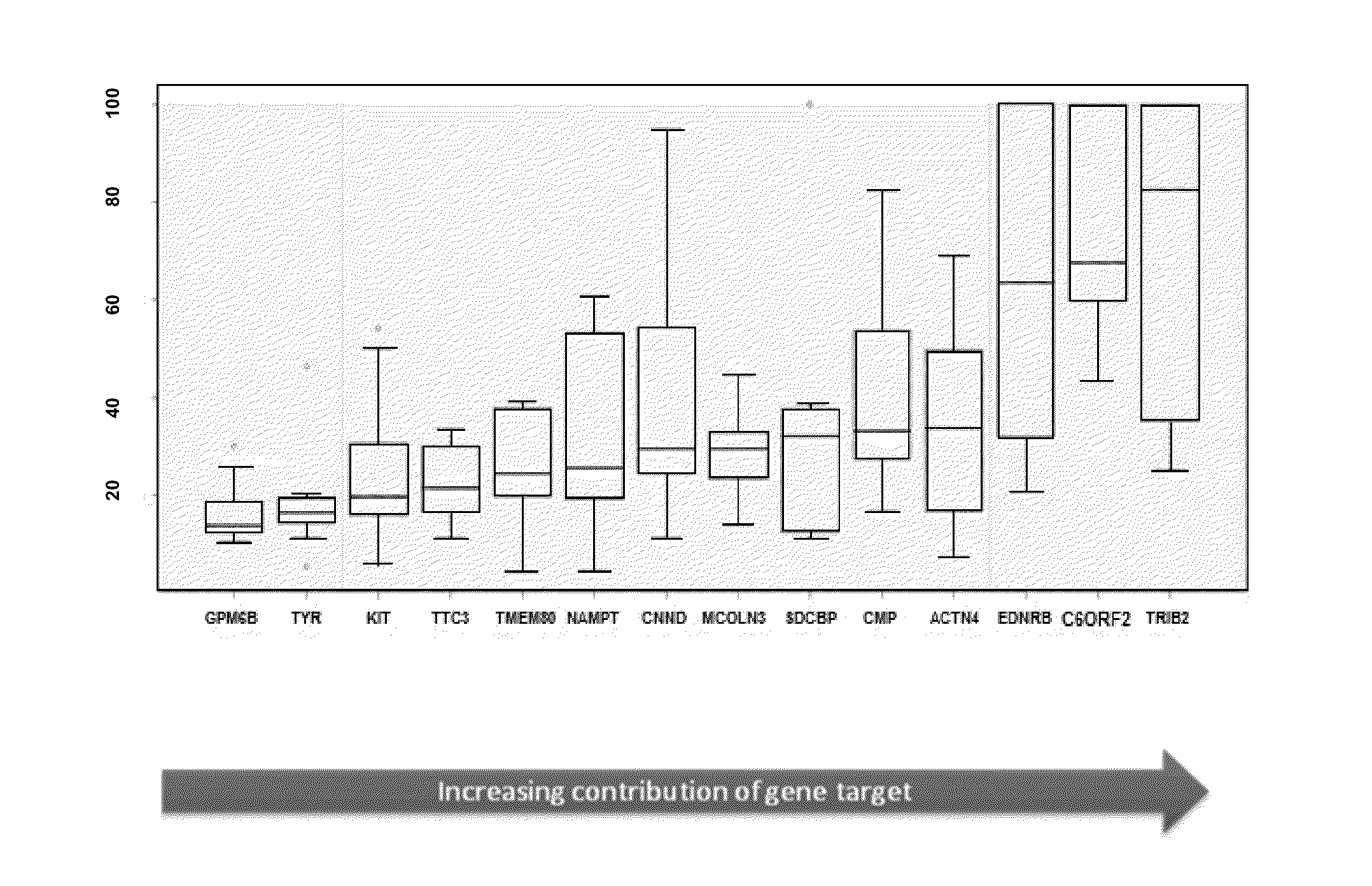
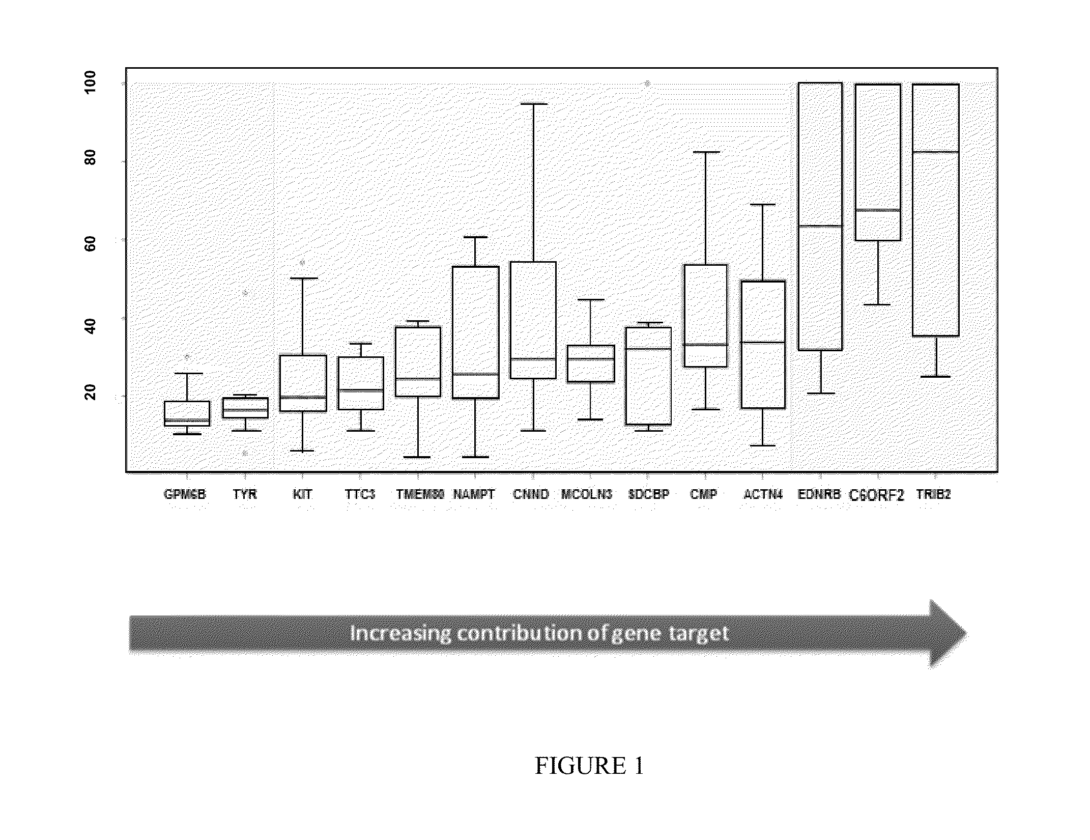
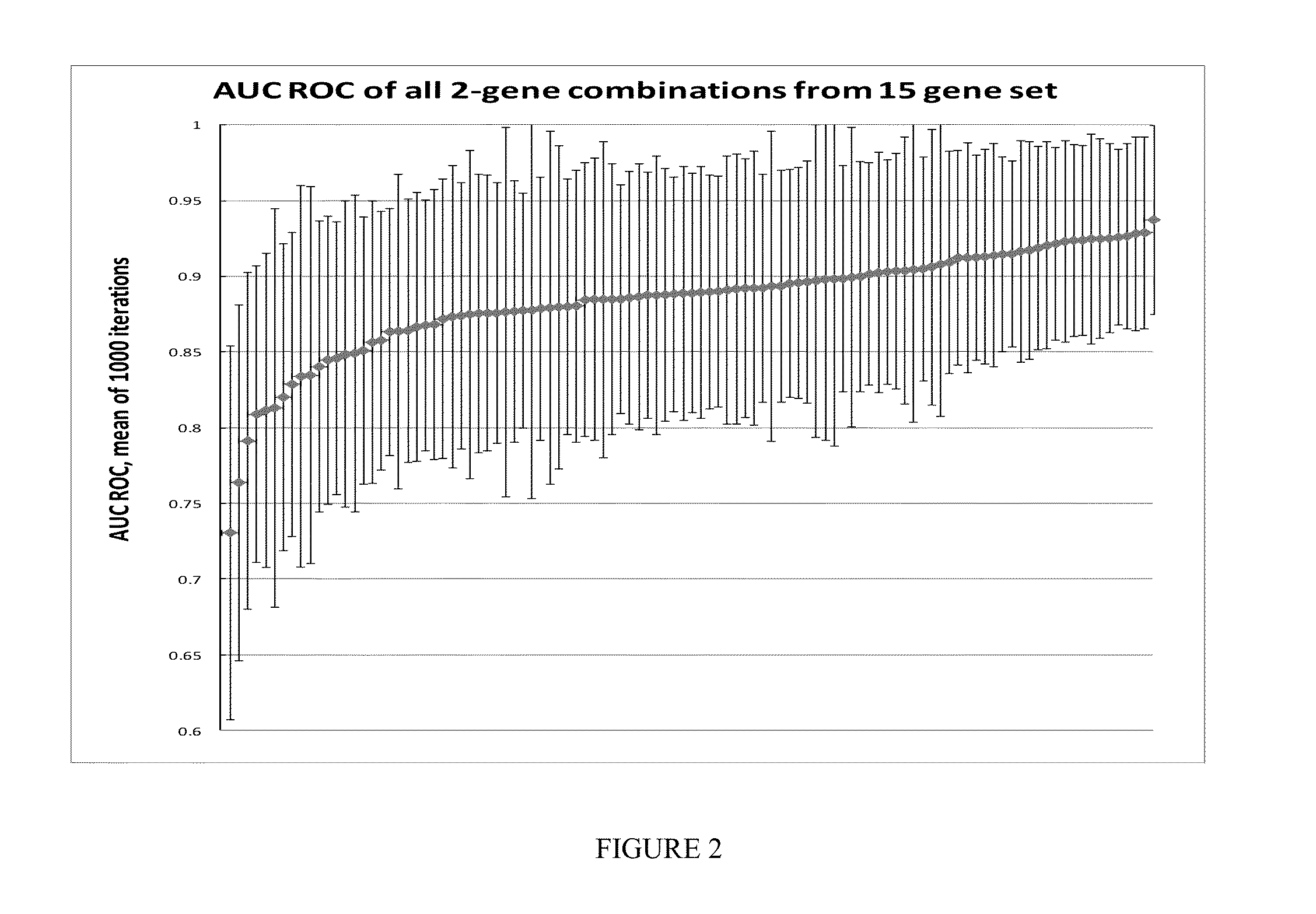
[0179]The OpenArray real-time PCR system (Life Technologies, Carlsbad, Calif.) was chosen for investigation. Each OA contained 48 sub-arrays (4×12). Each sub-array contained 18 target genes in triplicate, including genes procured from discovery studies to generate a melanoma classifier, and 3 internal genes (Wachsman et al., Brit. J. Dermatol 164:797 [2011]).

[0180]Latin Square Experimental Design and Method (See FIG. 3)

[0181]13 cloned human genes were used, including: ACTN4, B2M, LINC00518, CMIP, CNN2, EDNRB, GPM6B, KIT, MCOLN3, PRAME, SDCBP, TMEM80 and TRIB2.

[0182]Each of the 13 genes were pooled into groups and diluted to copies of 0, 64, 256, 1024, 4096, 1.6E+04, 6.6E+04, 2.6E+05, 1.0E+06, 4.2E+06, 1.7E+07, 6.7E+07 and 2.7E+08.

[0183]In every qPCR experiment, 13 groups of genes in 13 different copies were assayed in triplicate in the presence of ...


PUM

PropertyMeasurementUnit
depthaaaaaaaaaa
median survival timeaaaaaaaaaa

Abstract

The present invention provides methods for diagnosing melanoma and/or solar lentigo in a subject by analyzing nucleic acid molecules obtained from the subject. The present invention also provides methods for distinguishing melanoma from solar lentigo and/or dysplastic nevi and/or normal pigmented skin. The methods include analyzing expression or mutations in epidermal samples, of one or more skin markers. The methods can include the use of a microarray to analyze gene or protein profiles from a sample.

Description

CROSS-REFERENCE[0001]This application claims the benefit of U.S. Provisional Application No. 61 / 841,180 and U.S. Provisional Application No. 61 / 841,184 both filed Jun. 28, 2013, and entitled, “DIAGNOSIS OF MELANOMA BY NUCLEIC ACID ANALYSIS,” each of which are incorporated herein by reference in their entirety.BACKGROUND OF THE INVENTION[0002]Melanoma is a serious form of skin cancer in humans. It arises from the pigment cells (melanocytes), usually found in the skin. The incidence of melanoma is increasing at the fastest rate of all cancers in the United States with a lifetime risk of 1 in 68. Although melanoma accounts for only 4% of all dermatologic cancers, it is responsible for 80% of all deaths from skin cancers. The diagnosis of melanoma, when it is early stage disease, is key to its cure.[0003]The ability to cure melanoma in its earliest skin stage, melanoma in situ (MIS), is virtually 100% if the melanoma is adequately surgically excised. If the melanoma is caught in a later...

Claims


Application Information

Patent Timeline
Patent Type & Authority Applications(United States)
IPC IPC(8): G06F19/00C12Q1/68
CPCC12Q1/6886C12Q2600/158G06F19/34G06F19/345G16H50/20Y02A90/10
Inventor ALSOBROOK, JOHN P.PALMER, TARA J.
Owner DERMTECH INT
Why Eureka
Try Eureka