Method for Detecting Chromosome Structure and Gene Expression Simultaneously in Single Cells

a technology applied in the field of single cell chromosome structure detection, can solve the problems of limiting the scope of the experiment, difficult to obtain gene expression data, and lack of clear picture of chromosome structure and gene expression

Active Publication Date: 2014-01-23
THE TRUSTEES OF THE UNIV OF PENNSYLVANIA
View PDF2 Cites 6 Cited by
  • Summary
  • Abstract
  • Description
  • Claims
  • Application Information

AI Technical Summary

Problems solved by technology

However, each cell compacts these incredibly long polymeric molecules within the cell's nucleus, a space just a few microns across.
Unfortunately, there is a lack of a clear picture of chromosome structure and gene expression, which stems from the lack of effective tools for measuring genetic structure and function simultaneously at the single cell level.
In either case, gene expression data are difficult to obtain.
This approach is more direct, but studies

Method used


Image

  • Method for Detecting Chromosome Structure and Gene Expression Simultaneously in Single Cells
  • Method for Detecting Chromosome Structure and Gene Expression Simultaneously in Single Cells
  • Method for Detecting Chromosome Structure and Gene Expression Simultaneously in Single Cells

Examples


experimental examples

[0085]The invention is now described with reference to the following Examples. These Examples are provided for the purpose of illustration only and the invention should in no way be construed as being limited to these Examples, but rather should be construed to encompass any and all variations which become evident as a result of the teaching provided herein.

[0086]The following methods were used in performance of the experimental examples:

Cell Culture, Fixation, and Fluorescent In Situ Hybridization

[0087]Primary human foreskin fibroblasts (ATCC CRL 2097) or HeLa cells (gift from the lab of Phillip Sharp) were grown in Dulbeceo's modified eagle's medium with glutamax (DMEM, Life Technologies) supplemented with penicillin / streptomycin and 10% fetal bovine serum. Cells were enriched for G0 / G1 phase cells through a double-thymidine block (2 μg / mL thymidine in medium) procedure and releasing for an appropriate amount of time before fixation. To fix the cells, the protocol of Raj et al. (N...

example 1

Simultaneous Determination of Chromosome Structure and Gene Expression

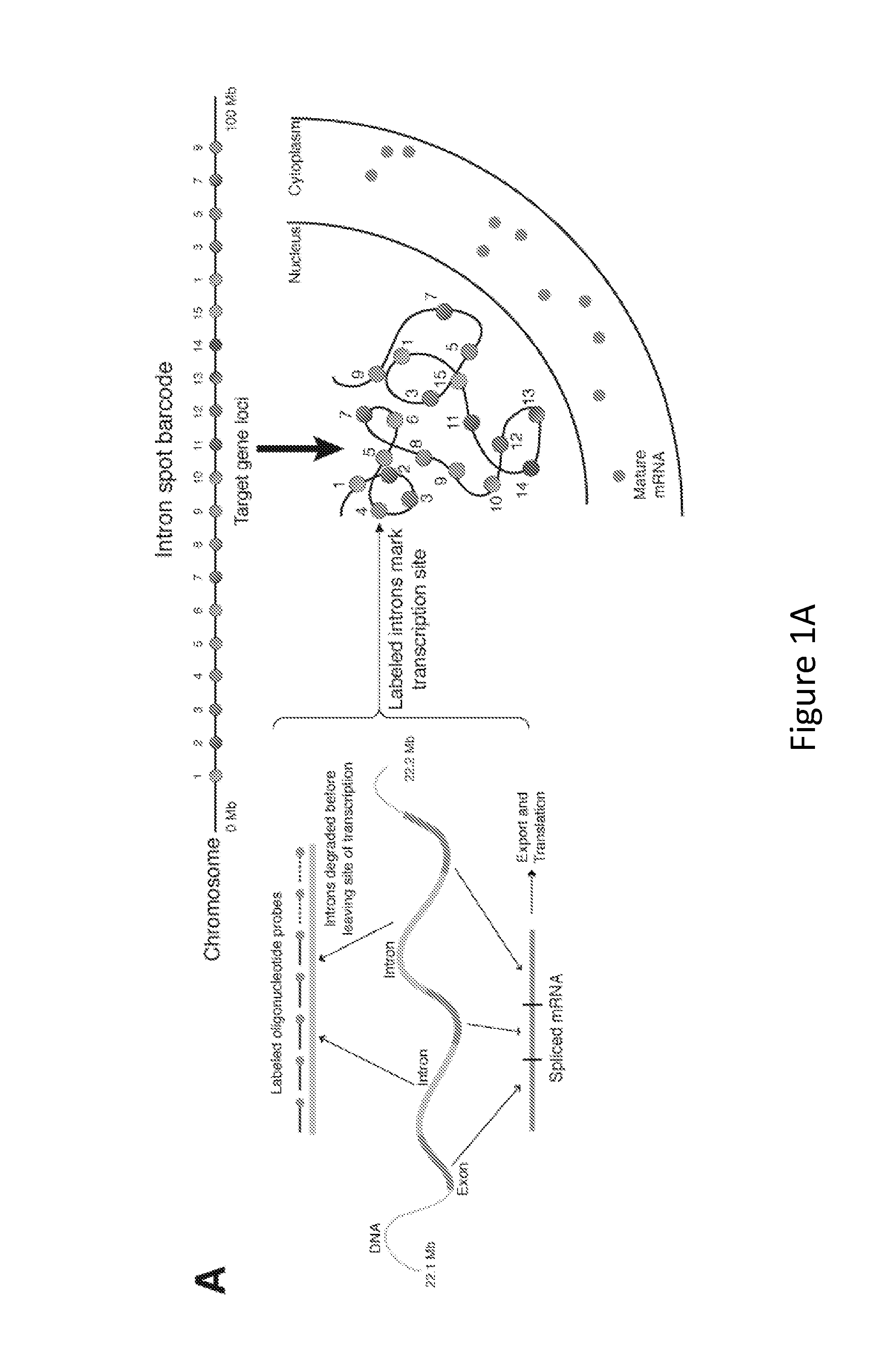
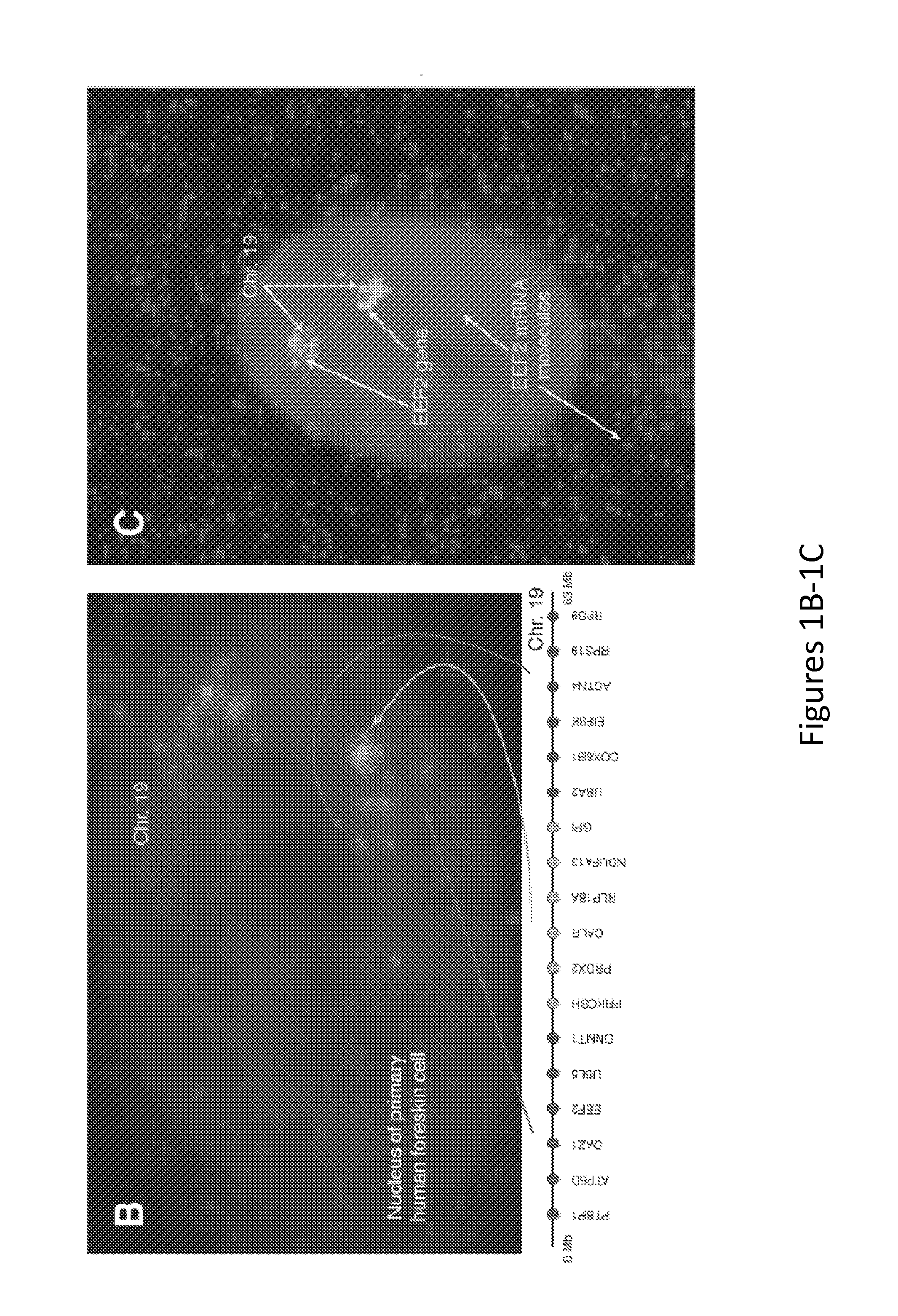
[0107]As depicted in FIG. 1B, 18 genes were targeted along chromosome 19, illuminating the chromosome robustly and with low background. Because the method of the present invention is based on RNA FISH, it is easy to combine with RNA FISH labeling mRNAs to simultaneously measure the expression of specific genes, as depicted in FIG. 1C. The introns themselves also yield critical gene expression information, since most genes' introns yield spots in fewer than 50% of cells. That said, the determination of overall chromosome structure via the methods of the present invention can additionally include the labeling of a large number of genes, thus imparting redundancy into the method, and may further include an RT-PCR screen to aid in the bioinformatic identification of abundant intron targets.

[0108]As depicted in FIG. 1B, distinctly labeled genes in three different regions of chromosome 19 reveal a remarkably ordered int...

example 2

Measuring Chromosome Structure and Gene Expression In Vivo

[0110]Many insights in systems biology have arisen from the use of time-lapse microscopy to understand the dynamics of cellular processes. For this reason, the methods of the present invention can also be applied to living cells to provide a dynamic view of chromosome structure and gene expression. To date, there is no way of looking at chromosome structure or gene expression in a non-perturbative (i.e., no genetic manipulation) manner via microscopy in single living cells.

[0111]Experiments can be performed to tackle the challenges of targeting nucleic acids in vivo. For example, considerations include hybridization kinetics, sequence design, oligonucleotide chemistry and delivery methods, amongst several others. RNA of a single intron from a highly expressed gene can be targeted by designing a series of 16-48 oligonucleotides, with the idea that the binding of a large number of oligonucleotides to a single location in the ce...


PUM

PropertyMeasurementUnit
Coloraaaaaaaaaa
Structureaaaaaaaaaa
Gene expression profileaaaaaaaaaa

Abstract

The present invention relates to systems and methods for measuring chromosome structure and gene expression. In one embodiment, the present invention includes an assay for determining the spatial organization of gene expression in the nucleus. The present invention also includes a method based on fluorescence in situ hybridization (FISH) that simultaneously yields information on the physical position and expression of individual genes. By lighting up a large number of targets on a particular chromosome using a bar-coding scheme, the large scale structure of an entire chromosome can be determined.

Description

CROSS-REFERENCE TO RELATED APPLICATIONS[0001]This application claims the benefit of priority from U.S. Provisional Application Ser. No. 61 / 439,623, filed on Feb. 4, 2011, the entire disclosure of which is incorporated by reference herein as if set forth herein in its entirety.BACKGROUND OF THE INVENTION[0002]The DNA in a single human cell, if fully elongated, would stretch for roughly two meters. However, each cell compacts these incredibly long polymeric molecules within the cell's nucleus, a space just a few microns across. Increasingly, researchers are finding that this compacted conformation is not just a random “ball of string”, but rather is highly organized and dynamically regulated (Cremer et al., 2001, Nat Rev Genet 2: 292-301). Developing a better understanding of this compacted form will provide key insights in how the spatial organization of genetic information in the nucleus affects its readout via gene expression.[0003]Gene expression involves the transcription of mRNA...

Claims


Application Information

Patent Timeline
IPC IPC(8): C12Q1/68
CPCC12Q1/6841C12Q2563/185C12Q2543/10C12Q2600/158
Inventor RAJ, ARJUNLEVESQUE, MARSHALL J.
Owner THE TRUSTEES OF THE UNIV OF PENNSYLVANIA
Why Eureka
Try Eureka