
Direct molecular diagnosis of fetal aneuploidy

a fetal aneuploidy and molecular diagnostic technology, applied in biochemistry apparatus and processes, fermentation, organic chemistry, etc., can solve the problems of increased anxiety of expectant parents, difficult to estimate the true incidence of fetal aneuploidy among all pregnancies, and increased maternal morbidity, so as to achieve the effect of more rapid performan

Active Publication Date: 2013-11-05
THE BOARD OF TRUSTEES OF THE LELAND STANFORD JUNIOR UNIV
View PDF10 Cites 3 Cited by
  • Summary
  • Abstract
  • Description
  • Claims
  • Application Information

AI Technical Summary

Benefits of technology

The present invention is about using digital analysis to get the numbers of different chromosomes in a sample of genomic DNA from a fetal sample. This is done by running a large number of reactions and analyzing the results statistically to eliminate random variation. The test is direct, meaning it doesn't require cell culture or other lengthy sample preparation, so it can be performed faster than previous methods. It can be done in 6 hours or 1-10 hours, depending on the protocol used. The only thing that's necessary is to extract the DNA from the sample and run enough cycles of amplification to produce a detectable result.

Problems solved by technology

The incidence of fetal aneuploidy and other chromosome abnormalities is approximately 9 per 1000 live births [1] It is difficult to estimate their true incidence among all pregnancies due to the strong association with fetal miscarriage and stillbirth.
This time delay may result in both increased anxiety for expectant parents, and greater maternal morbidity should pregnancy termination be desired in the setting of abnormal results.
All these techniques provide tools for detection of aneuploidy, however some are invasive and most of them tend to be lengthy, labor intensive and some are allele-dependent, so that the results depend on the underlying genetics of the population.
In conventional RT-PCR, for example, one threshold cycle corresponds to a 2-fold change in copy number, making it exceedingly challenging to measure smaller changes [6], such as a 1.5-fold increase in number of a trisomic chromosome as compared to a normal disomic chromosome.
However, all these previously described methods are cumbersome to implement, take a long time and require significant labor.

Method used


Image

  • Direct molecular diagnosis of fetal aneuploidy
  • Direct molecular diagnosis of fetal aneuploidy
  • Direct molecular diagnosis of fetal aneuploidy

Examples


example 1

Microfluidic Digital PCR for Detection of Aneuploidy

[0072]This example demonstrates that digital polymerase chain reaction (PCR) enables rapid, allele independent molecular detection of fetal aneuploidy.

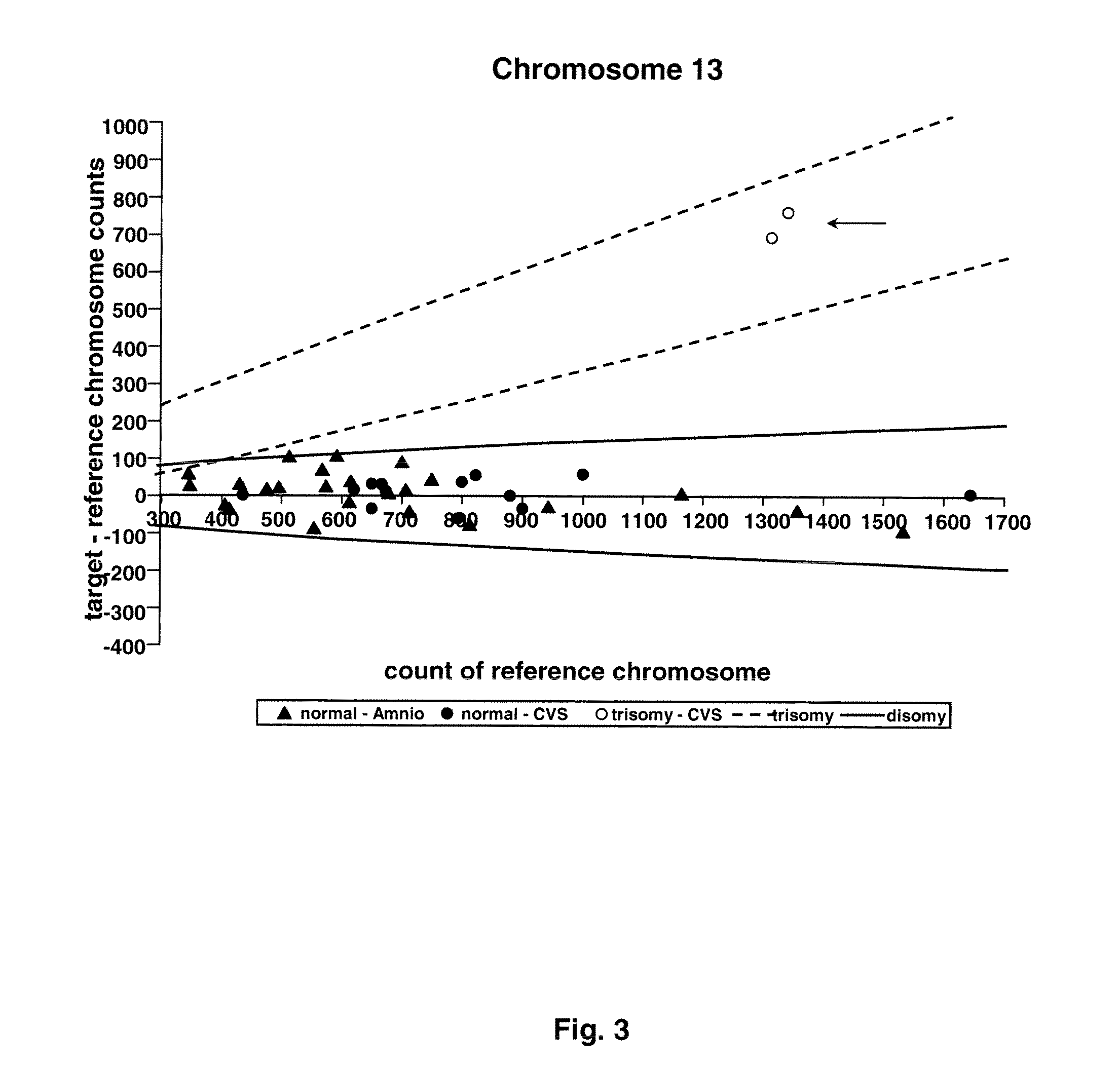
[0073]Twenty-four amniocentesis and 16 chorionic villus samples were used for microfluidic digital PCR analysis. Three thousand and sixty PCR reactions were performed for each of the target chromosomes (X, Y, 13, 18, and 21), and the number of single molecule amplifications was compared to a reference. The difference between target and reference chromosome counts was used to determine the ploidy of each of the target chromosomes.

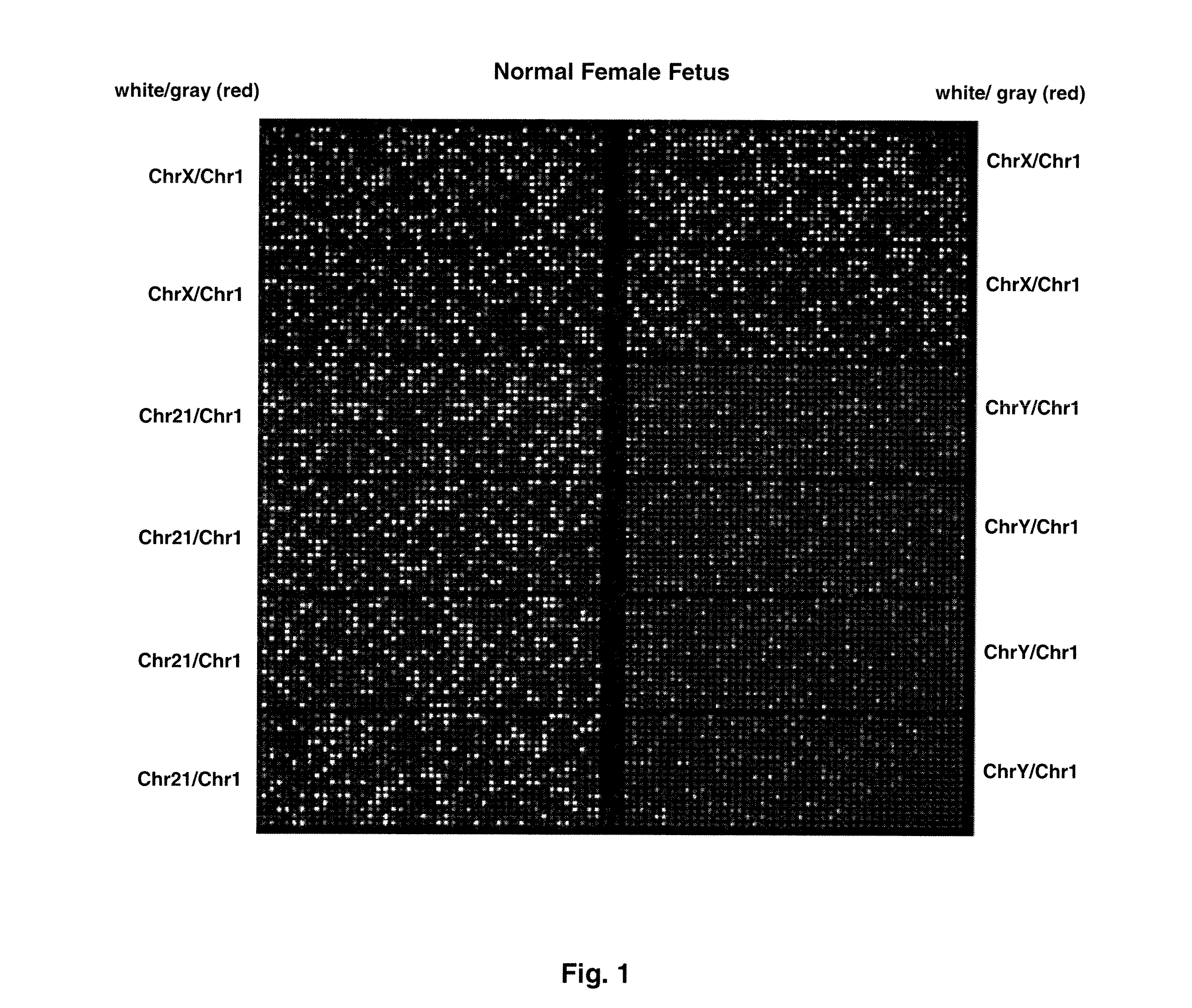
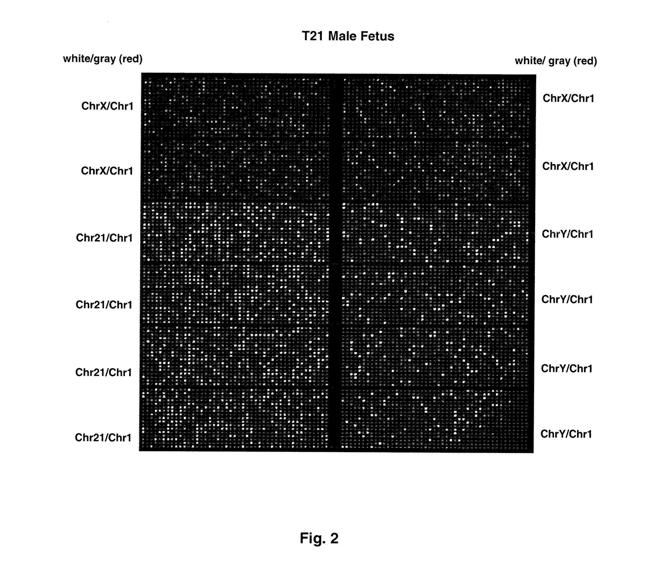
[0074]FIG. 1 shows sample false-color images of microfluidic digital PCR chips. The chips were 12.765 Digital Array microfluidic chips obtained from Fluidigm, South San Francisco, Calif.). Each chip has 12 panels, which are compartmentalized into 765 nanoliter chambers by micro-mechanical valves. Based on the estimation of DNA concentration with quantitative ...

example 2

Sample Collection and Preparation

[0078]Samples for detection of aneuploidy were obtained from pregnant women having clinically indicated amniocentesis or chorionic villus sampling (CVS).

[0079]In cases of amniocentesis, 1-2 mL from the clinical sample was submitted separately for digital PCR analysis. A total of 40 samples, consisting of 24 amniotic fluid and 16 CVS samples, were processed. One twin pregnancy and 1 triplet pregnancy were enrolled.

[0080]Amniotic fluid was centrifuged at 14,000 rpm. Supernatant was removed and the cell pellet was resuspended in phosphate buffered saline (PBS). Chorionic villi were suspended in PBS. Next, genomic DNA was extracted from amniotic fluid and chorionic villi with QIAamp DNA Blood Mini Kit (Qiagen, Valencia, Calif.) according to the manufacturer's instructions. The QIAamp DNA Blood Mini Kit simplifies isolation of DNA from blood and related body fluids with fast spin-column or vacuum procedures (see flowchart “QIAamp Spin Column Procedure”). ...

example 3

Digital and Microfluidical Digital PCR

[0081]Digital and microfluidical PCR assays were performed as follows.

[0082]Taqman PCR assay was designed to amplify 1 region on each of the following chromosomes: 1, 13, 18, 21, X, Y. Chromosome 1 was chosen to be the reference chromosome since it is not associated with any aneuploidy observed in ongoing pregnancies [9]. The assay of chromosome 1 contained a probe labeled with a HEX fluorophore, while the assays for the target chromosomes (13, 18, 21, X, Y) each contained a probe labeled with a FAM fluorophore. The amplicon of each assay was chosen to lie outside of the regions with known copy number variation in healthy individuals [10]. In particular, the amplicons of chromosomes 1, 13, and 18 cover ultraconserved regions [11], which are rarely found to be associated with copy number variation in healthy individuals [10]. The amplicons were all 80-90 bp in length to reduce any amplification bias. The sequences of the primers and probes are li...


PUM

PropertyMeasurementUnit
widthaaaaaaaaaa
widthaaaaaaaaaa
timeaaaaaaaaaa

Abstract

Methods and materials for detection of aneuploidy and other chromosomal abnormalities using fetal tissue are disclosed. Results can be obtained rapidly, without cell culture. The method uses digital PCR for amplification and detection of single target sequences, allowing an accurate count of a specific chromosome or chromosomal region. Specific polynucleic acid primers and probes are disclosed for chromosomes 1, 13, 18, 21, X and Y. These polynucleic acid sequences are chosen to be essentially invariant between individuals, so the test is not dependent on sequence differences between fetus and mother.

Description

FEDERALLY-SPONSORED RESEARCH OR DEVELOPMENT[0001]This invention was made with Government support under contract HG002644 awarded by the National Institutes of Health. The Government has certain rights in this invention.REFERENCE TO SEQUENCE LISTING, COMPUTER PROGRAM, OR COMPACT DISK[0002]Applicants assert that the text copy of the Sequence Listing is identical to the Sequence Listing in computer readable form found on the accompanying computer file. Applicants incorporate the contents of the sequence listing by reference in its entirety.BACKGROUND OF THE INVENTION[0003]1. Field of the Invention[0004]The present invention relates to the field of prenatal diagnosis of fetal aneuploidy.[0005]In particular, the invention relates to a method for rapid prenatal diagnosis and detection of fetal aneuploidy by using a microfluidic digital PCR (polymerase chain reaction) that enables rapid, allele independent molecular detection of fetal chromosomal aneuploidy utilizing uncultured amniocytes ...

Claims


Application Information

Patent Timeline
Patent Type & Authority Patents(United States)
IPC IPC(8): C12Q1/68C07H21/02C07H21/04
CPCC12Q1/6851C12Q1/6883C12Q2537/157C12Q2545/101C12Q2561/113C12Q2565/629C12Q2600/156C12Q2600/16
Inventor FAN, HEI-MUN CHRISTINAQUAKE, STEPHEN R.
Owner THE BOARD OF TRUSTEES OF THE LELAND STANFORD JUNIOR UNIV
Learn More