Robotic localizing aid for high intensity focused ultrasound delivery

a high-intensity, focused ultrasound technology, applied in ultrasonic/sonic/infrasonic image/data processing, applications, ultrasonic/sonic/infrasonic image/data processing, etc., can solve the problem of uncontrollable hemorrhage conditions, difficult dynamic change of intervening tissue properties, and inability to control bleeding, etc. problems, to achieve the effect of smooth and accurate delivery and stop bleeding

Inactive Publication Date: 2014-03-27
UNIVERSITY OF ROCHESTER
View PDF7 Cites 3 Cited by
  • Summary
  • Abstract
  • Description
  • Claims
  • Application Information

AI Technical Summary

Benefits of technology

[0019]It is another object of the invention to provide such accurate placement in a form which can be conveniently used in an operating room or a biopsy room.
[0021]To achieve the above and other objects, the present invention is directed to a holding structure to hold the biopsy needle and the HIFU transducer to allow the smooth and accurate delivery of the HIFU beam at the interface of the liver (or other organ), where bleeding will occur. The manipulator structure in at least some embodiments performs the biopsy and HIFU through the percutaneous approach. In addition, the procedure can be imaged by the ultrasound imaging probe. The HIFU probe can track the biopsy needle entry site even when the needle has been removed with the help of the robotic arm. The HIFU transducer a short pulse of HIFU in the needle tract to cause coagulation and stop bleeding. Targeting can be robotically controlled using data extracted from a conventional two-dimensional image taken via any suitable imaging technique (e.g., ultrasound, CAT, or MRI).

Problems solved by technology

Dynamic change of the intervening tissue properties is another challenge.
Uncontrolled hemorrhage conditions can be challenging in trauma patients, particularly for those with vascular, splenic, or hepatic injuries.
The inability to control bleeding might be due to the malfunction of the organs to produce platelets to stop the bleeding or due to blood diseases.
Failure to control hemorrhage can lead to insufficient organ perfusion (shock) and is the major cause of death in trauma victims.
Indeed, the major cause of mortality in combat casualties is often reported as exsanguination (bleeding to death).
In addition, conducting biopsy procedures onto the organs for diagnoses purposes can also cause the organ to bleed excessively.
Despite the availability of several hemostasis methods such as laser welding and argon beam coagulation, such methods do not arrest the bleeding problem effectively.
These existing cauterization methods such as electrocautery and laser can only deliver energy on the surface of tissues with a maximum penetration of 2-3 mm.
Furthermore, uncontrolled hemorrhage can be fatal.
However, HIFU as a tool for controlling hemorrhage is still in its infancy.
However, accurate aiming of the HIFU transducer relative to the location of the biopsy has not yet been achieved.
One particular problem is that once the biopsy has been performed, the hole left by the biopsy is often too small to be detected by medical imaging techniques such as ultrasound, CAT scanning, or MRI.

Method used


Image

  • Robotic localizing aid for high intensity focused ultrasound delivery
  • Robotic localizing aid for high intensity focused ultrasound delivery
  • Robotic localizing aid for high intensity focused ultrasound delivery

Examples


Embodiment Construction

[0033]A preferred embodiment will be described in detail with reference to the drawings, in which like reference numerals refer to like elements or steps throughout.

[0034]Shown in FIG. 1 is a section overview of the manipulator assembled onto the arm of the cart. In the system 100, the cart arm 102 supports the manipulator 104 over the body of the patent P and specifically over the patient's liver L.

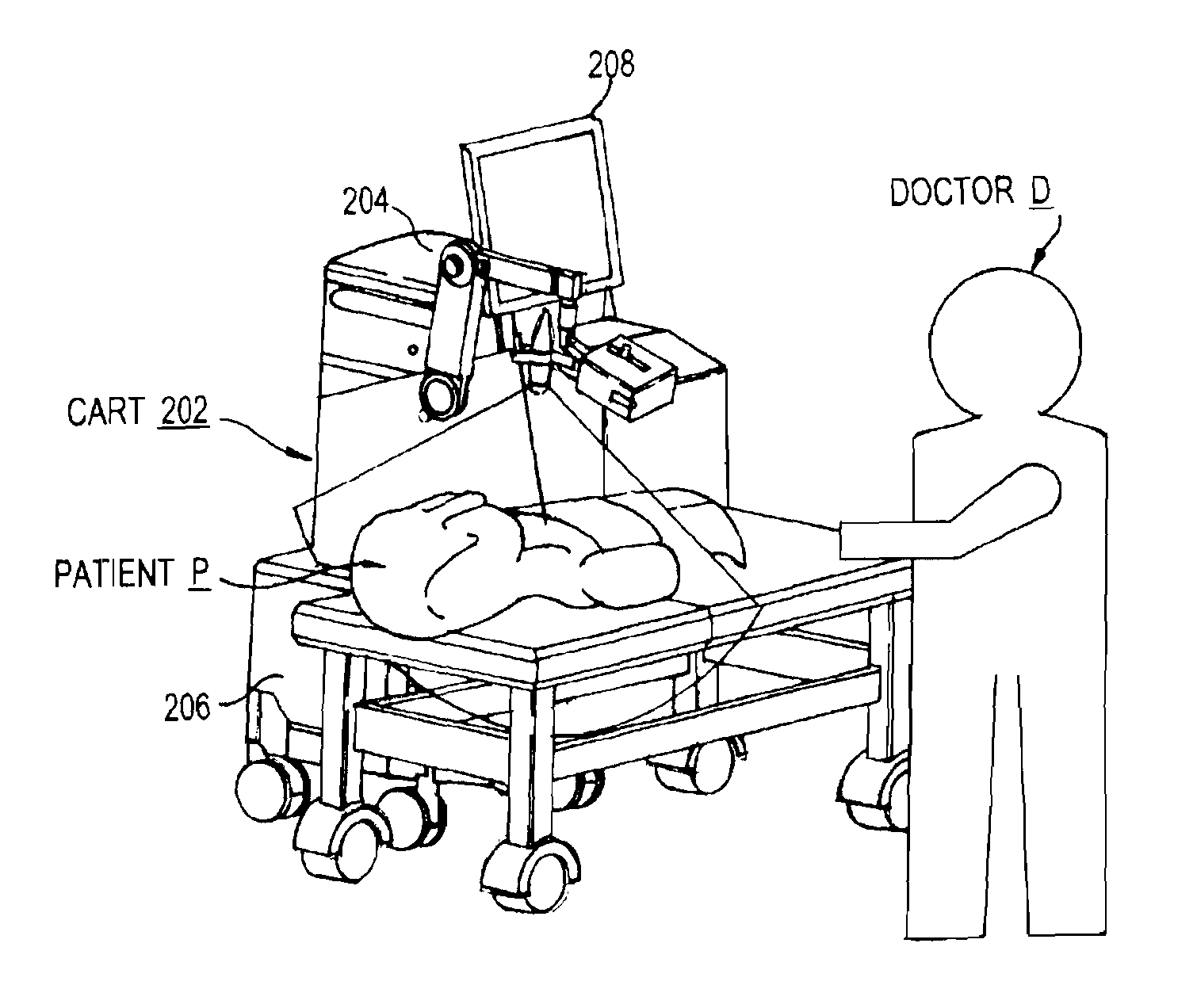
[0035]Shown in FIG. 2, the main components are lined up with the radiologist D beside the patient and facing the screens of the cart 202 and the ultrasound scanner 204 which is positioned on the right of the cart. The cart includes a base portion having a processor 206 for performing all of the processing disclosed herein, including automatic control, as well as a touch screen 208 for displaying images to the operator and for receiving inputs from the operator.

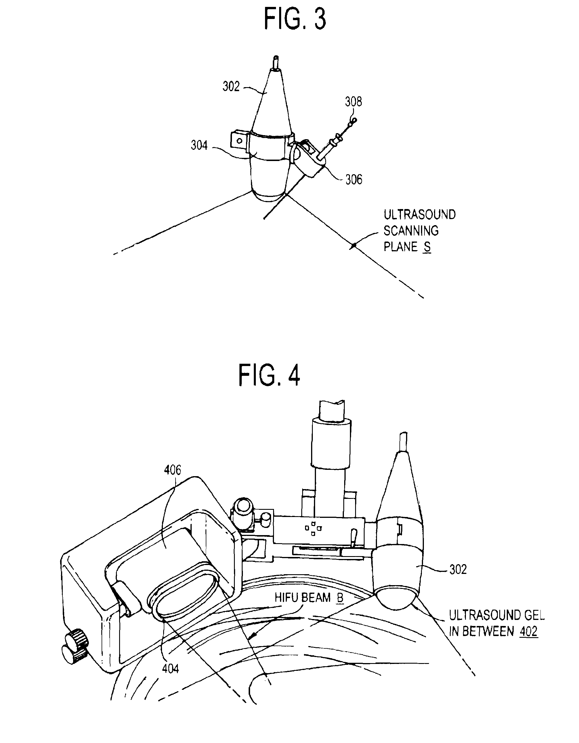
[0036]The sterilized needle holder is self aligned to the ultrasound probe holder as shown in FIG. 3. More specifically, the ultr...


PUM


Abstract

A holding structure holds a biopsy needle and a HIFU transducer to allow smooth and accurate delivery of the HIFU beam at the interface of the liver, where bleeding will occur. This manipulator structure performs the biopsy and HIFU through the percutaneous approach. In addition, the procedure is imaged by an imaging ultrasound probe. This imaging ultrasound probe can track the needle entry site even when needle has been removed. The HIFU transducer fires a short pulse of HIFU in the needle tract to cause coagulation and stop bleeding.

Description

REFERENCE TO RELATED APPLICATION[0001]The present application claims the benefit of U.S. Provisional Patent Application No. 60 / 087,087, filed Aug. 7, 2008, whose disclosure is hereby incorporated by reference in its entirety into the present disclosure.FIELD OF THE INVENTION[0002]The present invention relates to high-intensity focused ultrasound (HIFU) and more particularly to its delivery through a robotic controlled arm at a specified target point for hemostasis and therapeutic purposes.DESCRIPTION OF RELATED ART[0003]Ultrasound is a well-established imaging modality in the areas of medicine, biology and engineering. Its case of application and low cost make it the choice for medical imaging for most soft tissue diagnostic analysis. Ultrasound has been used to examine many parts of the body, including babies in the mother's womb, the vascular system, heart, organs within the abdominal cavity, urinary system, ovaries, brain and most recently, the muscles.[0004]Since the development...

Claims


Application Information

Patent Timeline
Patent Type & Authority Applications(United States)
IPC IPC(8): A61B10/02A61B8/08A61N7/02
CPCA61B10/0233A61B8/0841A61N7/02A61B8/4218A61B34/30A61B2090/378
Inventor DOGRA, VIKRAM S.NG, WAN SINGLIMM, YUNN SHIOW
Owner UNIVERSITY OF ROCHESTER
Why Eureka
Try Eureka