Computerized tomography (CT) fluoroscopy imaging system using a standard intensity ct scan with reduced intensity ct scan overlays

a fluoroscopy and computerized tomography technology, applied in computerized tomographs, medical science, diagnostics, etc., can solve the problems of significantly higher radiation emission during imaging than conventional 2d x-ray machines, and generally impossible to substantially continuously operate a conventional ct machine, and achieve the effect of reducing the intensity of ct scan overlays

Inactive Publication Date: 2014-04-24
NEUROLOGICA CORP
View PDF4 Cites 2 Cited by
  • Summary
  • Abstract
  • Description
  • Claims
  • Application Information

AI Technical Summary

Benefits of technology

[0009]In accordance with the present invention, there is provided a novel CT system capable of providing 3D fluoroscopy of an internal procedure site during a medical procedure so as to visualize patient anatomy, medical instruments, prosthesis, etc. during the medical procedure without subjecting the patient to unacceptable quantities of X-ray radiation. This new CT system provides 3D fluoroscopy of the internal procedure site using a standard intensity CT scan with reduced intensity CT scan overlays.

Problems solved by technology

Unfortunately, CT machines, since they must direct X-rays into the body from a variety of positions, emit significantly higher quantities of radiation during imaging than conventional 2D X-ray machines.
Therefore, it is generally impractical to operate a conventional CT machine substantially continuously during a medical procedure, whereby to provide 3D fluoroscopy during the medical procedure, since the quantity of radiation which would be emitted during the medical procedure is generally deemed unacceptable.
Furthermore, operating a conventional CT machine at significantly reduced X-ray intensities so as to provide 3D fluoroscopy is generally not a viable option, since scanning anatomy with significantly reduced X-ray intensities generally results in an inferior image, particularly where low-contrast (e.g., soft tissue) anatomy is involved.

Method used


Image

  • Computerized tomography (CT) fluoroscopy imaging system using a standard intensity ct scan with reduced intensity ct scan overlays
  • Computerized tomography (CT) fluoroscopy imaging system using a standard intensity ct scan with reduced intensity ct scan overlays
  • Computerized tomography (CT) fluoroscopy imaging system using a standard intensity ct scan with reduced intensity ct scan overlays

Examples


Embodiment Construction

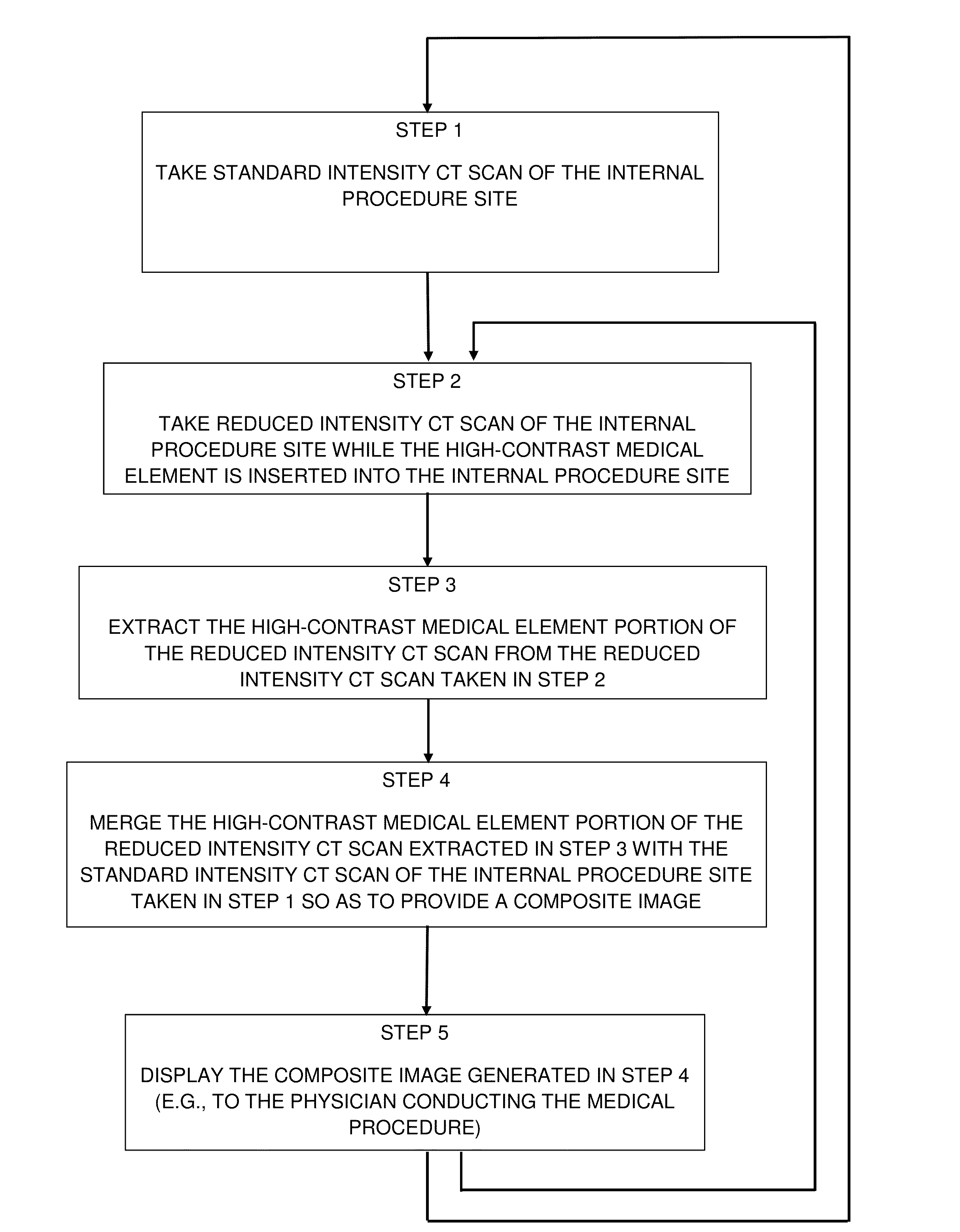
[0035]The present invention provides a novel CT system capable of providing 3D fluoroscopy of an internal procedure site during a medical procedure so as to visualize patient anatomy, medical instruments, prostheses, etc. during the medical procedure without subjecting the patient to unacceptable quantities of X-ray radiation. This new CT system provides 3D fluoroscopy of the internal procedure site using a standard intensity CT scan with reduced intensity CT scan overlays.

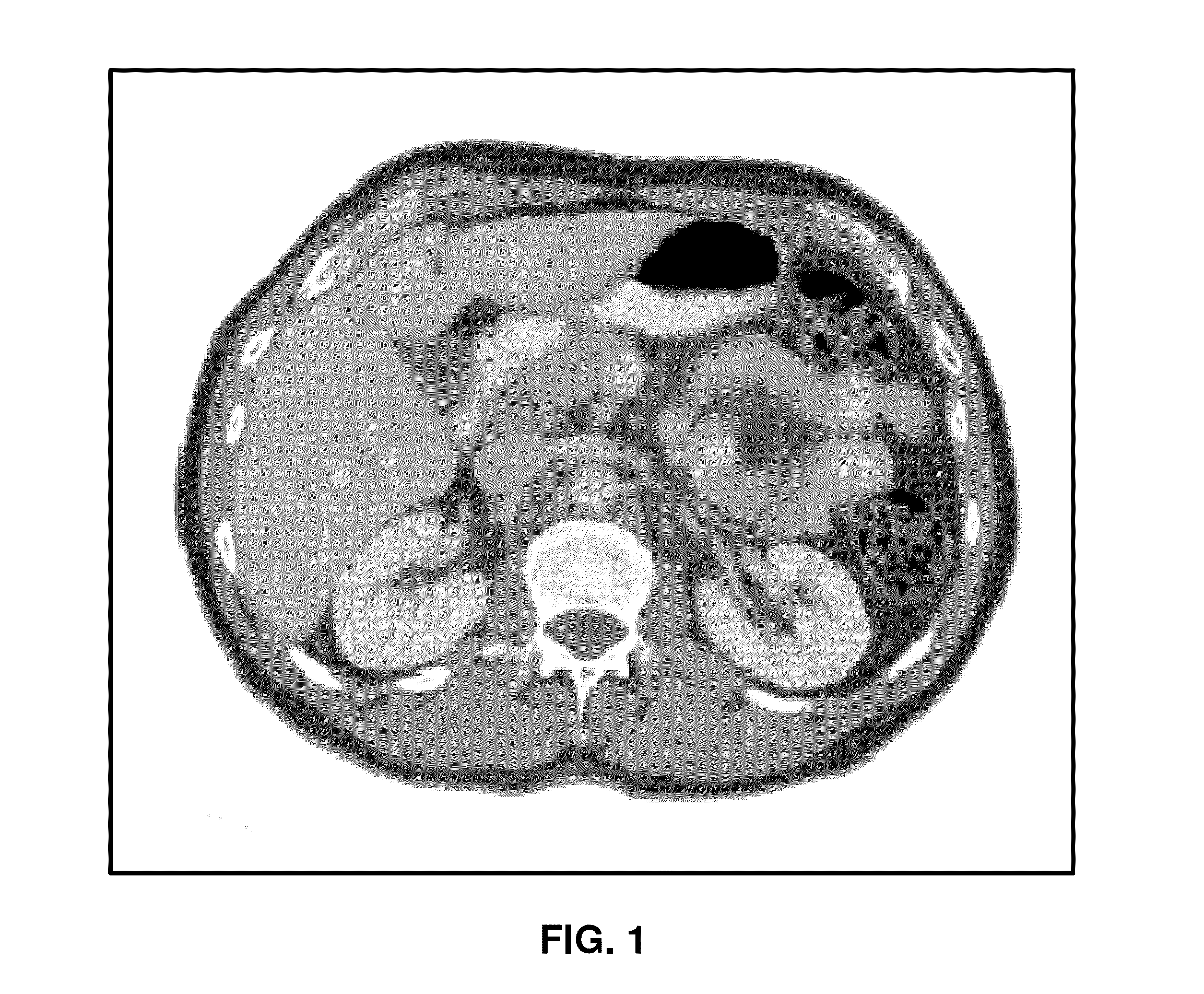
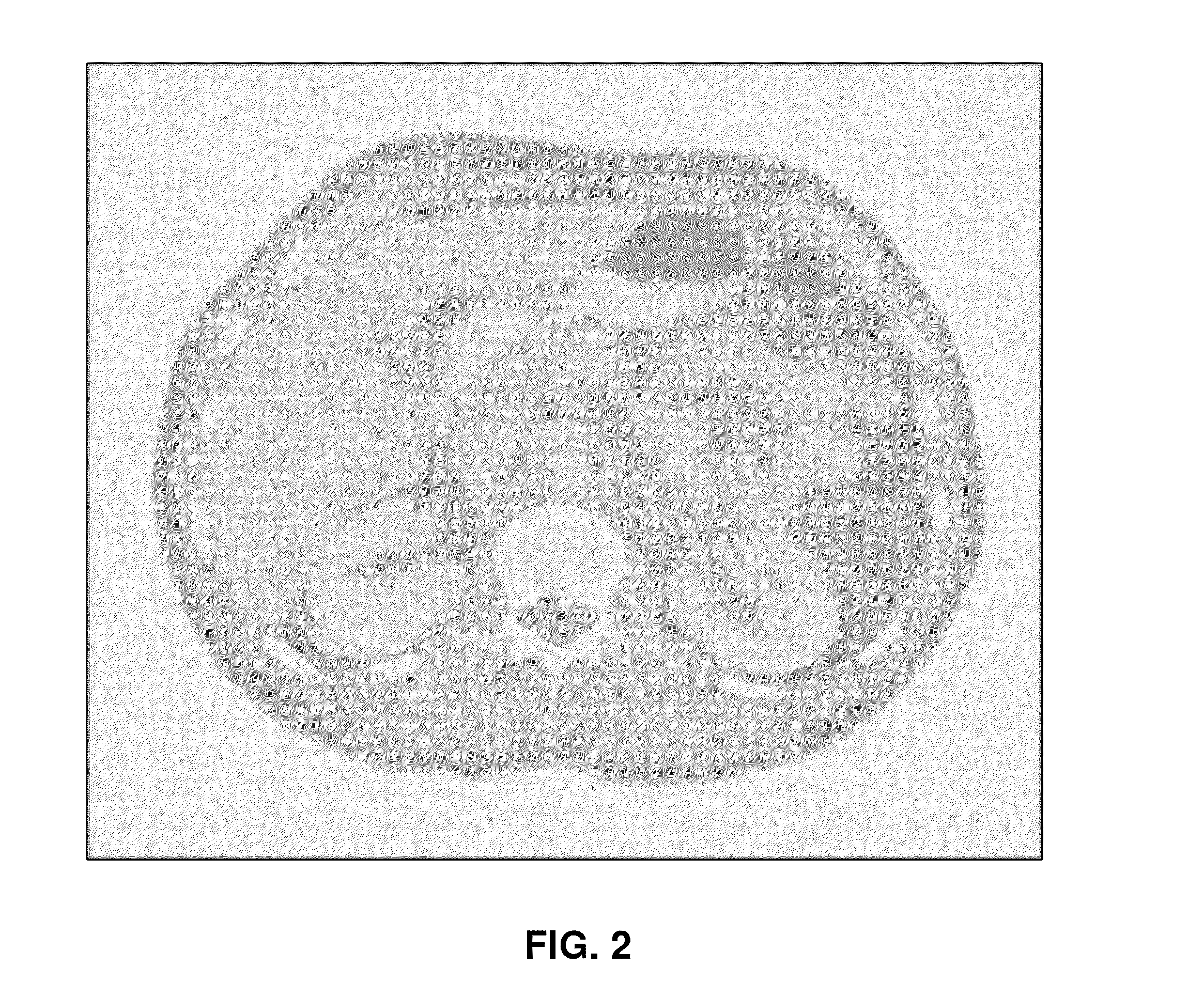
[0036]More particularly, it has been noted that certain medical elements (e.g., surgical instruments, prostheses, catheters, needles, injectable substances such as iodine, etc.) are relatively high-contrast elements which are capable of being accurately visualized using lower X-ray intensities than is generally necessary in order to accurately visualize low-contrast (e.g., soft tissue) anatomy. Thus, for example, FIG. 3 shows a high-contrast surgical instrument disposed within the anatomy imaged by a CT machine op...


PUM


Abstract

A method for providing CT fluoroscopy of an internal procedure site during a medical procedure so as to visualize patient anatomy and a high-contrast medical element during the procedure without subjecting the patient to unacceptable quantities of X-ray radiation, comprising: (1) taking a standard intensity CT scan of the internal procedure site; (2) taking a reduced intensity CT scan of the procedure site while the high-contrast medical element is inserted into the procedure site; (3) extracting the high-contrast medical element portion of the reduced intensity CT scan from the reduced intensity CT scan; (4) merging the high-contrast medical element portion of the reduced intensity CT scan extracted in Step 3 with the standard intensity CT scan of the procedure site taken in Step 1 so as to provide a composite CT scan; (5) displaying the composite CT scan; and (6) returning to either Step 1 or Step 2.

Description

REFERENCE TO PENDING PRIOR PATENT APPLICATION[0001]This patent application claims benefit of pending prior U.S. Provisional Patent Application Ser. No. 61 / 716,341, filed Oct. 19, 2012 by Eric Bailey for COMPUTERIZED TOMOGRAPHY (CT) FLUOROSCOPY IMAGING SYSTEM USING HIGH DOSE SCAN WITH LOW DOSE OVERLAY (Attorney's Docket No. NEUROLOGICA-10 PROV), which patent application is hereby incorporated herein by reference.FIELD OF THE INVENTION[0002]This invention relates to anatomical imaging systems in general, and more particularly to Computerized Tomography (CT) imaging systems.BACKGROUND OF THE INVENTION[0003]Computerized Tomography (CT) has emerged as a key imaging modality in the visualization of anatomy. CT scanners generally operate by directing X-rays into the body from a variety of positions, detecting the X-rays passing through the body, and then processing the detected X-rays so as to build a three-dimensional (3D) computer model of the patient's anatomy. This 3D computer model ca...

Claims


Application Information

Patent Timeline
Patent Type & Authority Applications(United States)
IPC IPC(8): A61B6/03A61B6/00
CPCA61B6/032A61B6/485A61B6/481A61B6/12A61B6/487A61B6/5235
Inventor BAILEY, ERIC
Owner NEUROLOGICA CORP
Why Eureka
Try Eureka