Endoscope system and operating method thereof

a technology of endoscope and operating method, which is applied in the field of endoscope system, can solve the problems of not being able to precisely reflect the oxygen saturation level in the display of abnormal areas on the screen, and achieve the effect of facilitating grasping sight, eliminating the time required for producing emphasized images, and high precision

Active Publication Date: 2014-06-05
FUJIFILM CORP
View PDF7 Cites 23 Cited by
  • Summary
  • Abstract
  • Description
  • Claims
  • Application Information

AI Technical Summary

Benefits of technology

[0017]According to the present invention, the emphasized image, in which the bright part of the abnormal area in which the calculation result of the biological function information calculating section is likely to be abnormal is further brightened for emphasis, is displayed on the display section. The abnormal area is displayed in the emphasized image so as to be just emphasized by the brightness to facilitate a grasp at sight, instead of determining a border with a normal area using a threshold value just as in the case of the Japanese Patent No. 2768936 and the U.S. Pat. No. 5,956,416. Therefore, it is possible to indicate the reliability of the biological function information, including the oxygen saturation level and the like, with high precision.
[0018]Note that, as for the biological function information image having the halation area that already exists, it is apparent that the calculation result of the halation area is abnormal. Thus, in the present invention, the biological function information image having the halation area is displayed on the display section as is, without producing the emphasized image. This eliminates time required for producing the emphasized image and improves the property of a moving image.

Problems solved by technology

However, although an area is judged to be the abnormal area, if the pixel value of the abnormal area is just slightly above the threshold value and the actual reliability of the oxygen saturation level is not low, the reliability of the oxygen saturation level is not precisely reflected in display of the abnormal area on the screen.

Method used


Image

  • Endoscope system and operating method thereof
  • Endoscope system and operating method thereof
  • Endoscope system and operating method thereof

Examples


first embodiment

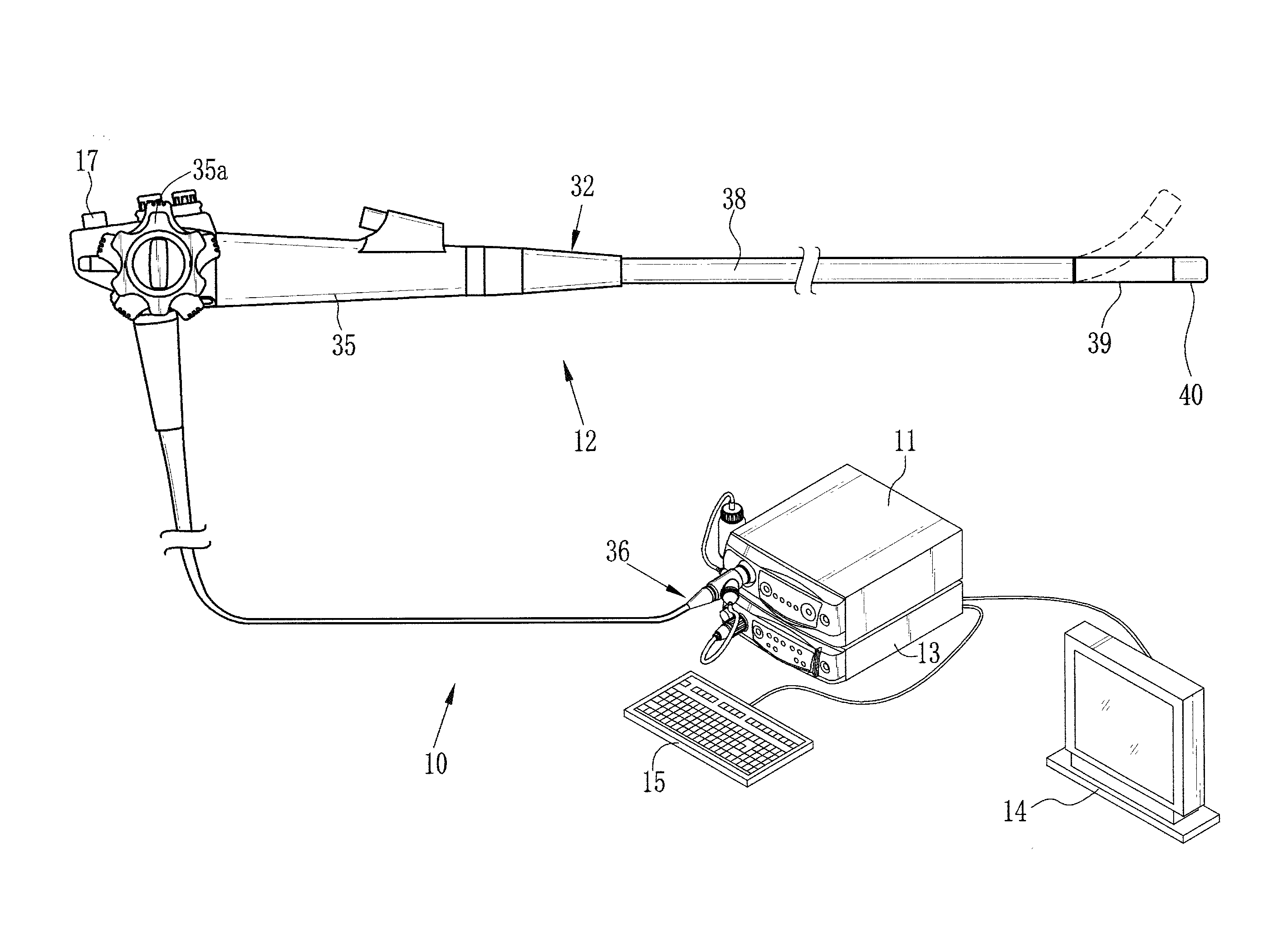
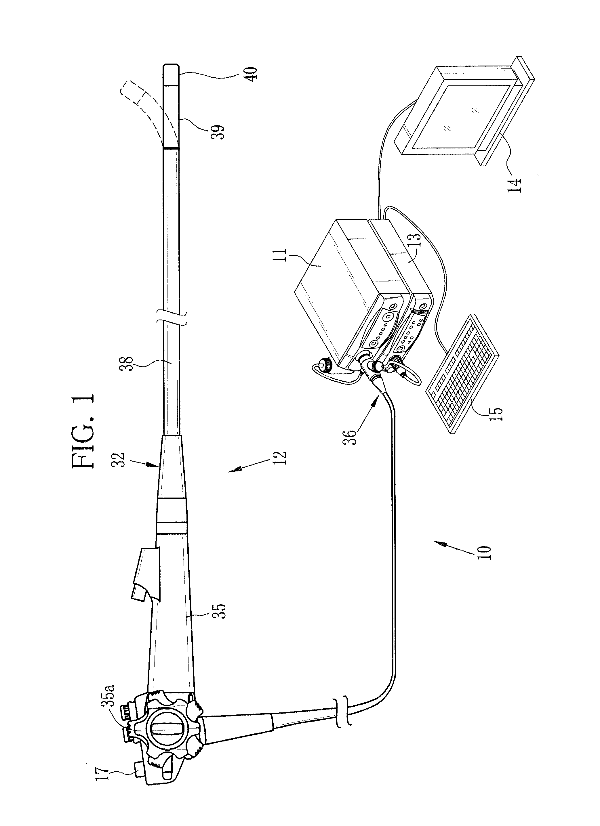
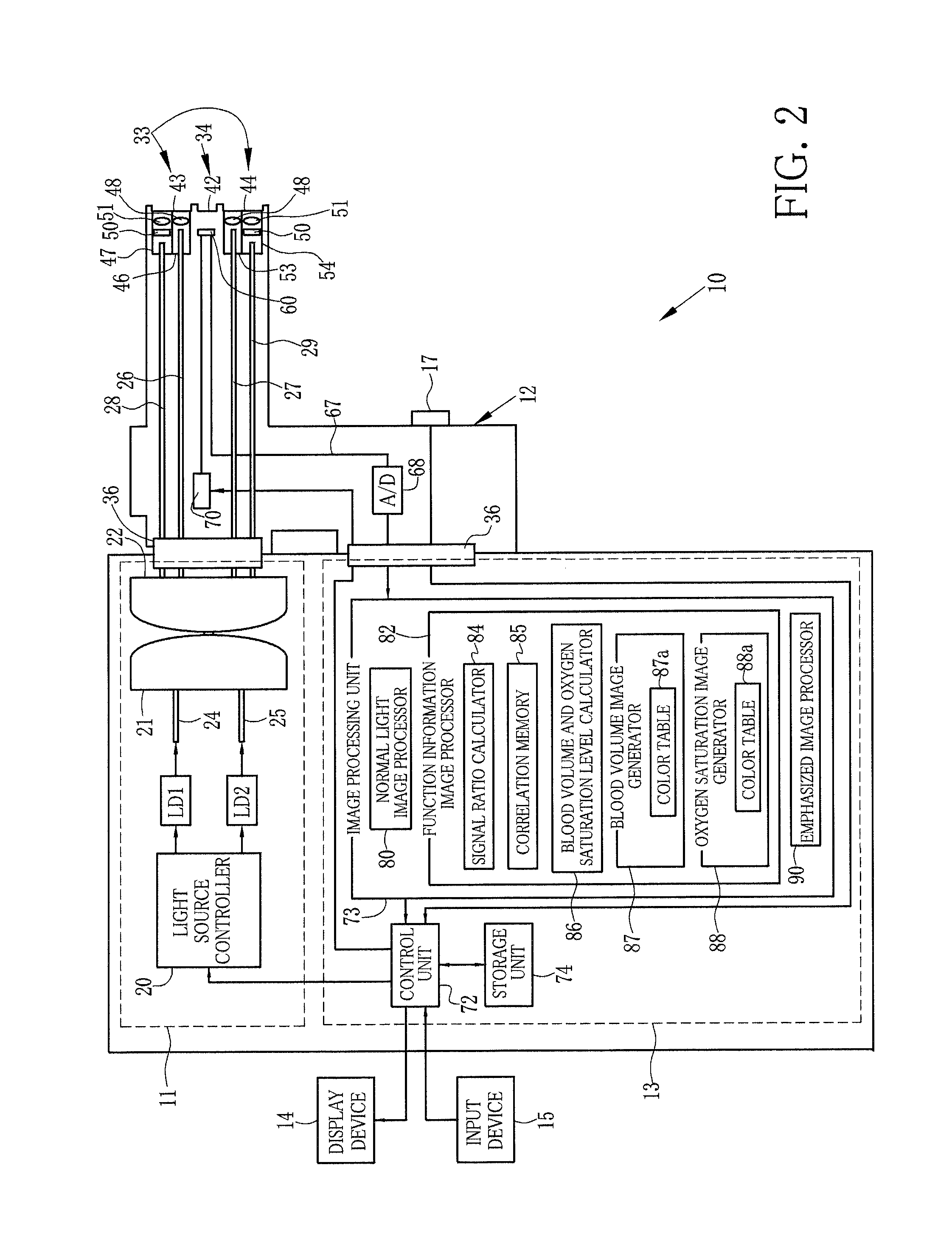
[0049]As shown in FIGS. 1 and 2, an endoscope system 10 is provided with a light source device 11 for emitting light in a predetermined wavelength band, an endoscope system 12 for imaging an area to be observed of an observation object while illuminating the observation object with the light from the light source device 11, a processor device 13 for processing an image signal obtained by the endoscope device 12, a display device 14 for displaying an image of the observation object based on the image signal processed by the processor device 13, and an input device 15 including a keyboard for inputting various types of information to the processor device 13 and the like.

[0050]The endoscope system 10 has a normal observation mode and a biological information observation mode. In the normal observation mode, a normal light image, being an image of the observation object under visible light whose wavelength range extends from blue to red, is displayed on the display device 14. In the bi...

second embodiment

[0101]In the above second embodiment, the first and second abnormal areas are emphasized by adjusting the luminance Y. However, a pixel value itself may be adjusted instead of the luminance Y. The first and second abnormal areas are emphasized by both the brightness and the darkness, but may be emphasized by only one of the brightness and the darkness.

[0102]In a third embodiment of the present invention, the illumination light needed for producing the blood volume image and the oxygen saturation image is produced using a white light source such as a xenon lamp and a rotating filter having a wavelength separation function. As shown in FIG. 19, an endoscope system 120 according to the third embodiment is provided with a broad band light source 121, a rotating filter 122, an optical fiber 123, and a rotation controller 124, instead of the laser light sources LD1 and LD2, the light source controller 20, and the combiner 21 of the first and second embodiments. The broad band light source...


PUM


Abstract

An observation object is imaged under irradiation with oxygen saturation level measurement light to obtain a first image signal, and the observation object is imaged under irradiation with white light to obtain a second image signal. A normal light image is produced from the second image signal. An oxygen saturation level is calculated from the first and second image signals. The calculated oxygen saturation level is imaged in an oxygen saturation image. By superimposing the normal light image on the oxygen saturation image, an emphasized oxygen saturation image is produced. In the emphasized oxygen saturation image, an abnormal area in which a calculation result of the oxygen saturation level is likely to be abnormal is emphasized by its brightness. The produced emphasized oxygen saturation image is displayed on a display device.

Description

BACKGROUND OF THE INVENTION[0001]1. Field of the Invention[0002]The present invention relates to an endoscope system for producing a biological function information image, which images biological function information including the amount of hemoglobin in blood, an oxygen saturation level, and the like, and an operating method thereof.[0003]2. Description Related to the Prior Art[0004]In recent medical care, an endoscope system that is provided with a light source device, an endoscope device, and a processor device is widely used. In an endoscopic diagnosis using this endoscope system, not only normal light observation that uses white light being broad band light as illumination light, but also special light observation that emphasizes a blood vessel of an observation object in display by using narrow band light in a narrowed wavelength band has become widespread.[0005]Besides the special light observation, it is performed to calculate biological function information, including the a...

Claims


Application Information

Patent Timeline
Patent Type & Authority Applications(United States)
IPC IPC(8): A61B1/00G06T5/00
CPCG06T5/008A61B1/00045A61B1/0005A61B1/0638A61B5/14551A61B5/1459G06T7/0012A61B5/489A61B1/063A61B1/0653G06T2207/10024G06T2207/10068G06T2207/10152G06T2207/20221G06T2207/30028G06T2207/10016A61B1/000094
Inventor SAITO, TAKAAKIYAMAGUCHI, HIROSHIIIDA, TAKAYUKI
Owner FUJIFILM CORP
Why Eureka
Try Eureka