Tumour cell and tissue culture

a tissue culture and tumor technology, applied in the field of tumor cell and tissue culture, can solve the problems of poor prognosis of many types of cancer, lack of satisfactory model system for gmb, and development of relevant model systems

Inactive Publication Date: 2014-06-05
ASTERAND UK
View PDF1 Cites 10 Cited by
  • Summary
  • Abstract
  • Description
  • Claims
  • Application Information

AI Technical Summary

Benefits of technology

[0021]The matrix cells form the organotypic environment in which the tumour cells grow. The term “organotypic culture” indicates that cells in the culture associate in a way that as closely as possible replicates the biochemical and physiological properties of the organ from which the cells are derived. In a “co-culture”, cells of at least two distinct types are cultured together. Co-cultured cells may be derived from different sources but can still associate together to replicate an in vivo situation. Co-cultures have properties that are derived from all cell types and from the interaction between the cells within the co-culture.
[0022]The inventors have surprisingly found that a 3D organotypic culture of matrix cells can grow and sustain tumour cells within the culture, so as to provide an in vitro 3D organotypic tumour cell culture according to the invention. The scaffold or matrix for tumour cell growth is provided primarily by the non-tumour cells themselves during culture. Surprisingly, both the dissociated matrix cells and the dissociated tumour cells, once mixed according to the methods of the invention, reorganise to form the organotypic co-cultures of the invention. Such formation of such organotypic tumour cell cultures has not previously been shown.
[0023]An advantage of the co-cultures of the present invention is that they allow interaction of tumour cells with other cell types that are relevant to in vivo tumour growth and pathology. The matrix cells of the invention provide a physiologically relevant environment for the tumour cells. Compacted matrix cells spontaneously reorganize into a complex 3D functional parenchyma over time. For example, compacted brain cells will lead to the formation of a tissue-like ...

Problems solved by technology

Despite aggressive treatment protocols, the prognosis for many types of cancer is still extremely poor.
However, a major challenge for the pharmaceutical industry remains the development of relevant model systems to profile compounds.
For example, although glioblastoma multiforme (GBM) is the most common and most aggressive type of primary brain tumour in humans, no satisfactory model system for GMB is available to date.
A large majority of novel anti-cancer agents fail in the clinic despite evidence of anti-tumour activity in conventional in vitro assays.
However, these models a...

Method used


Image

  • Tumour cell and tissue culture
  • Tumour cell and tissue culture
  • Tumour cell and tissue culture

Examples


example 1

Preparation of Human Tumour Cells

[0128]Human glioblastoma tumour cell lines, LN-18 (ATCC #CRL2610) were cultured and were transfected with either green fluorescent protein (GFP) expressing plasmids or red fluorescent protein (RFP) expressing plasmids. Cells were cultured and plated onto 6 well plates, at 1×10⁵ cells per well, grown overnight and checked to see ˜75% confluence. The cells were transfected with pEGFP-N3 (BD Biosciences cat no #6080-1) using Gene Juice (Novagen cat no #70967-EA) as per manufacturer's protocols. 24 hours after transfection, the cells were selected with Geneticin (GIBCO cat no #10131-019), a population of selected cells were used in the experiments. A population of selected cells were cultured over prolonged periods of time with occasional selection to maintain a population of transfected cells. The expression of fluorescent proteins by the tumour cells allows monitoring of the tumour cells for proliferation and morphological changes. The transfected tumo...

example 2

Preparation of Rat Brain Cells from Isolated Rat Cortex

[0129]The organotypic cultures were produced by preparing dissociated cells from isolated rat cortex. P0 Wistar rats were sacrificed and the brains removed. The cortices were dissected into a Petri dish and chopped into small pieces and put into cold EBSS (GIBCO cat no #24010). The tissue pieces were then triturated very gently. The resultant cell suspension was filtered through a 100 μm mesh filter fitting into a 50 ml falcon tube. The suspension was centrifuged at 1000 rpm / 3 minutes. The viable cells were counted using Trypan blue exclusion and the cell pellet resuspended to 50,000 cells / μl.

example 3

Establishment of 3D Organotypic Cultures from Dissociated Brain Cells

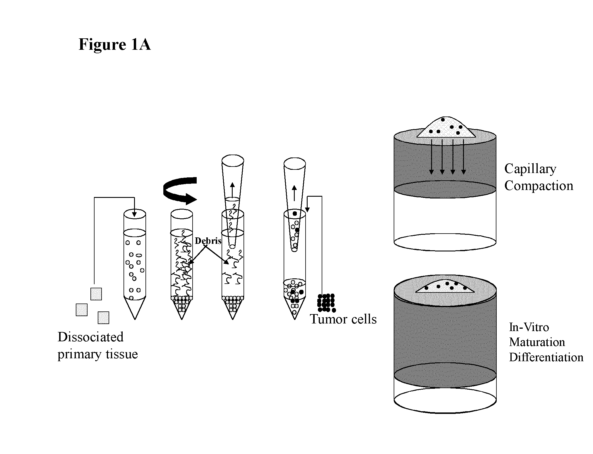
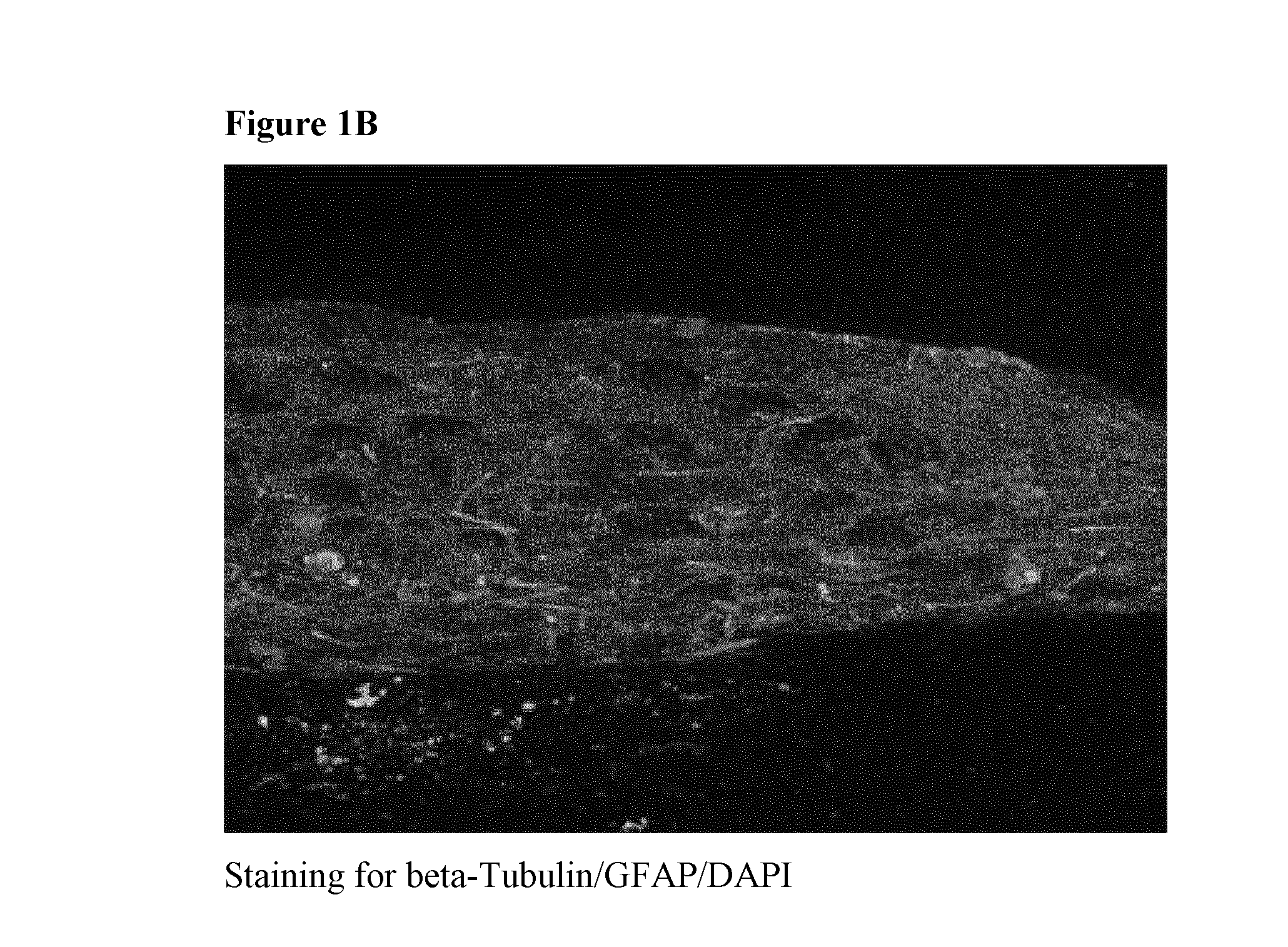
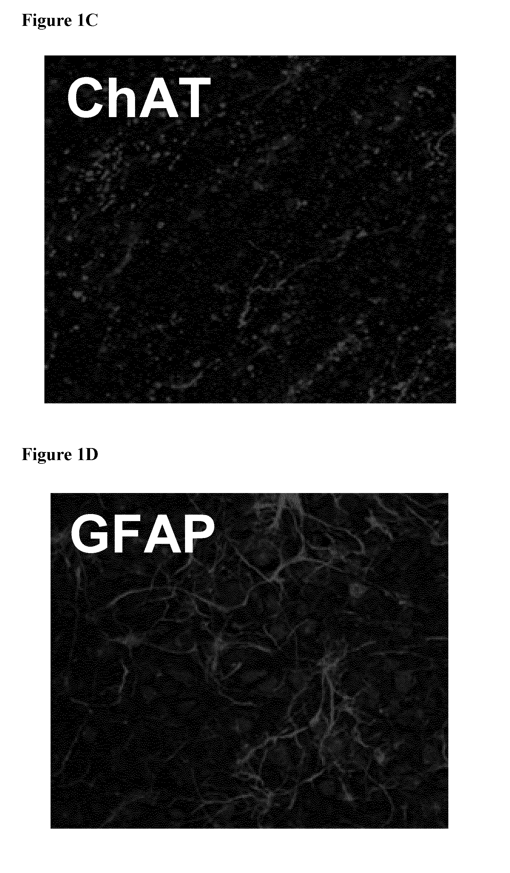
[0130]Dissociated rat brain cortices were allowed to re-aggregate on a semi-porous membrane at an air-liquid interface (cf. FIG. 1, right hand portion). Freshly dissociated brain cells from rat embryo were plated on hydrophilic PFTE membranes placed on media and allow to grow for 20 days. A transverse section of the cell masses obtained was processed for immunofluorescence using markers for neurons (beta-tubulin) and activated astrocytes (glial fibrillary acid protein, GFAP). As shown in FIG. 1B, the staining revealed that both neurons and astrocytes were present in such 3D cultures and that their physiological architecture was preserved (see also FIGS. 1 D, E, F). Cholinergic neurons and nerve terminals were subsequently visualized using an antibody directed against choline acetyltransferase (ChAT; FIG. 1C). As expected, ChAT staining was distributed throughout the whole brain co-culture with specific enrichment i...


PUM


Abstract

The present invention relates to in vitro three-dimensional organotypic cell co-culture. Cultures of the invention comprise tumour cells three-dimensionally disposed within a matrix of matrix cells which are distinct from the tumour cells, wherein the co-culture does not comprise a basement membrane. The cultures are useful for the study of cancers, for testing anti-tumour agent efficacy, and for high-throughput screening of candidate drugs.

Description

[0001]The present invention relates to in vitro tumour cell and tissue culture. In particular, the present invention relates to co-cultures, i.e., mixed cell cultures, comprising tumour cells and cells that are distinct from said tumour cells. The co-cultures of the invention are three-dimensional (3D) organotypic cultures. The invention relates to the formation of co-cultures by aggregation of dissociated cells. The tumour cells in the co-cultures of the invention may be derived from any source of tumour cells, including e.g. primary tumour tissue or tumour cell lines. Methods for preparing the cultures of the invention are also provided. Cultures of the invention are useful for the study of cancer, cancer therapy and cancer diagnostics. For example, cultures of the invention are useful in methods for screening for anti-cancer agents and for testing the efficacy of anti-cancer agents. The methods and cultures of the invention are well suited to applications in a high-throughput for...

Claims


Application Information

Patent Timeline
IPC IPC(8): G01N33/50
CPCC12N5/0693G01N33/5011G01N33/15G01N33/50G01N33/5014
Inventor SUNDSTROM, LARSBIGGS, THELMAFOREMAN, JANET
Owner ASTERAND UK
Why Eureka
Try Eureka