Assays, methods and compositions for diagnosing cancer

a technology of cancer and composition, applied in the field of conditions, can solve the problems of loss of mmr protein expression, high microsatellite instability (msi-h), and no large patient cohort verification of these methods, and achieve the effect of high level of assay multiplexing and scalable automation

Inactive Publication Date: 2014-08-28
GO PATH GLOBAL
View PDF2 Cites 5 Cited by
  • Summary
  • Abstract
  • Description
  • Claims
  • Application Information

AI Technical Summary

Benefits of technology

[0012]A method and single-tube assay are disclosed for identification and quantitative analysis of differentially methylated MLH1 promoter DNA sequences that are associated with some cancers and neoplastic diseases in general in an individual by obtaining a biological sample comprising DNA from the individual, detecting the presence of and measuring the level of methylated MLH1 promoter sequences, and comparing the presence of and level of methylation in the sample to a normalization reference level of “normal” beta-actin gene promoters which is amplified in the same single-tube reaction, wherein a difference in the level or pattern of methylation of the sample compared to the normalization reference level identifies abnormally methylated MLH1 promoter sequences associated with such cancers and other neoplastic diseases, including but not limited to some CRCs, endometrial cancers and gastric cancers. In a further embodiment, a single-tube assay for determining the presence of neoplastic disease in a subject is disclosed, the assay comprising: isolating a single-stranded DNA encoding MLH1 from a biological sample taken from the subject using the probe of the invention, wherein the biological sample is selected from tissue, urine, stool, saliva, blood and serum; treating the single-stranded DNA with bisulfite; amplifying the DNA using the primers of the invention, and determining the level of methylation of the MLH1 promoter region of the single stranded DNA, wherein the presence of MLH1 promoter methylation is an indication of the presence of neoplastic disease in the subject. In an alternative embodiment, the method and single-tube assay can be combined with a miniaturized array platform that allows for a high level of assay multiplexing and scalable automation for sample handling and data processing. Genomic probe and corresponding primers are also disclosed, that are useful in the methods of the invention as they enable detection of differentially methylated genomic MLH1 promoter sequences currently associated with colorectal cancers, endometrial carcinoma and gastric cancer although it may be associated with other cancers and other phenotypes in the future.

Problems solved by technology

LS is caused by germline mutations in DNA mismatch repair (MMR) genes (MLH1, MSH2, MSH6, PMS2), which lead to high microsatellite instability (MSI-H) and loss of MMR protein expression.
However, these methods either are non-quantitative, or they use primers and probes not detecting exclusively methylated MLH1 DNA, or primers and probes not selectively targeting the promoter genomic region critical for MLH1 expression.
Moreover, none of these methods has been verified in large patient cohorts, as discussed in M. T. Parsons et al.
These limitations prevent scientists and clinicians from rendering an unambiguous interpretation of the MLH1 methylation status of tested tumors.
However, we have found that neither method provides a consistently clear cut-off for unambiguous determination of MLH1 methylation in a group of CRC tumors with known information for MLH1 protein expression and BRAF mutations.
However, the currently available methods for MLH1 DNA methylation frequently generate inconsistent results among different studies and therefore cannot be applied for diagnostic purposes.
Furthermore, there is no FDA-approved standard test for MLH1 methylation in clinical use today.
Additionally, the selection of patients for genetic testing to diagnose LS is challenging in clinical practice.
As a result, a significant number of LS patients are overlooked and many patients without mismatch repair gene mutations are sent for genetic testing, leading to an unnecessary increase in inconvenience for the patients and in laboratory cost.
However, the currently available methods either are non-quantitative, or they use primers and probes not detecting exclusively methylated MLH1 DNA, or primers and probes not selectively targeting the promoter genomic region critical for MLH1 expression.
These limitations prevent an unambiguous interpretation of the MLH1 methylation status of tested tumors and result in false positives.

Method used


Image

  • Assays, methods and compositions for diagnosing cancer
  • Assays, methods and compositions for diagnosing cancer
  • Assays, methods and compositions for diagnosing cancer

Examples


example 1

Primers and Probe

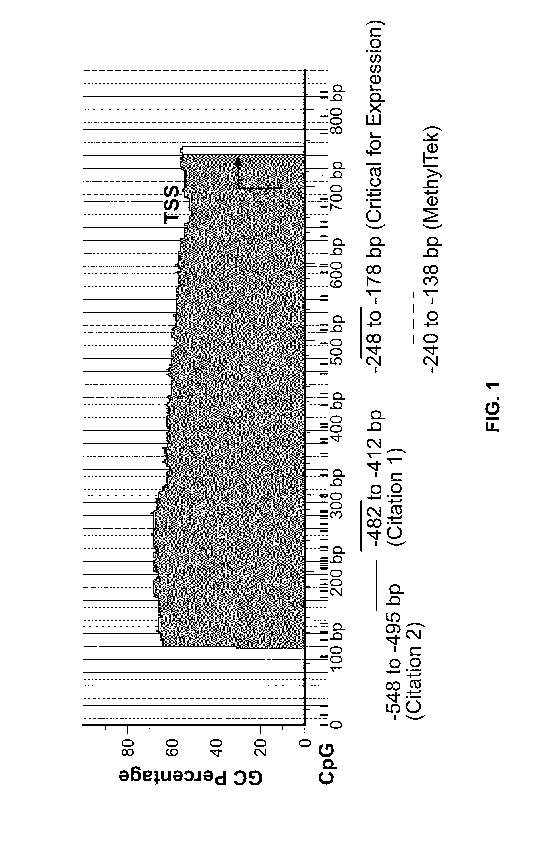
[0032]Various embodiments are disclosed that enable the identification of reliable MLH1 methylation markers for the improved diagnosis and prediction of the susceptibility, diagnosis and staging of neoplastic disease, including CRC. To develop a reliable assay for accurately detecting MLH1 DNA methylation, a novel quantitative real-time system with primers and probe was designed for amplifying exclusively methylated MLH1 DNA. These primers and probe specifically target the region of the MLH1 promoter region critical for its expression, as identified in G. Deng et al. (1999). As discussed in detail below, the assay has been found to provide an accurate determination of MLH1 methylation status in CRC tissue.

[0033]Although several methods have been developed for MLH1 DNA methylation, these are mainly for research purposes and none of them has been successfully developed into an accepted standard assay for clinical molecular diagnostic use. A comprehensive study has ide...

example 2

Assay for Detecting MLH1 Methylation

[0039]The assay for detecting MLH1 DNA methylation combines amplification of MLH1 methylation and ACTB normalization control in a novel one-tube system. This design minimizes the amplification bias between MLH1 and the ACTB control due to variations from pipetting and amplification efficiency in different PCR reactions. The assay comprises the primers and 6-FAM / TAMRA probe for MLH1 methylation selected from those set forth in FIG. 4 (although other probes and primers may be further developed), as well as primers and probes for the ACTB control. The probes for detection of MLH1 methylation and the ACTB control are labeled with different reporter dyes—e.g., 6-FAM and HEX, respectively. However, any suitable reporters now known or hereafter developed is within the scope of the invention. Exemplary primers for the ACTB control are known in the art. For example, VIC / TAMRA labeled probes are commercially available from Applied Biosystems (ABI) and Life ...

example 3

Test Quantitation

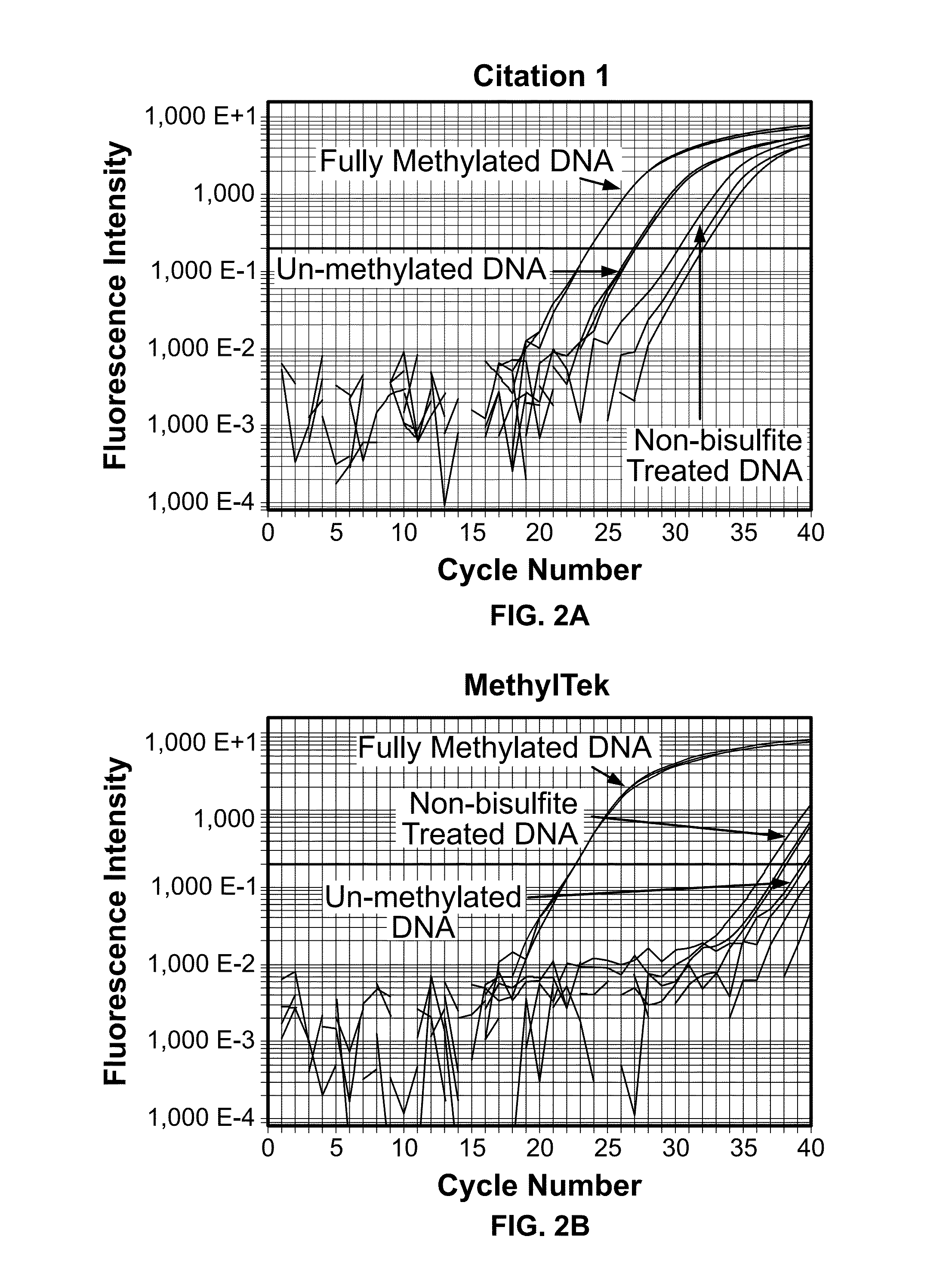
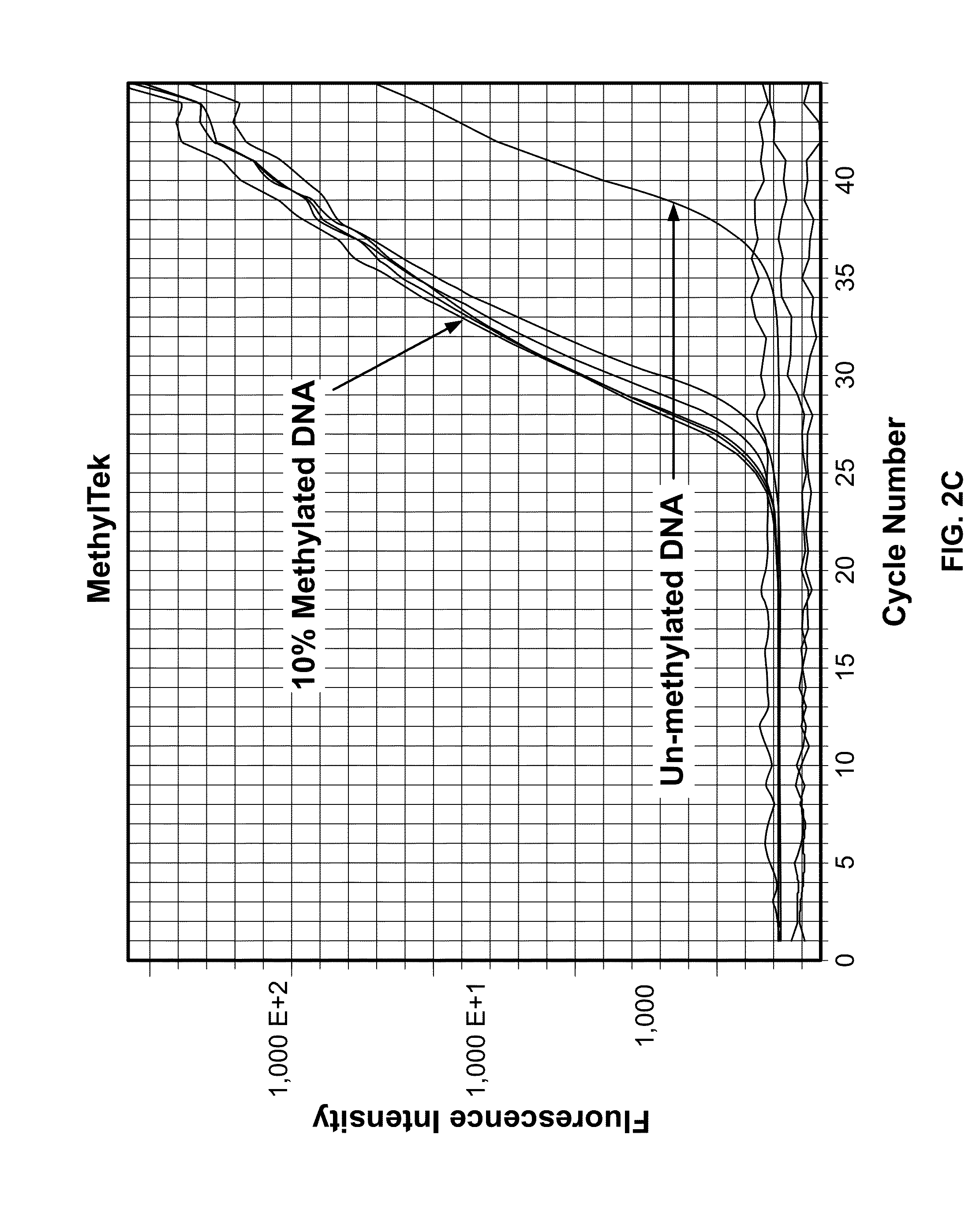
[0041]The assay was tested for the ability to selectively detect methylated DNA. The assay was performed as described in Example 2, using the forward primer MLH1-qMSPJHF1 (Seq. ID No. 1), the reverse primer MLH1-qMSPJHR1&2 (Seq. ID No. 2), and the probe MLH1-qMSPJHP (Seq. ID No. 4), as shown in FIG. 4. In vitro methylated lymphocyte DNA was used as a positive control. Non-methylated or non-bisulfite treated lymphocyte DNA were used as negative controls.

[0042]The assay was found to be highly specific and sensitive in detecting MLH1 DNA methylation in comparison to the highly cited, prior art method of Bettstetter (2007) (FIG. 1). As shown in FIG. 2A, the method of Bettstetter (2007) had only 8-fold difference (3 cycles) in selectively detecting in vitro fully methylated DNA over non-methylated DNA. Moreover, this method was also found to nonspecifically amplify bisulfite unconverted DNA.

[0043]In contrast to the prior art methods, the present assay showed more than 10...


PUM

PropertyMeasurementUnit
fluorescenceaaaaaaaaaa
fluorescence intensityaaaaaaaaaa
, real time PCRaaaaaaaaaa

Abstract

The present invention provides a method and single-tube assay for identification and quantitative analysis of differentially methylated MLH1 promoter sequences that are associated with certain types of cancer in an individual by obtaining a biological sample comprising DNA from the individual, detecting the presence of and measuring the level of methylated MLH1 promoter sequences, and comparing the presence of and level of methylation in the sample to a normalization reference of “normal” beta-actin gene promoters, wherein a difference in the level or pattern of MLH1 methylation of the sample compared to the Actin gene reference level identifies abnormally methylated MLH1 promoter sequences associated with cancer.

Description

[0001]This application claims the benefit of U.S. Provisional Application No. 61 / 771,061, filed Feb. 28, 2013, which is hereby incorporated by reference in its entirety.BACKGROUND OF THE INVENTION[0002]The present invention relates to conditions characterized by differentially methylated MLH1 promoter sequences and, in particular, to diagnostic and prognostic methods that exploit the presence of such DNA sequences that exhibit altered MLH1 promoter sequences.[0003]In one embodiment, the invention may be used as a diagnostic for certain cancers and non-cancerous neoplastic diseases having hypermethylation of the MLH1 promoter, including but not limited to colorectal disease, endometrial carcinoma and gastric cancer.[0004]For example, Lynch syndrome (LS) is a hereditary form of colorectal cancer (CRC) and is responsible for 2-5% of newly diagnosed patients with CRC. LS is caused by germline mutations in DNA mismatch repair (MMR) genes (MLH1, MSH2, MSH6, PMS2), which lead to high micro...

Claims


Application Information

Patent Timeline
Patent Type & Authority Applications(United States)
IPC IPC(8): C12Q1/68
CPCC12Q1/6886C12Q2600/112
Inventor LU, JIM Z.GUO, ZHONGMIN
Owner GO PATH GLOBAL
Why Eureka
Try Eureka