
System and method for attenuation correction in emission computed tomography

a computed tomography and emission correction technology, applied in tomography, instruments, applications, etc., can solve the problems of significant diagnostic error, hampered image quality of emission computed tomography system such as combined pet/ct or spect/ct, and respiratory misalignmen

Inactive Publication Date: 2014-09-18
UNIVERSITY OF MACAU
View PDF5 Cites 18 Cited by
  • Summary
  • Abstract
  • Description
  • Claims
  • Application Information

AI Technical Summary

Benefits of technology

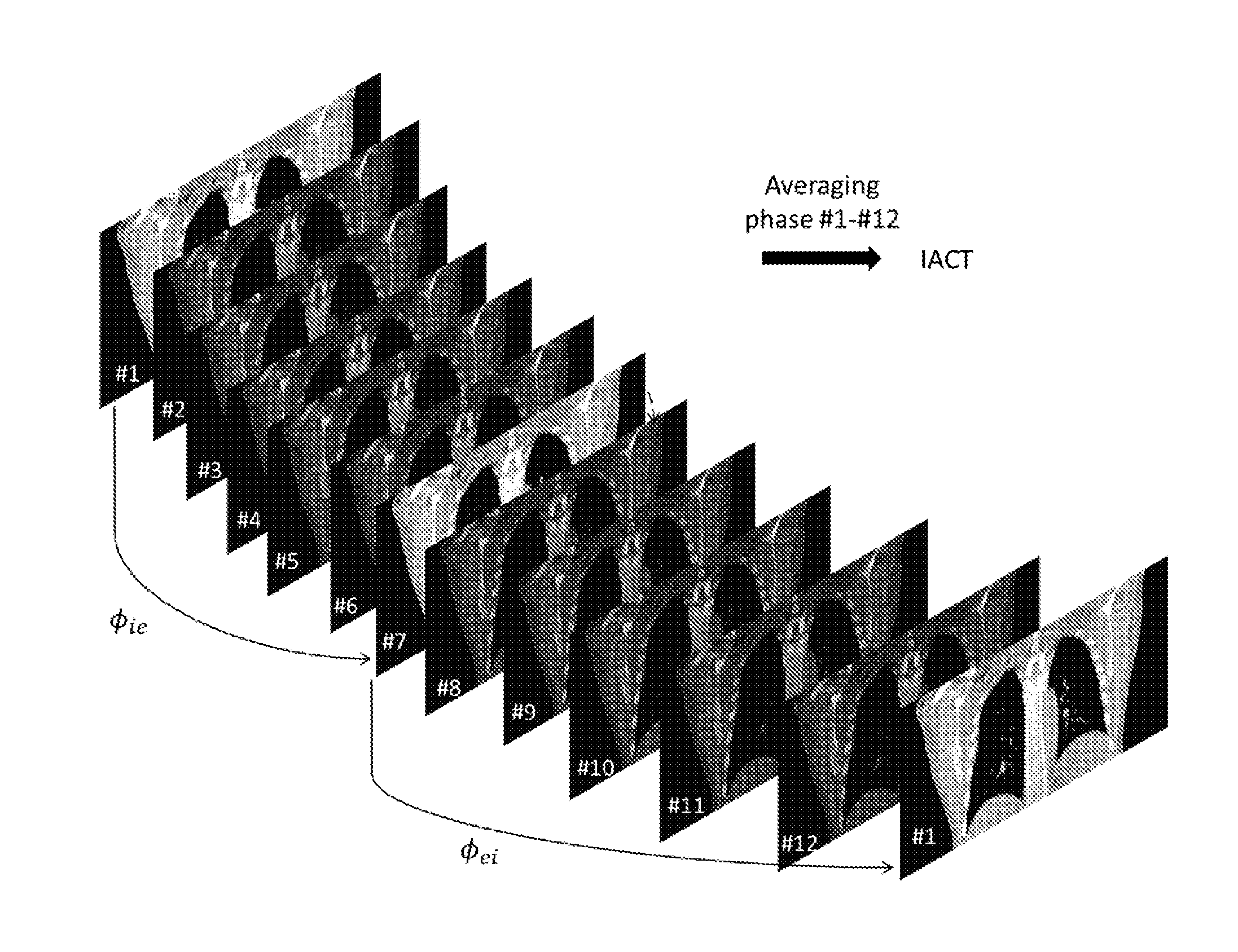
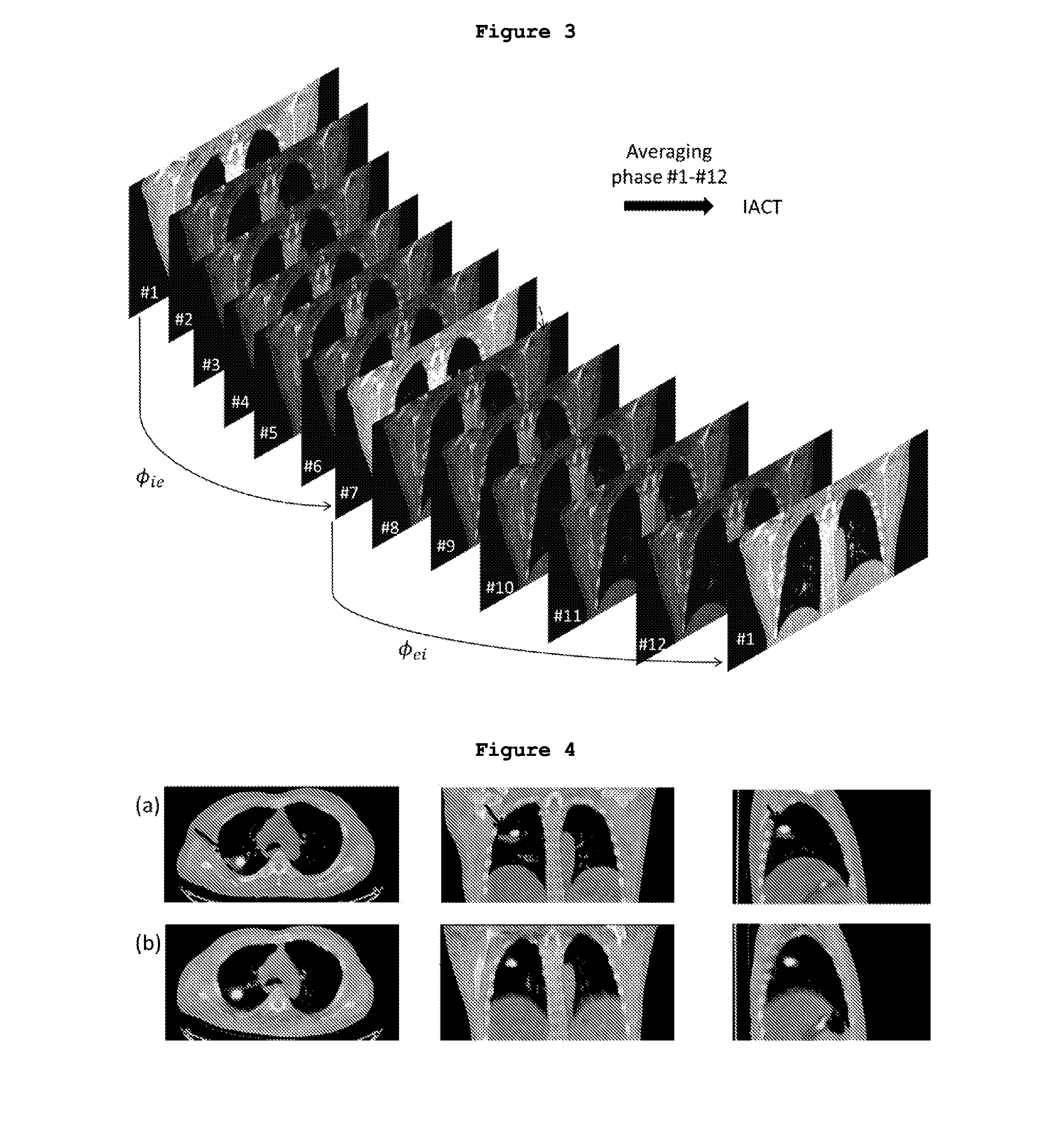
The present invention provides a method for attenuation correction in reconstructed emission computed tomography (ECT) images. This is achieved by generating an Interpolated Average CT (IACT) image based on one or more CT images acquired during momentsary suspension of breathing motion at specific phases in the breathing cycle of a subject by means of a breathing control device. An ECT image is then acquired from the same section of the subject and corrected for attenuation using the IACT image as an attenuation map in the ECT reconstruction that comprises the use of a reconstruction algorithm. This results in reconstructed ECT images with attenuation correction. The present invention also provides a system for generating Interpolated Average CT image of a subject that includes an active breathing controller for momentarily suspending breathing motion of a subject at one or more specific phases in a breathing cycle, a computing device for identifying one or more specific phases in the breathing cycle and a CT scanner for acquiring one or more CT images when the subject's breathing motion is momentarily suspended by the ABC.

Problems solved by technology

Image quality of emission computed tomography systems such as combined PET / CT or SPECT / CT is hampered by respiratory misalignments and artifacts.
The problem of respiratory artifacts has been most closely studied for myocardial perfusion PET / CT, while more than 40% of the studies have artifactual defects in the cardiac region when no steps are taken to address the PET / CT alignment, causing significant diagnostic error (Gould et al., 2007).
Mismatched attenuation correction (AC) can also cause SUV overestimation for lower lung tumors that are located close to the liver dome, leading to complicated SUV errors (Liu et al., 2009).
All these PET / CT mismatching artifacts will lead to inaccurate localization and quantification of tumors, hence potential misdiagnoses (Osman et al., 2003; Allen-Auerbach et al., 2006; von Schulthess et al., 2006; Pan et al., 2013).
This method is not practical for all patients since it requires patients' compliance and may not be feasible for patients with limited pulmonary function (Senan et al., 2004).
The main problem of CACT is relatively high radiation dose.
However, its main disadvantages are the high dose from the potential 4D CT, increased acquisition and postprocessing time.

Method used


Image

  • System and method for attenuation correction in  emission computed tomography
  • System and method for attenuation correction in  emission computed tomography
  • System and method for attenuation correction in  emission computed tomography

Examples


example 1

Active Breathing Controller (ABC) Design

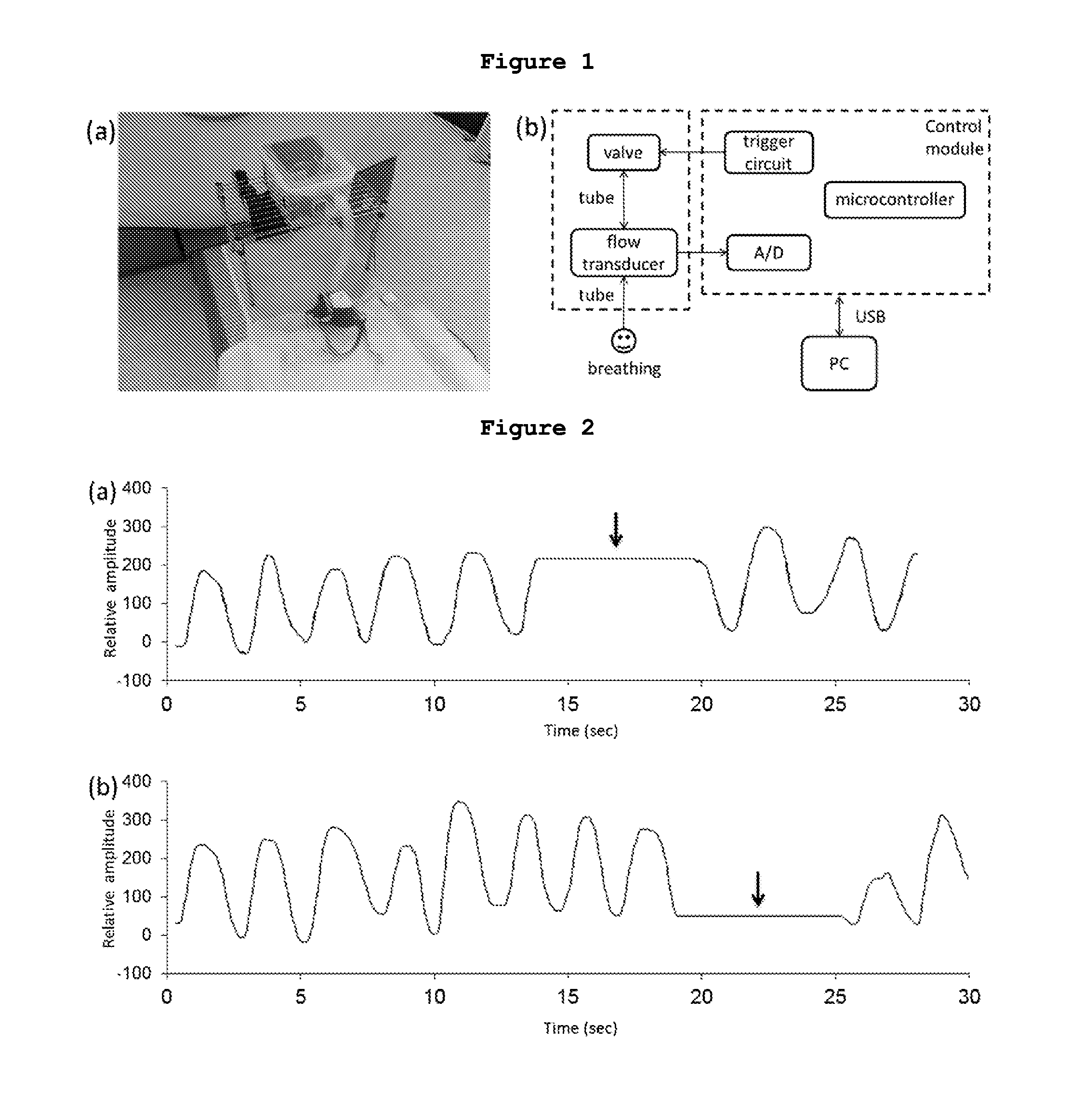
[0097]The ABC used in this example integrates a spirometer, an air mask / mouth piece, and a tube-valve system as shown in FIG. 1. Patients were asked to perform mouth-breathing using the air mask or mouth piece that is connected to the tube. The flow sensor inserted in the tube detected real-time breathing flow rate of the patients and sampled the signal to the microcontroller, which further preprocessed the signal and sent it to a computer through a USB connector. An acquisition program based on C++ was developed to process the input signal and control the switching of the valve in the tube. The program can detect the change of the air flow direction according to the flow rate measurement to locate the end-inspiration and end-expiration phases, while the trigger circuit can then automatically control the closing of the valve located in the end of tube to suspend the patients' breathing. Hence, the operator only needs to notify the program when...

example 2

Clinical Setup

[0118]In this Example, two normal volunteers were recruited and images were acquired using a PET / CT scanner (Discovery VCT, GE Medical Systems, Milwaukee, Wis., USA). The patients were injected with 328 MBq and 406 MBq of 18F-FDG respectively and scanned 1 hour post injection. Thoracic PET data were acquired for 2 bed positions with 3 minutes per bed position. One standard helical CT (HCT), two extra end-inspiration and end-expiration breath-hold helical CTs were obtained using ABC for IACT were performed for each subject (FIG. 8). The standard HCT acquisition settings were: 120 kV, smart mA (range 30-150 mA) helical mode, 0.984:1 pitch, 0.5 s gantry rotation and total 4.4 s scan time. For IACT, two breath hold CTs were acquired at 120 kV, 10 mA helical modes, 0.984:1 pitch, 0.5 s gantry rotation time and a total of 4.4 s acquisition time for each scan.

IACT Generation

[0119]B-spline, a deformable image registration algorithm, was applied to calculate the deformation vec...

example 3

[0126]In this example, 4D Extended Cardiac Torso (XCAT) Phantom is used for realistically model the anatomy, activity distribution of a patient injected with 18F-FDG, and the respiratory motions. An analytical projector and OS-EM reconstruction algorithm provided by STIR (Software for Tomographic Image Reconstruction) was used for modelling a GE Discovery STE PET Scanner. The respiratory cycle was divided into 13 phases starting from the end-inspiration phase, and two maximum respiratory motion amplitudes of 2 cm and 3 cm were modeled. The attenuation maps representing CACT, IACT, HCTin and HCTex are shown in FIG. 11 (a) to (d). Thoracic spherical lesions having diameters of 10 mm and 20 mm are placed at 4 different locations individually at lower left lung, lower right lung, middle right lung and upper right lung as shown in FIG. 12 (a) to (d). The target-to-background ratios (TBR) of 4:1 and 8:1 was used for respiratory amplitude of 2 cm while the TBR of 6:1 and 12:1 was used for ...


PUM


Abstract

The present invention relates to systems and methods for attenuation correction to improve reconstructed image quality and quantitative accuracy and reduce radiation dose in emission computed tomography. In one embodiment, the present invention provides an interpolated average CT (IACT) method and breathing control devices.

Description

[0001]This application claims the benefit of U.S. 61 / 787,572 filed Mar. 15, 2013. The entire disclosure of the preceding application is hereby incorporated by reference into this application. Throughout this application, various references or publications are cited. Disclosures of these references or publications in their entireties are hereby incorporated by reference into this application in order to more fully describe the state of the art to which this invention pertains.FIELD OF THE INVENTION[0002]The present invention relates to systems and methods for attenuation correction to improve reconstructed image quality and quantitative accuracy and reduce radiation dose in emission computed tomography, and relates particularly to systems and methods which employ an interpolated average CT (IACT) method and breathing control devices.BACKGROUND OF THE INVENTION[0003]Image quality of emission computed tomography systems such as combined PET / CT or SPECT / CT is hampered by respiratory mis...

Claims


Application Information

Patent Timeline
Patent Type & Authority Applications(United States)
IPC IPC(8): A61B6/00A61B6/03G06T11/00
CPCA61B6/5264G06T11/005A61B6/037A61B6/032A61B6/503A61B6/5205A61B6/5247A61B6/5258A61B6/583G06T2211/464
Inventor MOK, SENG PENGSUN, TAO
Owner UNIVERSITY OF MACAU
Learn More