Cell separation method

a cell and cell technology, applied in the field of immunology, can solve the problems of time-consuming and laborious centrifugation steps, limited methods to small amounts of cells, and includes a time-consuming and laborious centrifugation step, and achieve the effect of increasing the volume of the supernatant and rapid sedimentation of the cells

Active Publication Date: 2014-10-09
MILTENYI BIOTEC B V & CO KG
View PDF6 Cites 13 Cited by
  • Summary
  • Abstract
  • Description
  • Claims
  • Application Information

AI Technical Summary

Benefits of technology

[0019]It is an advantage of the present invention that there is no need for exerting centrifugal forces for rapid sedimentation of the cells. Preferentially, the method exploits the synergistic effect of applying gravity sedimentation (1×g) and magnetic forces only. Preferentially the magnetic source is positioned to the side of the container containing the sample to be processed. This arrangement results in two forces acting on the cells, one is the gravity sedimentation to the bottom and the other is the magnetic force to the side of the container containing the sample to be processed. But the position of the magnetic source or sources may vary resulting in different directions in which the forces act on the cells.
[0020]In addition unexpectedly, it was found that the sedimentation of cells in a sample is accelerated if simultaneously (1) the erythrocytes are aggregated by an erythrocyte aggregation reagent such as HPMC-15 and (2) one or more undesired cellular components are bound specifically by magnetic particles having a size between 100 and 1400 nm, wherein a magnetic field gradient is applied to the magnetic particles, immobilizing the cells bound to the magnetic particles.
[0021]In addition surprisingly, it was found that the shape of the cell pellet can be influenced by the selection of the antigen recognizing moiety which is coupled to a magnetic particle and by the choice of the position of the magnetic source relative to the container containing the sample to be processed. If an antigen recognizing moiety, which recognizes a surface protein of one or more undesired cellular components is used, but which does not recognize a surface protein of erythrocytes, the pellet is separated into two parts. Only a slight transition is generated in this case if the magnetic source is positioned on the side of the container containing the sample to be processed as illustrated in FIG. 13. The use of one or more antigen recognizing moieties which recognize a surface protein of erythrocytes and also a surface protein of one or more undesired other cellular components, e.g. CD36, results in a magnetically enforced sedimentation of all cellular components which are desired to be sedimented and / or immobilized. If the magnetic source is positioned in the same way as shown in FIG. 13 the pellet forms under such conditions a different shape, resembling an exponential function curve resulting in a smooth transition as illustrated in FIG. 14. This or similar shapes of pellets are advantageous for removing the supernatant if e.g. a pipette is used resulting in an increased volume of the supernatant which can be recovered (Example 27).
[0022]The compositions of the present invention comprise i) an erythrocyte aggregation reagent and ii) a set of one or more mono- and / or multi-specific magnetic particles with at least one antigen recognizing moiety coupled to the particles, wherein said particles with said at least one antigen recognizing moiety specifically bind to at least one antigen specific for one or more undesired cellular components.

Problems solved by technology

But these methods are limited to small amounts of cells only.
All density separation techniques have the same basic limitations: they can not separate subpopulations of cells with overlapping density distributions such as lymphocyte subsets and they include time-consuming and laborious centrifugation steps.
The disadvantage of this method is that it includes a time-consuming and laborious centrifugation step.
This method suffers from the time-consuming process of more than 1 hour until the desired cell are available for further processing steps.
In addition disadvantageously is the rather low recovery of desired cells.

Method used


Image

  • Cell separation method
  • Cell separation method
  • Cell separation method

Examples


example 1

Size of Particles

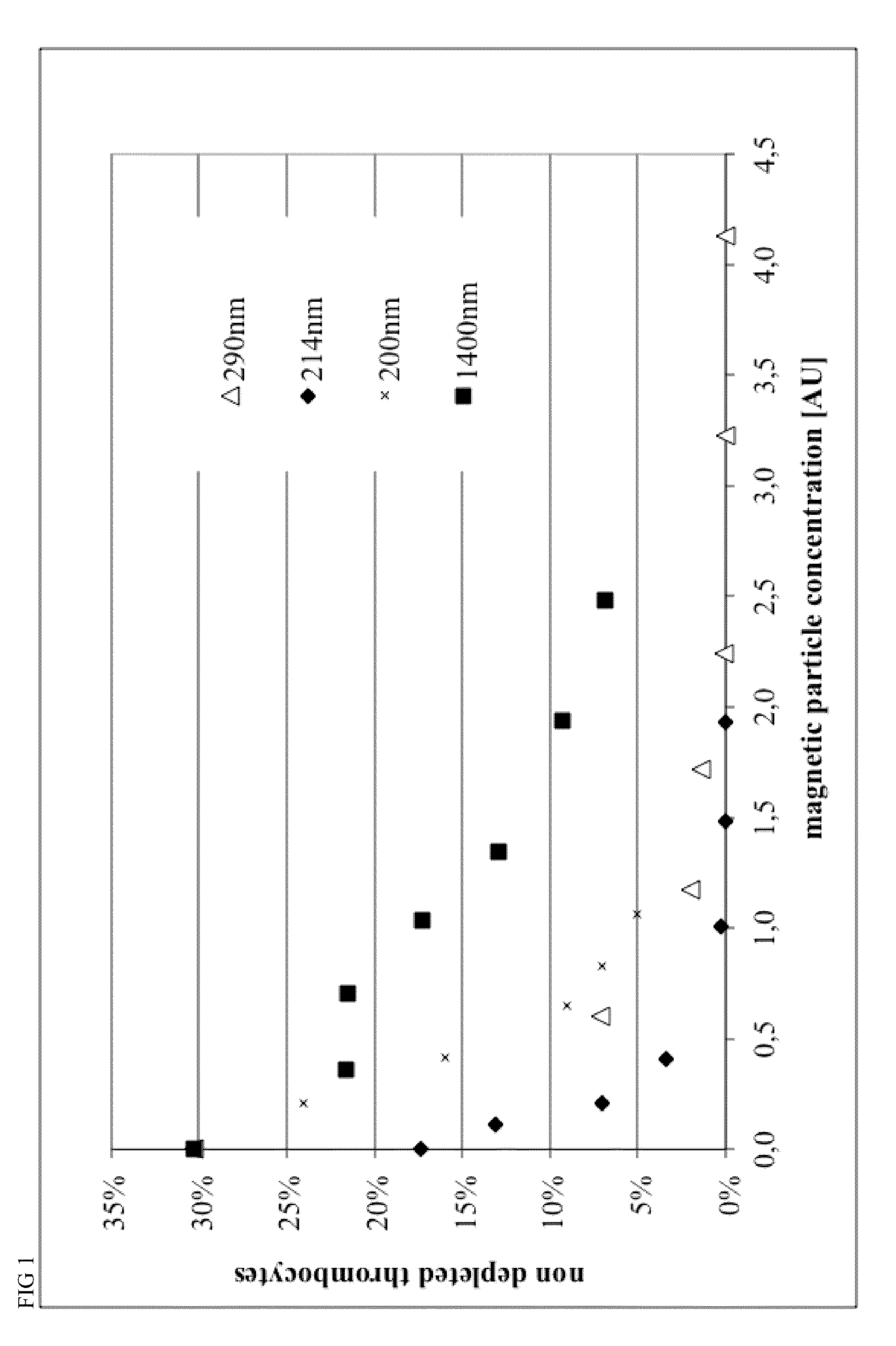
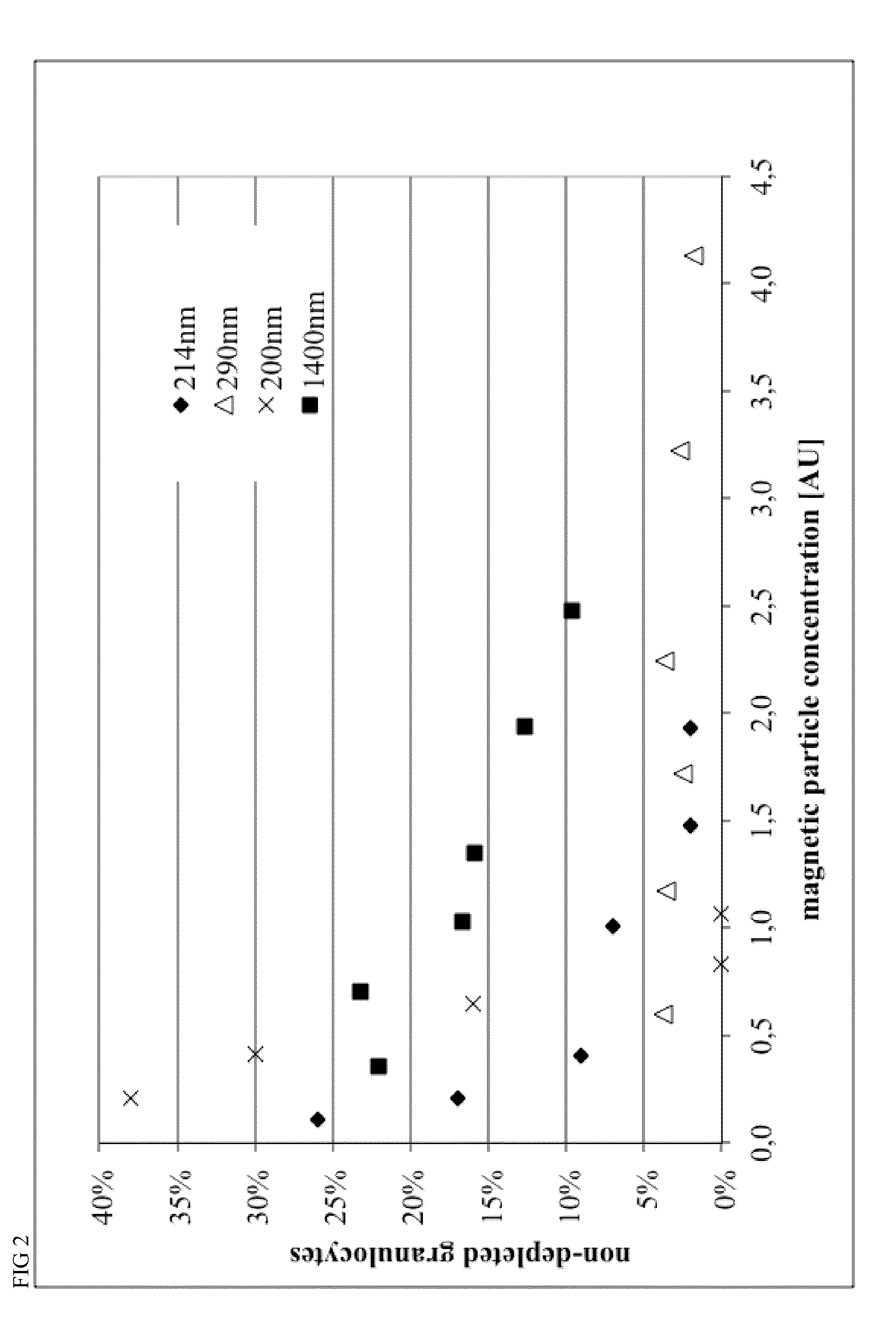
[0124]Magnetic beads were manufactured with different process parameters according to Example 4, resulting in different size of the particles. Particle size was characterized by Beckman Coulter Delsa Nano instrument. Monoclonal antibodies recognizing CD15 and CD61 antigens were covalently conjugated to magnetic beads in a 1:1 ratio, resulting in 40 ug antibody per mL of bead suspension at a concentration of OD450=10.

[0125]Different amounts of conjugated beads and 0.2 mL of a 2% stock solution of hydroxypropylmethylcellulose were given to 1 mL of Buffy Coat from human whole blood within a 5 mL FACS tube, mixed for 15 minutes within MACSmix™ Tube rotator (Miltenyi Biotec) and placed in a MACSiMag™ Separator for 20 minutes. Supernatant was completely removed using a pipette, transferred to a FACS Tube, cells were counted using a Sysmex KX21 hematology analyzer and cells were analyzed using a MACSquant Analyzer flow cytometer (Miltenyi Biotec).

[0126]Erythrocytes, thromb...

example 2

Mono-Specific Particles

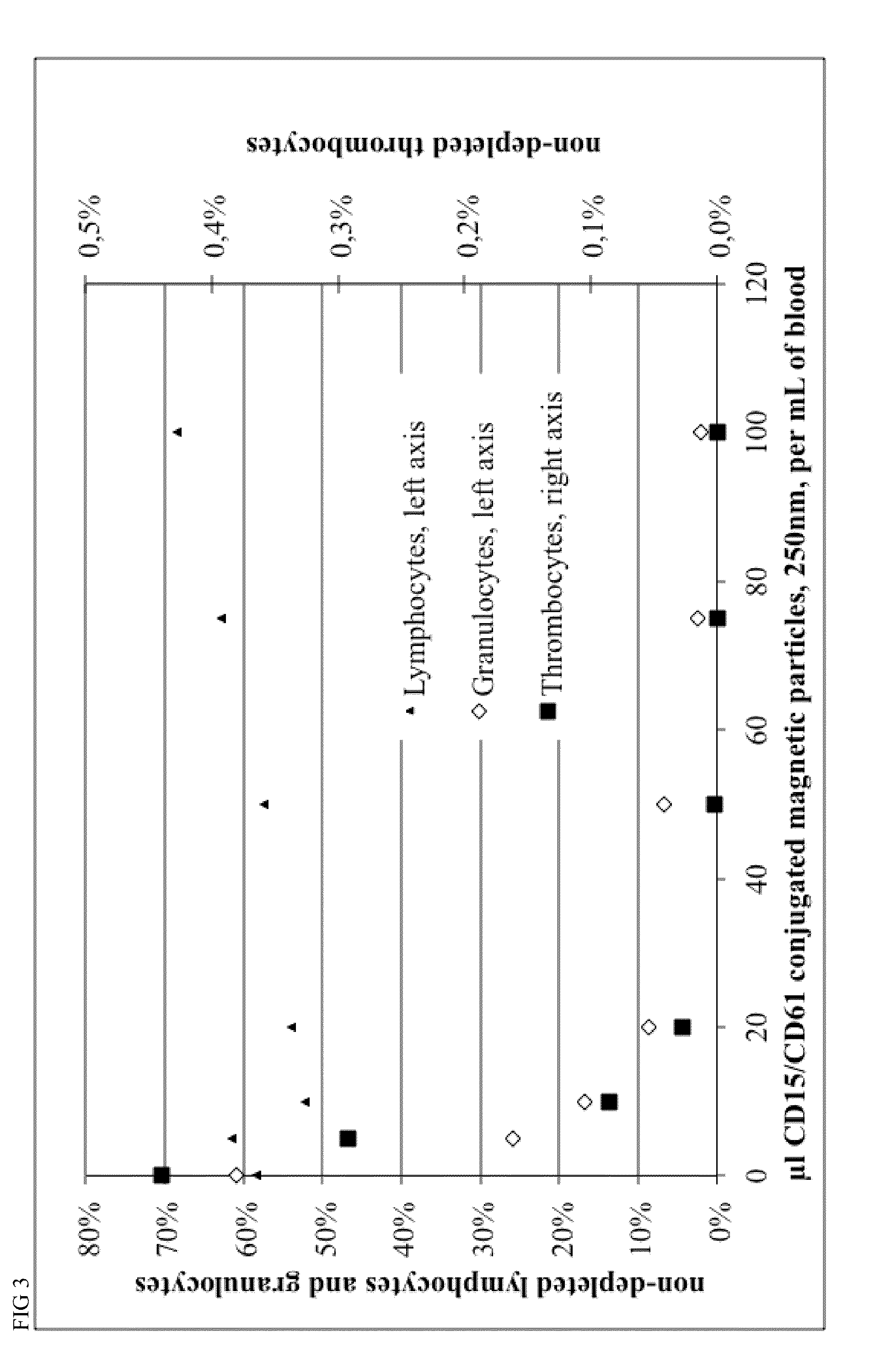
[0128]Monoclonal antibodies recognizing CD15 and CD61 were individually conjugated to magnetic beads (Example 4, average diameter 100 nm). Bead conjugated antibodies were given to 1.3 mL of whole blood, 0.7 mL of PBS buffer and 200 ul of HPMC15 stock solution. Granulocytes and platelets, respectively or combined, were removed for 98% to >99%, (see FIG. 5).

[0129]Monoclonal antibodies recognizing CD19 and CD56 were individually conjugated to magnetic beads (Example 4, average diameter 220 um). Bead conjugated antibodies were given to 1.3 mL of whole blood, 0.7 mL of PBS buffer and 200 ul of HPMC15 stock solution. B cells and NK cells respectively were removed for >98% (see FIG. 6 and FIG. 7).

example 3

Multi-Specific Particles

[0130]Either 2, 3 ,4, 6 or 8 different antibodies were conjugated to either 100 nm or 200 nm magnetic beads (see Example 4) in previously evaluated antibody ratios. Antibody ratios were determined by titrating mono-specific conjugates and determining individually the lowest amount of antibody sufficient for complete removal of a cell subset. The individual antibody concentrations were used to calculate ratios. Bead conjugated antibodies were given to 1.3 mL of whole blood, 0.7 mL of PBS buffer and 200 ul of 2% HPMC15 stock solution, mixed for 15 minutes within MACSmix™ Tube rotator (Miltenyi Biotec) and placed in a MACSiMag™ Separator for 20 minutes. Supernatant was completely removed using a pipette, transferred to a FACS Tube, cells were counted using a Sysmex KX21 hematology analyzer and cells were analyzed using a MACSquant Analyzer flow cytometer (Miltenyi Biotec). More than 92% of the cell populations analyzed were depleted, see FIG. 8.


PUM

PropertyMeasurementUnit
sizeaaaaaaaaaa
sizeaaaaaaaaaa
timeaaaaaaaaaa

Abstract

The present invention provides methods and compositions for separating cells from a sample containing erythrocytes. The method is for recovering desired cells from a sample containing the desired cells, erythrocytes and undesired cells comprising: a) contacting the sample with a composition, said composition comprising: i) an erythrocytes aggregation reagent ii) at least one antigen recognizing moiety coupled to a magnetic particle, wherein said particle with said at least one antigen recognizing moiety specifically binds to at least one antigen specific for one or more undesired cellular components; b) applying simultaneously i) gravity sedimentation for sedimentation of erythrocytes and ii) a magnetic field gradient to said sample for immobilizing said magnetic particle generating a pellet and a supernatant phase, and c) recovering the desired cells from the supernatant phase. Compositions for the use within the present method are also disclosed.

Description

FIELD OF INVENTION[0001]The present invention relates generally to the field of immunology, in particular to processes and compositions for separation of cells.BACKGROUND OF THE INVENTION[0002]Cell separation methods are wide spread in scientific and clinical laboratories for research, diagnostics, or clinical applications. Most strategies of cell separation are based on different physical properties such as size and density.[0003]Normally one has to get rid of the erythrocytes before dealing with the cells of interests, the leukocytes. This can be done among other methods by separation of cells using e.g. gradient density centrifugation, peripheral blood mononuclear cells (PBMC) sample preparation or by erythrocyte lysis, all well known methods in the art.[0004]After the elimination of erythrocytes methods using solid phase particles attached to antigen binding moieties, which recognize surface antigens on cells, can be used to separate white blood cells into subpopulations. Such m...

Claims


Application Information

Patent Timeline
Patent Type & Authority Applications(United States)
IPC IPC(8): G01N1/34
CPCB01D21/00C12Q1/02C12N5/0087B03C1/01G01N1/34B03C2201/26B01D21/0009B01D21/262B03C1/288
Inventor KAULING, BURGUNDHUPPERT, VOLKER
Owner MILTENYI BIOTEC B V & CO KG
Why Eureka
Try Eureka