
Methods for Use of Neural Stem Cell Compositions for Treatment of Central Nervous System Lesions

Active Publication Date: 2014-10-16
RGT UNIV OF CALIFORNIA +1
View PDF0 Cites 4 Cited by
  • Summary
  • Abstract
  • Description
  • Claims
  • Application Information

AI Technical Summary

Benefits of technology

The patent text describes a new method for treating spinal cord injuries using neural stem cells. These cells can be injected into the spinal cord and can grow to form connections with other nervous system cells. The treatment involves adding a growth factor to the stem cells, which helps them to survive and extend their connections. This method has been found to be effective in promoting spinal cord healing and restoring function.

Problems solved by technology

However, by themselves, post-natal neuronal cells cannot induce the extensive structural and functional recoveries provided according to the protocol of the invention.

Method used


Image

  • Methods for Use of Neural Stem Cell Compositions for Treatment of Central Nervous System Lesions
  • Methods for Use of Neural Stem Cell Compositions for Treatment of Central Nervous System Lesions
  • Methods for Use of Neural Stem Cell Compositions for Treatment of Central Nervous System Lesions

Examples


example i

Animal Model of Spinal Cord Injury

[0076]Adult female Fischer 344 rats (160-200 g, n=27) were subjects of this study. NIH guidelines for laboratory animal care and safety were strictly followed. Animals had free access to food and water throughout the study. All surgery was done under deep anesthesia using a combination (2 ml / kg) of ketamine (25 mg / ml), xylazine (1.3 gm / ml) and acepromazine (0.25 mg / ml). Adult female Fischer 344 rats underwent C5 lateral hemisection or T3 complete transection.

example ii

Preparation of Embryonic Cell Grafts

[0077]Embryonic day 14 (E14) spinal cord from transgenic Fischer 344-Tg (EGFP) rats ubiquitously express GFP under the ubiquitin C promoter and provided donor tissue for grafting (Rat Resource and Research Center, University of Missouri, Columbia, Mo.). Following C5 or T3 dorsal laminectomy, the dura was cut longitudinally and retracted. A 2-mm-long block of spinal cord was cut and removed using a combination of iridectomy scissors and microaspiration, with visual verification to ensure complete transection ventrally and laterally. E14 spinal cord from Fischer 344 rats expressing GFP was freshly dissected and dissociated.

[0078]Dissociated E14 cells were re-suspended in a fibrin matrix (25 mg / ml fibrinogen and 25 U / ml thrombin). Growth factors were added to the cell / adhesive suspension: BDNF (50 μg / ml), neurotrophin-3 (NT-3; 50 μg / ml), platelet-derived growth factor (PDGF-AA; 10 μg / ml), insulin-like growth factor 1 (IGF-1; 10 μg / ml), epidermal grow...

example iii

Grafting of Pluripotent Neural Stem Cells Derived from Embryonic Spinal Cord

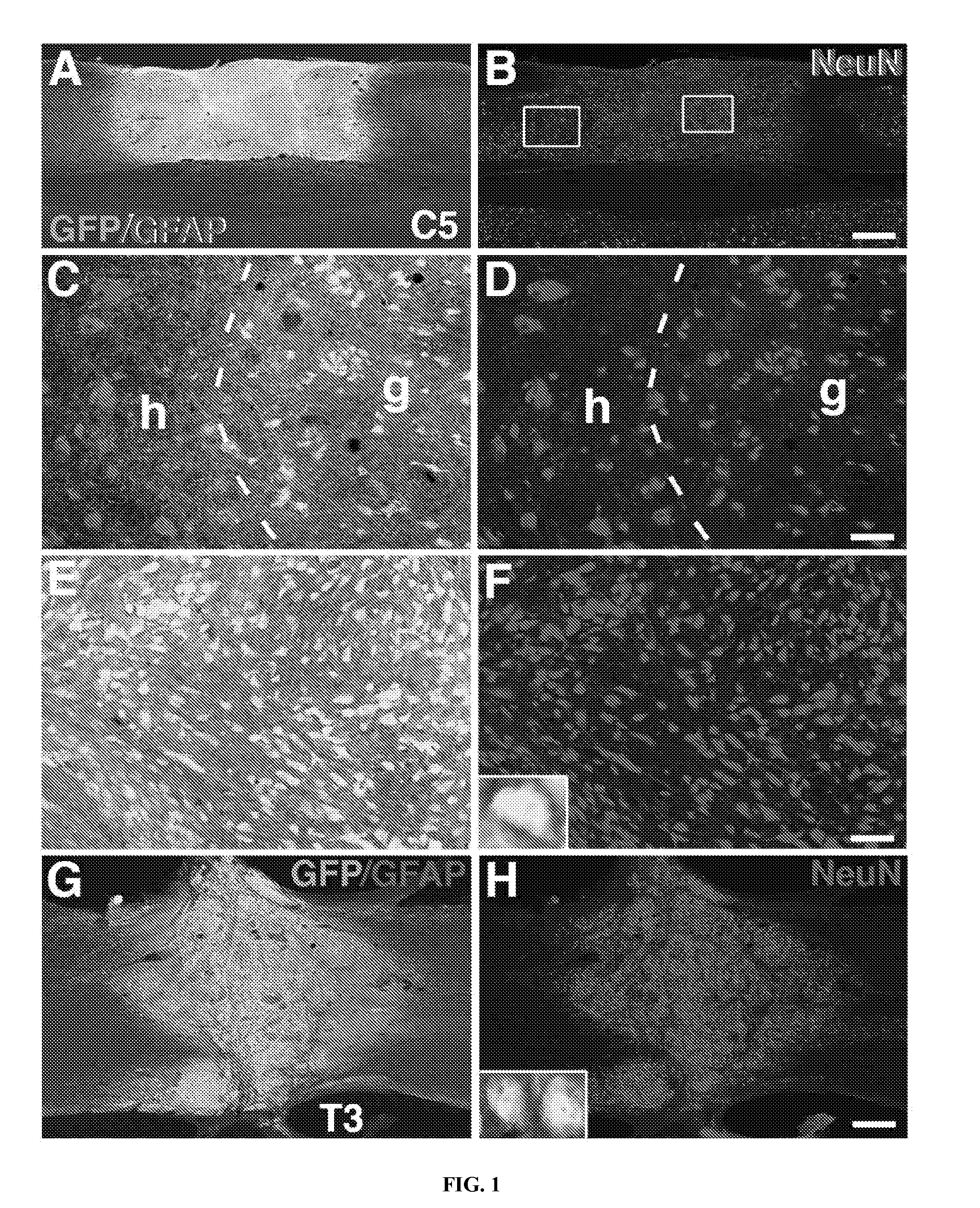
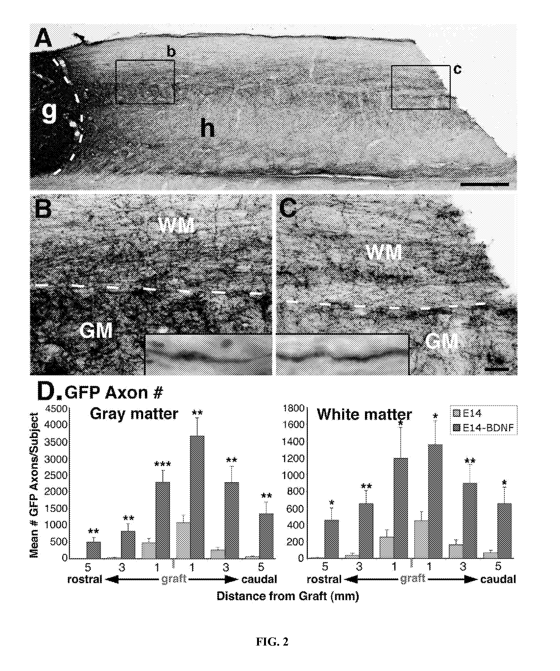
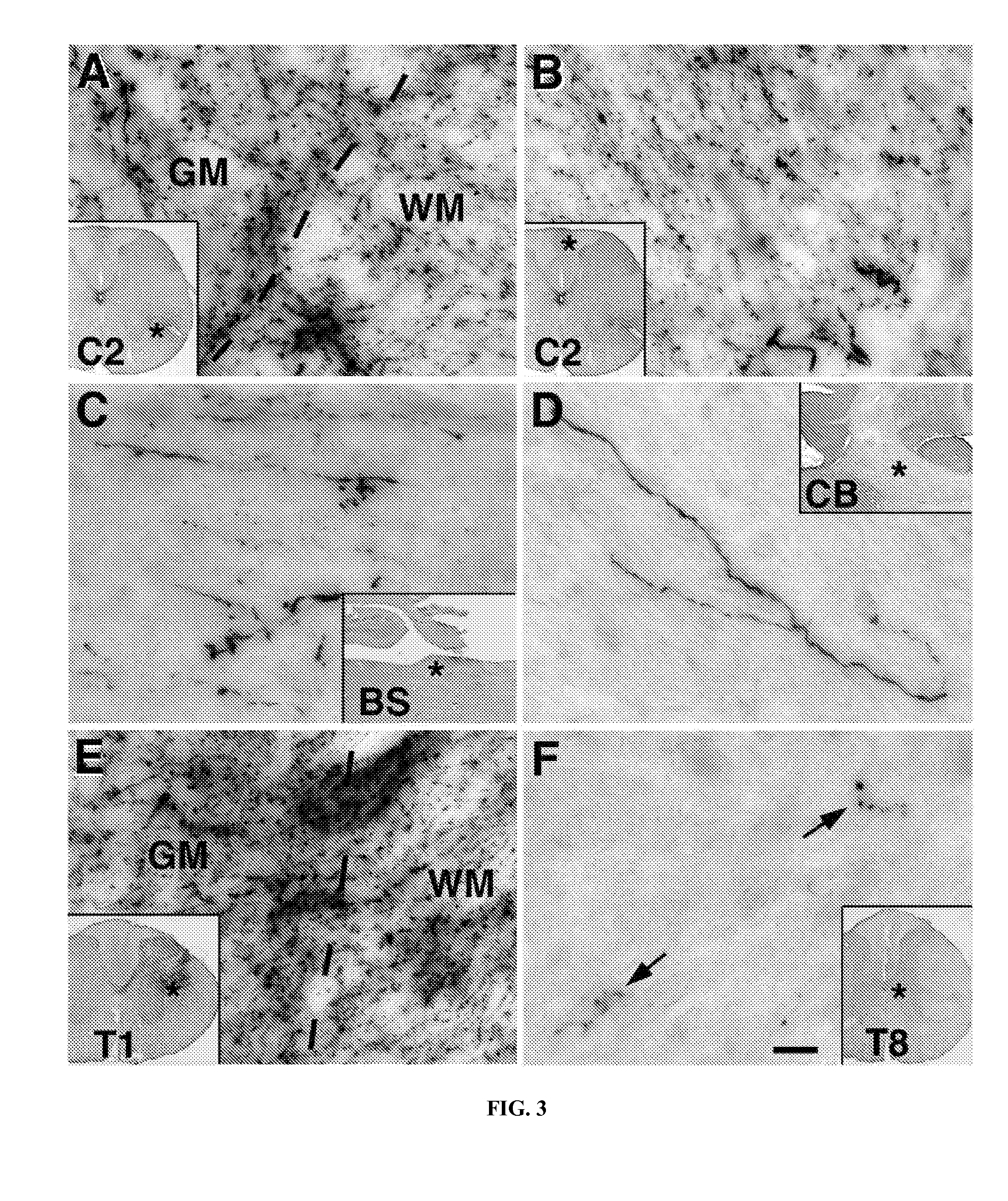
[0079]Spinal cord tissue derived from genetically modified green fluorescent protein (GFP)—expressing rat embryos was grafted to the adult lesioned spinal cord. Under the ubiquitin C promoter, “green” rats express the GFP reporter gene in every cell, providing an unprecedented opportunity to track the fate, integration, extension and differentiation of grafted cells types within the inhibitory milieu of the adult injured spinal cord.

[0080]Cell suspensions of embryonic day 14 (E14) spinal cords from Fischer 344 rats expressing GFP under the ubiquitin C promoter were grafted into either acute mid-cervical (C5) lateral hemisection lesion sites or, two weeks after injury, into upper thoracic (T3) complete transection lesion sites, in non-transgenic F344 rats.

[0081]16 adult rats underwent C5 hemisection lesions and were divided into two groups. One group of 6 rats received E14 GFP-expressing grafts only (n=6), wh...


PUM

PropertyMeasurementUnit
Massaaaaaaaaaa
Massaaaaaaaaaa
Compositionaaaaaaaaaa

Abstract

Methods for inducing non-embryonic lesioned central nervous system neurons to survive, integrate, extend axons over long distances, induce intra-lesion ingrowth of neurons into the lesion from host tissue and form synapses in vivo. Pluripotent neural stem cells are grafted into the lesioned CNS tissue within a tissue adhesive suspension, optionally in the presence of growth factors. No modification of the neuronal regenerative inhibitory environment of the CNS is necessary.

Description

BACKGROUND OF THE INVENTION[0001]1. Field of the Invention[0002]The invention relates to methods for treating central nervous system lesions in a mammal. The invention further relates to a method for using neural stem cells to induce formation of functional synaptic junctions and corticospinal regeneration in the spinal cord.[0003]2. Background Information[0004]The mammalian spinal cord shows little spontaneous recovery after injury. Furthermore, although regeneration of damaged spinal cord tissues (e.g., axons and neurons) can sometimes be induced to a degree through treatment, formation of functional synaptic junctions between axonal termini and adjacent neurons remains elusive.[0005]The degree of motor function loss varies with the identity of the damaged tissue and the extent of damage incurred, as well as with species. For example, the rubrospinal tract influences movement through direct and reciprocal spinal motor projections that reflect activity of the rubro-cortico-cerebell...

Claims


Application Information

Patent Timeline
IPC IPC(8): A61K38/18A61K35/30
CPCA61K38/18A61K38/185A61K35/30A61K2300/00A61P21/00A61P25/00
Inventor LU, PAULTUSZYNSKI, MARK H.
Owner RGT UNIV OF CALIFORNIA
Learn More