Device for the treatment of an ocular disease

a technology for ocular diseases and injection devices, which is applied in the direction of pump control, internal electrodes, therapy, etc., can solve the problems of poor vision recovery after retinal detachment, poor treatment effect, and inability to ensure that submacular injection is not risky, and achieves the effect of efficient electroporation

Inactive Publication Date: 2014-10-23
INST NAT DE LA SANTE & DE LA RECHERCHE MEDICALE (INSERM)
View PDF6 Cites 11 Cited by
  • Summary
  • Abstract
  • Description
  • Claims
  • Application Information

AI Technical Summary

Benefits of technology

[0015]As will be described in more detail later in the description, the inventors have discovered that such an injection device is very efficient for electroporation in the suprachoroidal space, in particular because it provides a plurality of injection needles spread on the first contact surface, allowing an homogenous distribution of the pharmaceutical composition, in p...

Problems solved by technology

Such a macular detachment may be associated with vision threatening.
Indeed, it is well known that poor vision recovery after retinal detachment is correlated with macular detachment.
In diseased eyes, knowing the uncertainty of central vision recovery after macular detachment, it is difficult to ensure that sub...

Method used


Image

  • Device for the treatment of an ocular disease
  • Device for the treatment of an ocular disease
  • Device for the treatment of an ocular disease

Examples


Embodiment Construction

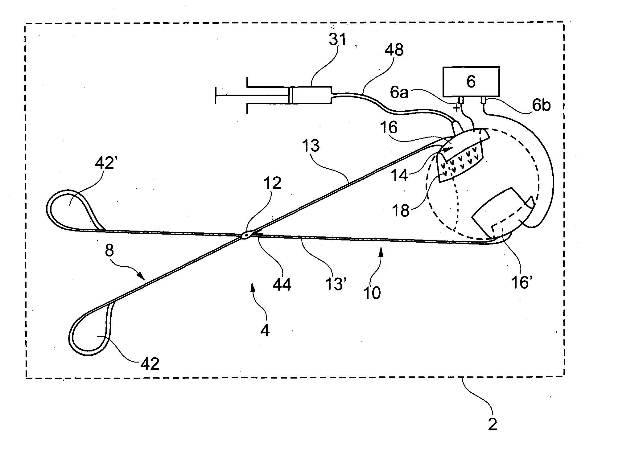
[0071]The electroporation device 2 shown in FIG. 1 comprises a scissor-like injection device 4 according to the invention, and an electrical generator 6.

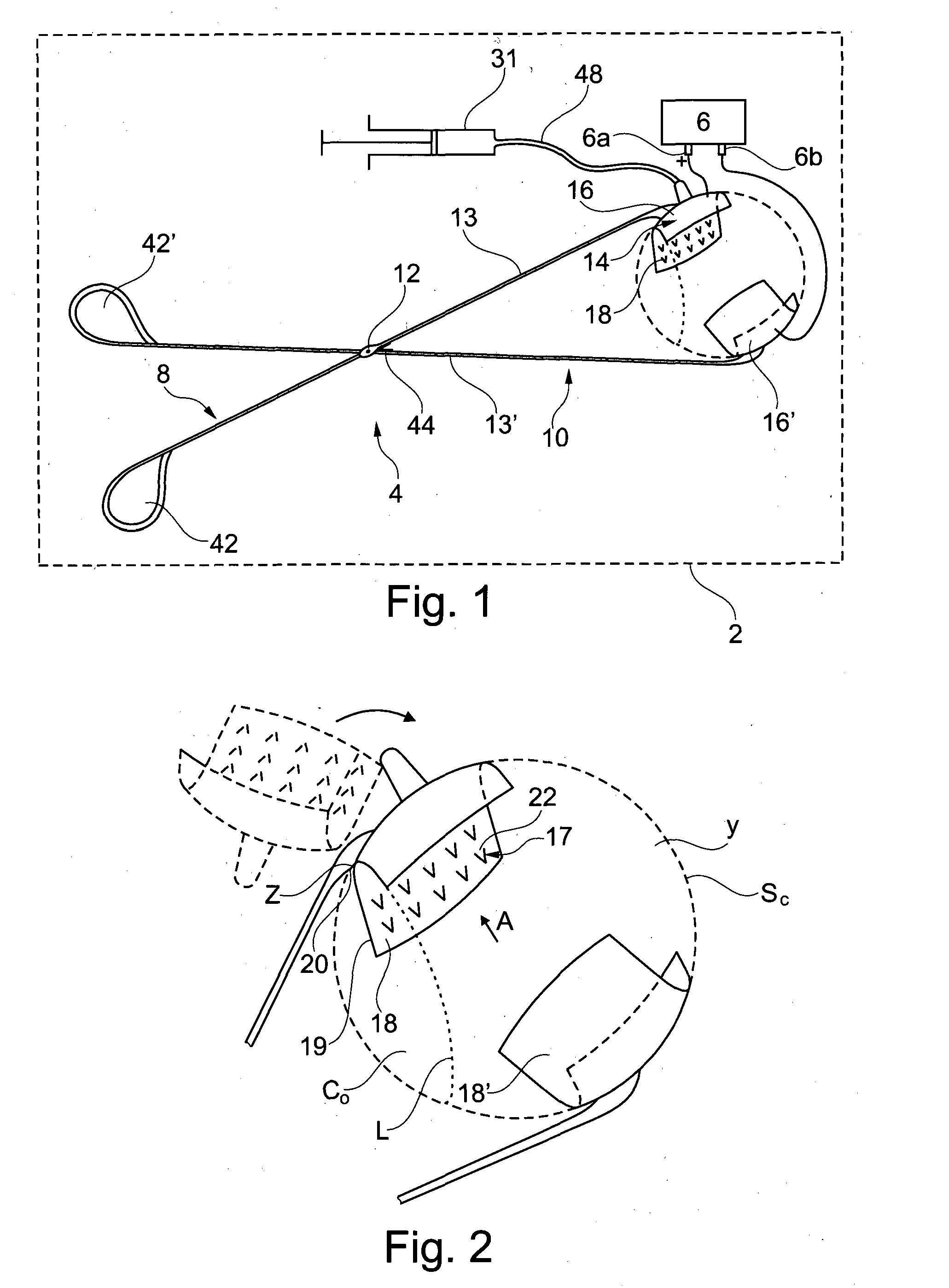
[0072]As shown in FIGS. 2 and 3, the injection device 4 comprises a first arm 8 and a second arm 10, rotationally mounted on the first arm 8, around an axis 12. The first arm 8 and the second arm 10 are used as first and second electrodes of the electroporation device 2, respectively, the first arm being also used for the injection.

First Arm

[0073]The first arm 8 comprises a first proximal part 13, and a first support 14, preferably made of a conductive material, presenting a first cup 16 provided with a set of injection needles 17.

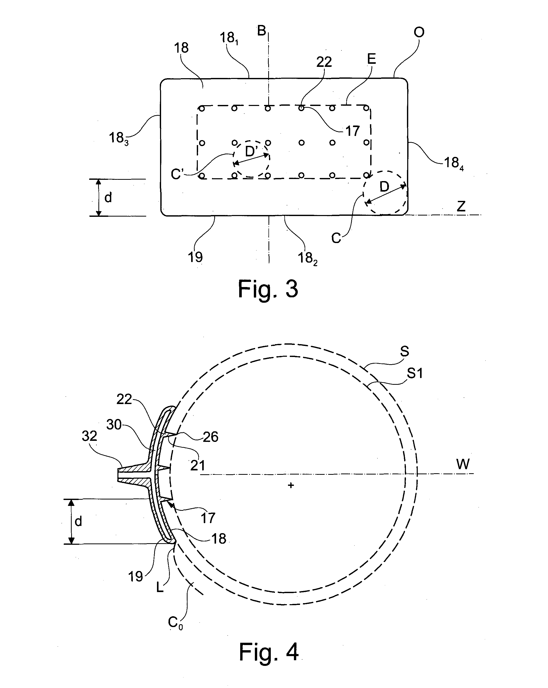
[0074]The first support 14 is rotationally mounted on the first proximal part 13 around an axis Z. The axis Z is substantially perpendicular to the general direction of the injection needles 17.

[0075]The distance between any injection needle 17 and the axis Z is preferably more than 4 mm, preferably more t...


PUM


Abstract

The present invention relates to an injection device comprising: —a first support (14) having a cup-shaped first contact surface (18) intented to come into contact with a first region of an outside surface of an eye, —a set of at least four injection needles (17) in fluid communication with each other and protruding from said first contact surface (18) at respective insertion points (22) so that the distance between the distal end (26) of any of said injection needles to said first contact surface is between 0.6 mm and 1.3 mm, the insertion points of the injection needles on the first contact surface being spread on said first contact surface so that the diameter (D′) of the largest circle (C′) that it is possible to include completely in the convex surface (E) defined by said insertion points on a front view of said first contact surface, without any insertion point being included in said circle, is less than 8 mm.

Description

FIELD OF THE INVENTION[0001]The present invention relates to an injection device for the treatment of an ocular disease in a subject.BACKGROUND OF THE INVENTION[0002]In recent years, there have been exciting new advances for the treatment of ocular diseases such as age-related macular degeneration and diabetic retinopathy, using biotherapies. Because the eye is a small, confined organ, isolated by barriers, it has been identified as an organ of choice for local gene therapy.[0003]For example, hereditary retinal dystrophies are due to mutations in gene encoding proteins in photoreceptors (cones and rods) or in retinal pigment epithelial cells (RPE). Whilst gene replacement in photoreceptor cells is still under pre-clinical evaluation, the most striking advances in this field have been made for RPE65 gene replacement in RPE cells, for the treatment of Leber congenital amaurosis (LCA). Not only was it shown that viral gene transfer in the RPE was feasible and efficient in animal models...

Claims


Application Information

Patent Timeline
IPC IPC(8): A61F9/00
CPCA61F9/0026A61F9/0017A61N1/0526A61N1/327F04C2270/0421
Inventor BEHAR-COHEN, FRANCINE
Owner INST NAT DE LA SANTE & DE LA RECHERCHE MEDICALE (INSERM)
Why Eureka
Try Eureka