System and method for pelvic floor repair
a pelvic floor and system technology, applied in the field of pelvic floor system and method, can solve the problems of high degree of skill, high rate of complications, and difficulty in accessing the vaginal wall to the ssl
- Summary
- Abstract
- Description
- Claims
- Application Information
AI Technical Summary
Benefits of technology
Problems solved by technology
Method used
Image
Examples
example 1
Ultrasound Imaging of the Ischial Spines and Sacrospinous Ligaments
[0087]The anatomy of the pelvic floor of women suffering from POP was imaged using ultrasound in order to find out if ultrasound imaging can be used to identify the SSL and Ischial Spine. Doppler imaging was also used to identify blood vessels at the area of the SSL.
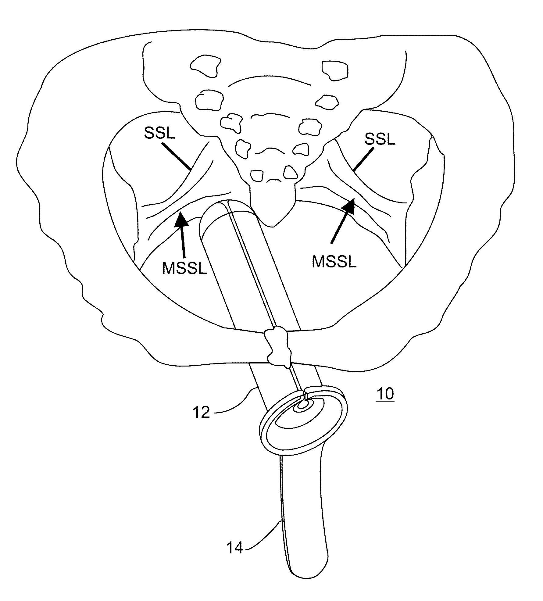
[0088]As is shown in FIG. 3, the SSL includes dense connective tissue and contributes to the stability of the bony pelvis. It attaches to the ischial spine laterally and lower part of the sacrum and coccyx medially. The sacrospinous, along with the sacrotuberous ligament, divides the sciatic notches of the ischium into the lesser and greater sciatic foramen (GSF). The internal pudendal and inferior gluteal vessels, sciatic nerve, and other branches of the sacral nerve plexus pass through the GSF in close proximity to the ischial spines and SSL.
[0089]On the superior or pelvic surface of the SSL lies the coccygeus muscle, which together with the levator ani...
example 2
Cadaver Study
[0096]A cadaver study was undertaken in order to identify and measure the distance and path between anatomical landmarks within the posterior pelvis and the vaginal apex.
Materials and Methods
[0097]An abdominal dissection was performed on a formalin-embedded human cadaver while in in a dorso-supine position. The vaginal apex was identified and marked and an ultrasound probe was inserted into the vaginal canal. Anatomical landmarks within the posterior pelvis including the MSSL, the MSTL, the ischial spine, the sacrum, the rectum, the pudendal bundle and the iliac vessel were identified and their distance from the apex was measured using a ruler (FIGS. 5a-b).
Results
[0098]The results are presented in Table 2 below.
TABLE 2AnatomicalDistance from US probelandmarktip (in cm)commentsMSSL3.5MSTL4.5angled 45-60 downwardsIschial spine4.5Sacrum3.5Rectum2.5Pudendal bundle4.5Iliac vessels5.5
PUM
 Abstract
Description
Claims
Application Information
 - R&D
- Intellectual Property
- Life Sciences
- Materials
- Tech Scout
- Unparalleled Data Quality
- Higher Quality Content
- 60% Fewer Hallucinations
Browse by: Latest US Patents, China's latest patents, Technical Efficacy Thesaurus, Application Domain, Technology Topic, Popular Technical Reports.
© 2025 PatSnap. All rights reserved.Legal|Privacy policy|Modern Slavery Act Transparency Statement|Sitemap|About US| Contact US: help@patsnap.com


