Selective Plasma Activation for Medical Implants and Wound Healing Devices

a technology of selective plasma activation and medical implants, applied in the field of devices, can solve the problems of reduced biocompatibility of imds with the body, infection and capsular contraction remain significant clinical problems, and the biocompatibility of the implant is limited by the reduction of biocompatibility

Inactive Publication Date: 2014-12-25
CENT HOSPITALIER UNIV VAUDOIS C H U V
View PDF1 Cites 14 Cited by
  • Summary
  • Abstract
  • Description
  • Claims
  • Application Information

AI Technical Summary

Benefits of technology

[0015]In contrast to prior cell-adhesive surfaces, in various embodiments, the present invention provides a surface with activated small (e.g., microscale) areas in order to control the size of the focal adhesions for each cell, with an effect on cell differentiation.

Problems solved by technology

One of the major problems associated with all types of implants is biocompatibility of the implant with the body, and in particular with the tissue adjacent to the site of the implant.
Infection and capsular contraction remain significant clinical problems.
IMDs are limited by their reduced biocompatibility: the body recognizes the implant as a foreign material and builds scar tissue around it.
Lack of biocompatibility leads to reduced performance of the devices, pain to the patients, high re-operation rates, and augmenting the costs and risks significantly.
When sensors, electrodes or drug delivery parts are embedded in scar tissue the function of devices such as pacemakers, implantable defibrillators, insulin pumps, neurostimulators is affected with life threatening consequences.
For example, despite attempts to design implantable biosensors for glucose and other monitoring functions, none developed to date provide pain-free, reliable and continuous monitoring.
One reason is that current implantable sensors suffer from a progressive loss of function after relatively short periods of time in vivo.
Fibrosis results in loss of blood vessels at the site of implantation and, therefore, in a reduced access to blood glucose levels.
These factors can also interfere with the function of other implants and implantable devices, such as insulin pumps, pacemakers, artificial joints, and artificial organs.
While suitable for bone or tooth implants, this approach is not useful in more complex prosthetic devices or in biosensors, which requires use of a variety of materials.
While suitable for prostheses such as breast implants, such coatings are not practical for prosthetic devices or biosensors having complex geometries.
Such systemic administration can result in side effects such as generalized immunosuppression, bloating, and psychiatric problems, especially over the long term.
The cell adhesive pattern does not allow fibroblasts to exert forces on the surface where they attach through the focal adhesions and prevent them from becoming myofibroblasts (the main cell responsible for fibrosis and contraction).
This process reduced the fibrotic tissue around the implant but the process is expensive and involves multiple steps taking hours to complete.
Such general activated surfaces using relatively large stencil patterns do not control focal adhesions.
Thus, current devices cannot prevent encapsulation of the implanted device by fibrotic overgrowth.

Method used


Image

  • Selective Plasma Activation for Medical Implants and Wound Healing Devices
  • Selective Plasma Activation for Medical Implants and Wound Healing Devices
  • Selective Plasma Activation for Medical Implants and Wound Healing Devices

Examples


examples

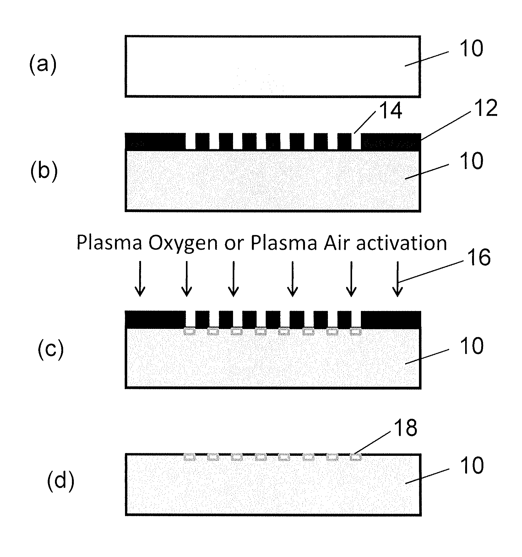
[0118]The invention describes a surface with anti-fibrotic properties. Anti-fibrotic is defined as having characteristics reducing the presence and development of myofibroblasts, cells responsible for fibrotic tissue formation and contraction. The bio-physical properties of the surface are sufficient to allow fibroblasts to attach, but impair myofibroblasts development.

[0119]Myofibroblast development is defined by an in vitro assay in which fibroblasts are seeded on the surface and myofibroblast differentiation is induced with TGF-β1. Myofibroblasts are defined as cells positive for α-SMA.

[0120]To identify the ideal properties of the antifibrotic properties several surface activation patterns were tested by the above mentioned assay. On silicone, selective activation of specific islet size (length of 4 μm and width of 2 μm) and distribution with a regular distance between the islets of 5 μm reduced 4-fold the differentiation of human dermal fibroblasts to myofibroblasts compared to ...


PUM

PropertyMeasurementUnit
widthaaaaaaaaaa
widthaaaaaaaaaa
distanceaaaaaaaaaa

Abstract

A treated surface of a device (10) for implantation or for application as a wound dressing, comprises an array of plasma-activated hydrophilic cell-adhesive areas (18) having the ability to reduce fibrous reaction, is in the form of an array of islets of activation. Each islet of activation (18) has a length which is less than 6 μm, a width which is less than 2 μm and the distance between islets is preferably from 4 μm to 6 μm. The islets of activation (18) are surrounded by non-activated, non-adhesive, hydrophobic areas.

Description

FIELD OF THE INVENTION[0001]This invention relates to devices for implantation or for application as a wound dressing and is particularly concerned with treating the surfaces of such devices for increasing cell adhesion and decreasing biofouling and fibrosis (encapsulation). The invention relates to the treated device surfaces, the devices with such treated surfaces, and methods and apparatus for treating the device surfaces.BACKGROUND OF THE INVENTION[0002]Implantable medical devices (IMDs) are rapidly revolutionizing medicine.[0003]Implantable artificial materials and devices, such as drug delivery systems, pacemakers, artificial joints, and organs play an important role in health care today. In addition to these devices, implantable monitoring devices or “biosensors” have great potential for improving both the quality of care and quality of life.[0004]One of the major problems associated with all types of implants is biocompatibility of the implant with the body, and in particula...

Claims


Application Information

Patent Timeline
Patent Type & Authority Applications(United States)
IPC IPC(8): A61L15/16A61L15/44A61L27/28
CPCA61L2400/18A61L15/42A61L2300/23A61L15/44A61L27/50A61L27/28A61L15/16A61L2300/252
Inventor PIETRAMAGGIORI, GIORGIOMAJD, HICHAM
Owner CENT HOSPITALIER UNIV VAUDOIS C H U V
Why Eureka
Try Eureka