Eukaryotic Cells with Artificial Endosymbionts for Multimodal Detection

a technology of endosymbionts and eukaryotic cells, applied in the field of endosymbiosis, eukaryotic cells engineered with artificial endosymbionts, magnetotactic bacteria, etc., can solve the problem that none of these alterations are heritable to daughter cells

Inactive Publication Date: 2015-01-08
BELL BIOSYST
View PDF2 Cites 19 Cited by
  • Summary
  • Abstract
  • Description
  • Claims
  • Application Information

AI Technical Summary

Benefits of technology

[0011]The invention also relates to methods of using multimodal detection to characterize the eukaryotic cell containing the single-celled organism. In the methods of the invention, eukaryotic cells with single-celled organisms have multiple phenotypes that relate to the single-celled organism. In an embodiment, the multiple phenotypes allow for detection of the eukaryotic cells using multimodal observation methods. In an embodiment, multimodal detection is used for non-invasive in vivo imaging of the eukaryotic cells. Each method or modality of imaging can allow visualization of different aspects of anatomy and physiology, and combining these provides greater information about the eukaryotic cells in an in vivo environment.

Problems solved by technology

However, none of these alterations are heritable to daughter cells.

Method used


Image

  • Eukaryotic Cells with Artificial Endosymbionts for Multimodal Detection
  • Eukaryotic Cells with Artificial Endosymbionts for Multimodal Detection
  • Eukaryotic Cells with Artificial Endosymbionts for Multimodal Detection

Examples


example 1

Microinjection of gfp+AMB-1 into Murine Cells

[0174]A. Construction of gfp+AMB-1.

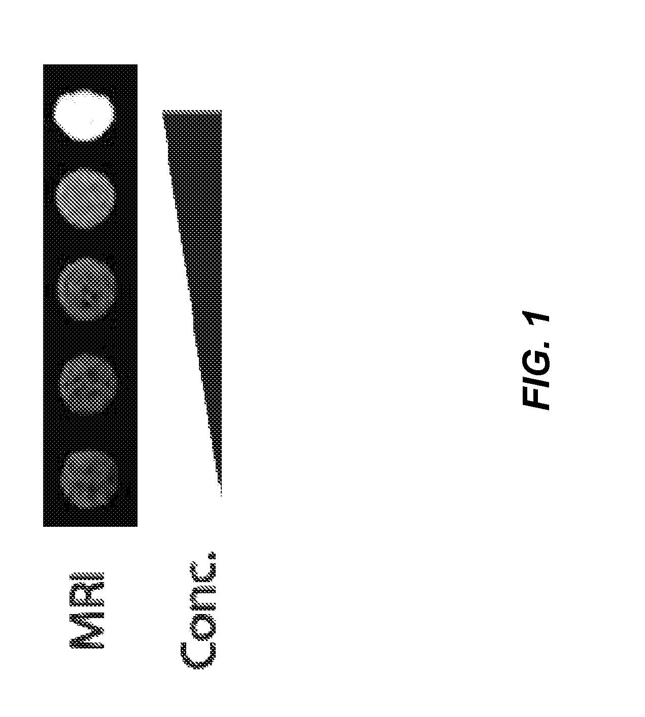
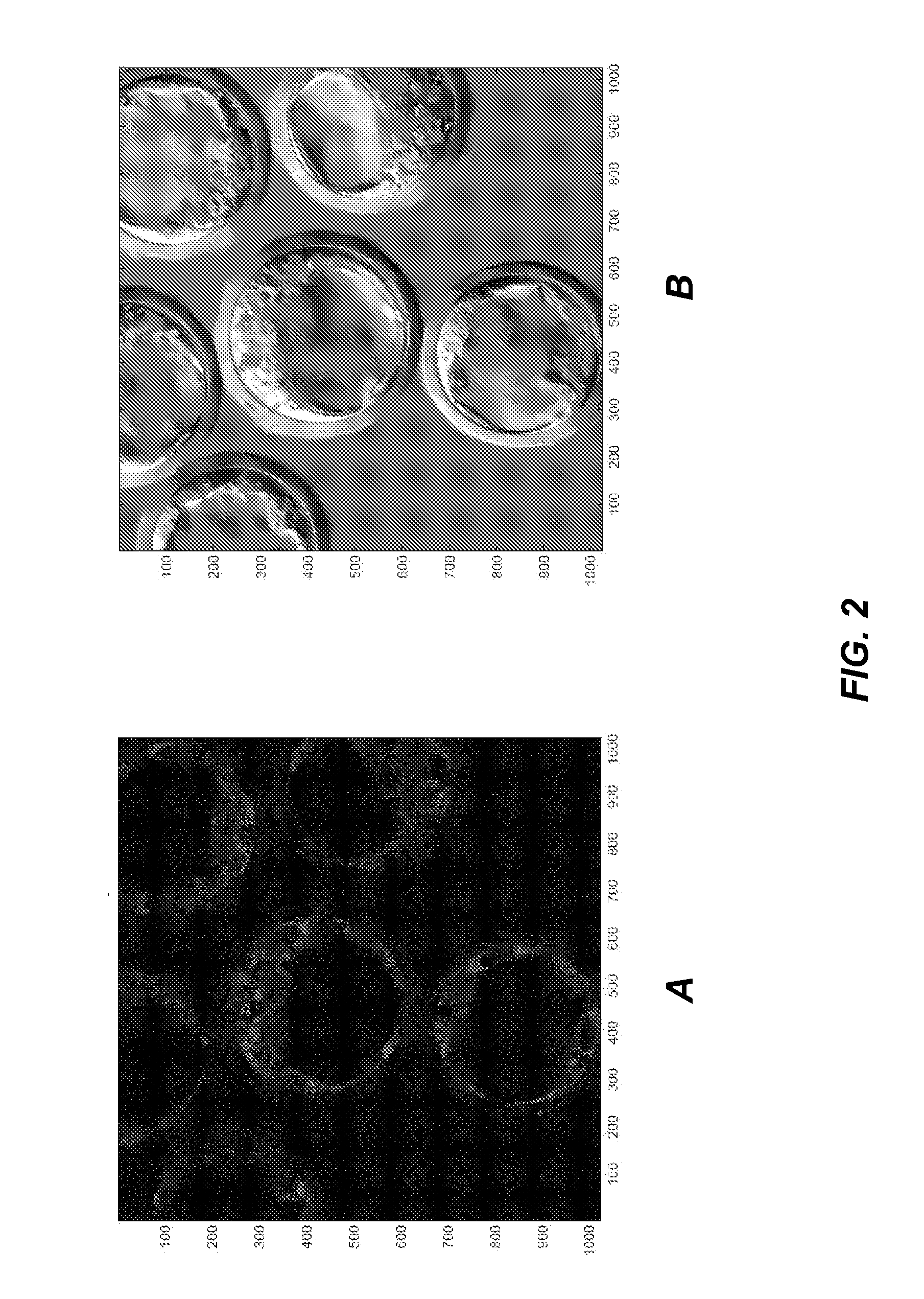
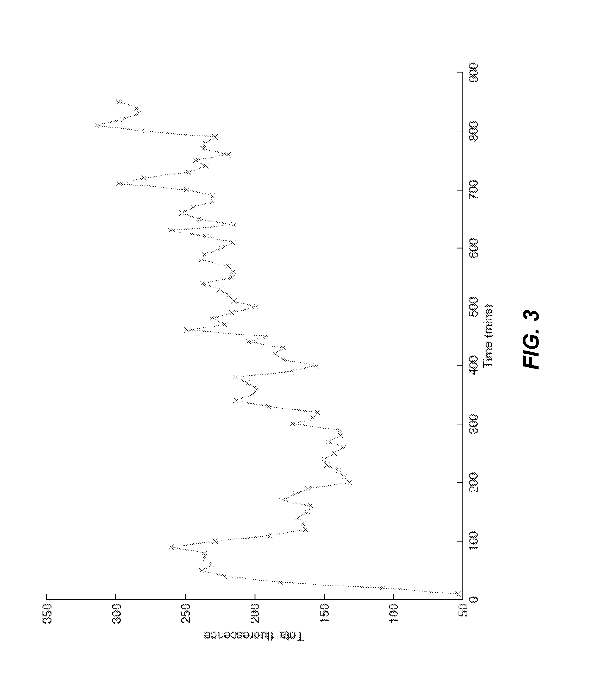
[0175]Expression vectors for eGFP, one including a Shine-Dalgarno sequence upstream of the gfp gene and one without a Shine Dalgarno, sequence were cloned into cryptic broad host range vector pBBR1MCS-2 (Kovach, M. E. et al., “Four new derivatives of the broad-host-range cloning vector pBBR1MCS, carrying different antibiotic-resistance cassettes,”Gene 166, 175-176, (1995), incorporated herein by reference in its entirety for all purposes). AMB-1 (ATCC 700264) was transformed with this construct (see, e.g., Matsunaga, T. et al., “Complete genome sequence of the facultative anaerobic magnetotactic bacterium Magnetospirillum sp. strain AMB-1,” DNA Res. 12, 157-166 (2005); Burgess J. G., et al., “Evolutionary relationships among Magnetospirillum strains inferred from phylogenetic analysis of 16S rDNA sequences,”J Bacteriol. 175:6689-6694 (1993); Matsunaga T, et al., “Gene transfer in magnetic bacteria: trans...

example 2

Phagocytic Entry of AMB-1

[0184]Receptor mediated: The inlAB gene is amplified from L. monocytogenes genomic DNA (ATCC 19114) and is inserted into pBBR1MCS-5, the gentamicin cognate of pBBR1MCS-2 (see Kovach, M. E., et al., “Four new derivatives of the broad-host-range cloning vector pBBR1MCS, carrying different antibiotic-resistance cassettes,”Gene 166, 175-176, (1995)), and gfp+inlAB+ AMB-1 is generated. The gfp+inlAB+ AMB-1 is co-cultured with eukaryotic host cells, including common epithelial tumor cell lines Coco-2, MDA-MB-231 and MCF7, non-epithelial tumor cell lines, such as HT-1080 and HL60, and murine stem cells. Fluorescent microscopy and FACS are used to monitor and quantify internalization and intracellular location.

[0185]Expression of pore-forming haemolysin (hlyA) in AMB-1 is achieved through amplification of hlyA from L. monocytogenes genomic DNA (ATCC 19114). The amplified hlyA is inserted into pBBR1MCS-3 (the tetracycline cognate of pBBR1MCS-2), which is then used to...

example 3

Regulation of AMB-1 Growth

[0187]Regulation of AMB-1 growth in embryonic stem cells can be regulated as follows. Coleoptericin-A (ColA) is amplified from total Sitophilus oryzae cDNA. Expression of ColA in beetles of genus Sitophilus regulates titers of γ-Protobacterium, which has naturally developed close symbiotic relationship the beetles, and resides in specific cells called bacteriocytes. (Login, F. H. et al., “Antimicrobial peptides keep insect endosymbionts under control,”Science 334(6054):362-365 (2011), incorporated herein by reference in its entirety for all purposes).

[0188]Murine embryonic stem cells comprising gfp+ AMB-1 are treated using a neural differentiation protocol. MTB expression levels are quantified using qPCR and fluorescent microscopy. Amplified colA is then expressed in the gfp+ AMB-1 embryonic stem cells. A promoter is selected to provide optimal ColA expression levels.


PUM

PropertyMeasurementUnit
doubling timeaaaaaaaaaa
magnetic field amplitudeaaaaaaaaaa
magnetic field frequenciesaaaaaaaaaa

Abstract

The present invention is directed generally to eukaryotic cells comprising single-celled organisms that are introduced into the eukaryotic cell through human intervention and which transfer to daughter cells of the eukaryotic cell, and methods of introducing such single-celled organisms into eukaryotic cells. The invention provides single-celled organisms that introduce a phenotype to eukaryotic cells that is maintained in daughter cells. The invention additionally provides eukaryotic cells containing magnetic bacteria. The invention further provides eukaryotic cells engineered with single-celled organisms to allow for multimodal observation of the eukaryotic cells. Each imaging method (or modality) allows the visualization of different aspects of anatomy and physiology, and combining these allows the imager to learn more about the subject being imaged.

Description

CROSS-REFERENCES TO RELATED APPLICATIONS[0001]This application is a continuation-in-part of U.S. application Ser. No. 13 / 838,717, filed Mar. 15, 2013, which is a continuation-in-part of U.S. application Ser. No. 13 / 374,799, filed on Jan. 13, 2012, and which is a continuation-in-part of PCT / US2013 / 021414, filed on Jan. 14, 2013, which is a continuation-in-part of U.S. application Ser. No. 13 / 374,799, filed on Jan. 13, 2012, the entire contents of which applications are hereby incorporated by reference in its entirety for all purposes.FIELD OF THE INVENTION[0002]The present invention relates generally to the field of endosymbiosis, eukaryotic cells engineered with artificial endosymbionts, and magnetotactic bacteria. In particular, the invention provides single-celled organisms such as artificial endosymbionts, including magnetotactic bacteria, eukaryotic cells to host those single-celled organisms, methods of using eukaryotic cells containing single-celled organisms, and methods of i...

Claims


Application Information

Patent Timeline
Patent Type & Authority Applications(United States)
IPC IPC(8): G01N33/50C12Q1/02
CPCG01N33/5005G01N2469/00C12Q1/02B82Y5/00B82Y15/00C12N15/03A61K51/1203A01K2207/05C12N2510/00A61K49/0097A61K49/1896A01K67/0275A61N2/12A61B5/05C12N5/16C12N5/163A61K49/08C12N5/166C12Q1/6897G01N33/5091
Inventor BELL, III, CALEB B.BAZAROV, ALEXEY
Owner BELL BIOSYST
Why Eureka
Try Eureka