Ligand having three finger structure and a method for detecting a molecule by using thereof
a technology of ligands and finger structures, applied in the field of ligands having three finger structures, can solve the problems of time and extra work for purifying antibodies, often not obtained antibodies,
- Summary
- Abstract
- Description
- Claims
- Application Information
AI Technical Summary
Benefits of technology
Problems solved by technology
Method used
Image
Examples
example 1
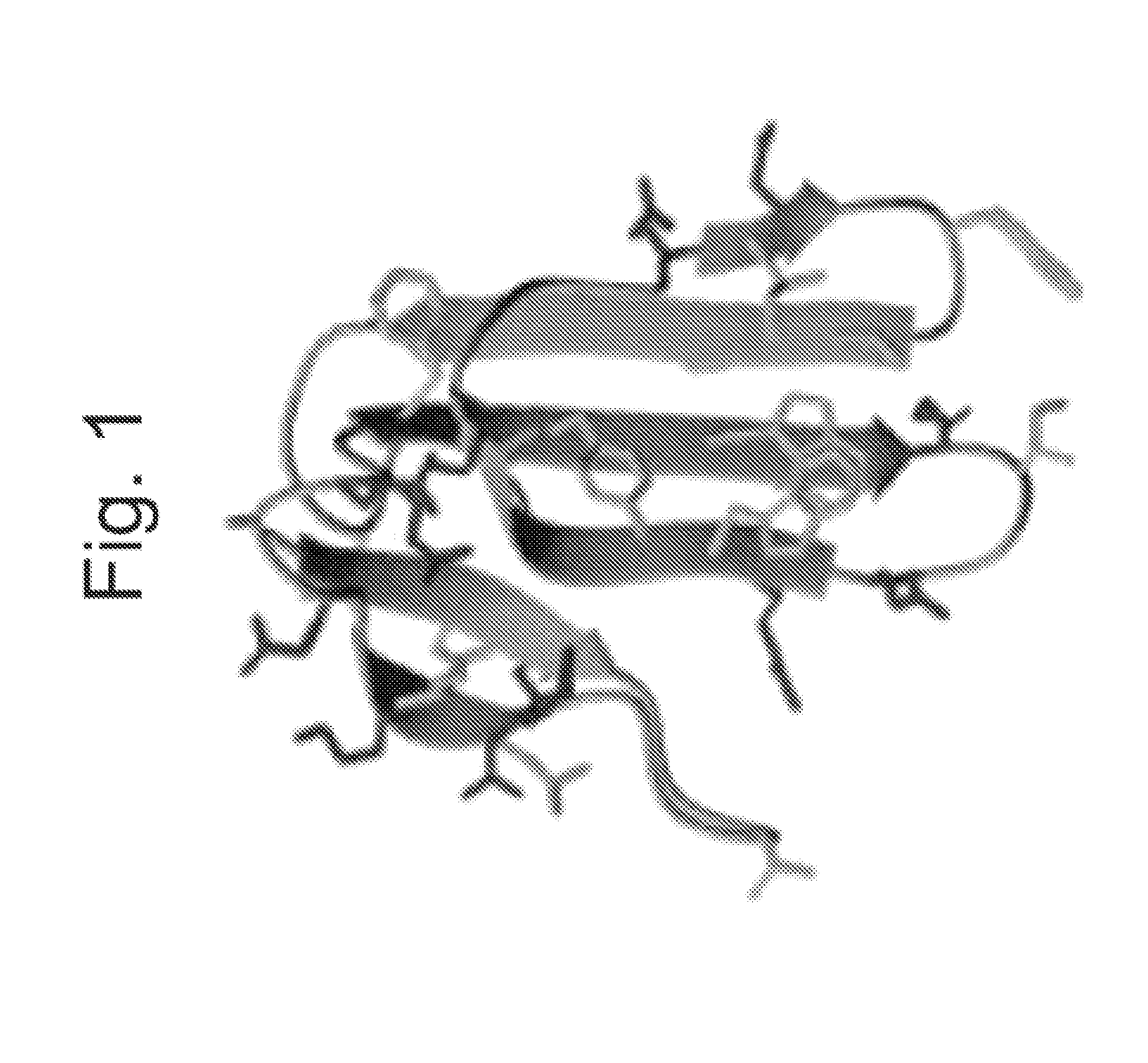
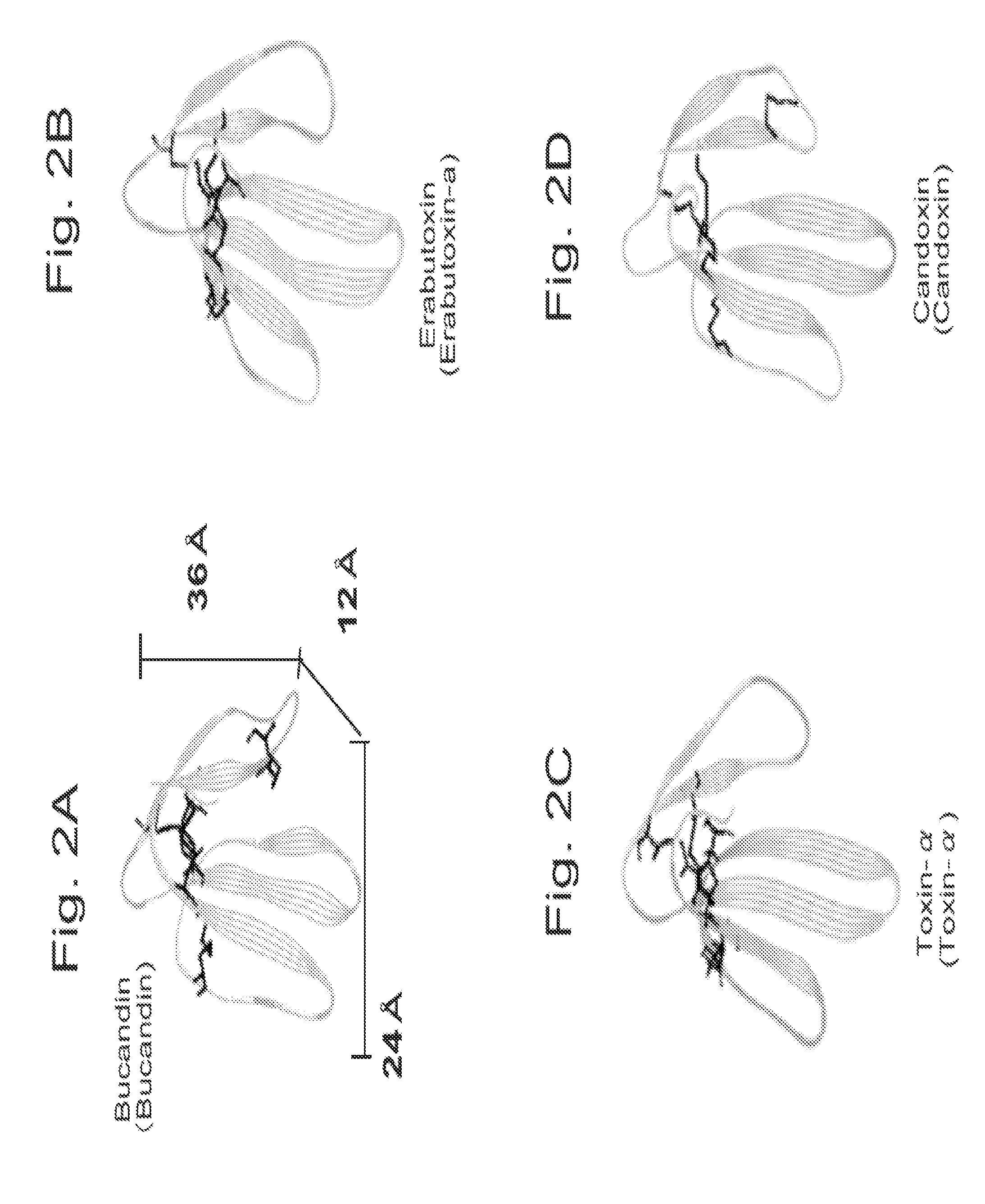
Manufacture of the Ligand by Using Bucandin
[0134]Bucandin used in the present invention was purchased from Operon Biotechnology Inc. In order to manufacture the construct, T7-UTR fragment comprising T7-promoter, cap site, Xenopus globulin untranslated sequence (UTR) and translation start site, spacer (GGGS) 2, His 6-tag, spacer (GGGS) and Y-tag sequence were purchased from Geneworkd Inc.
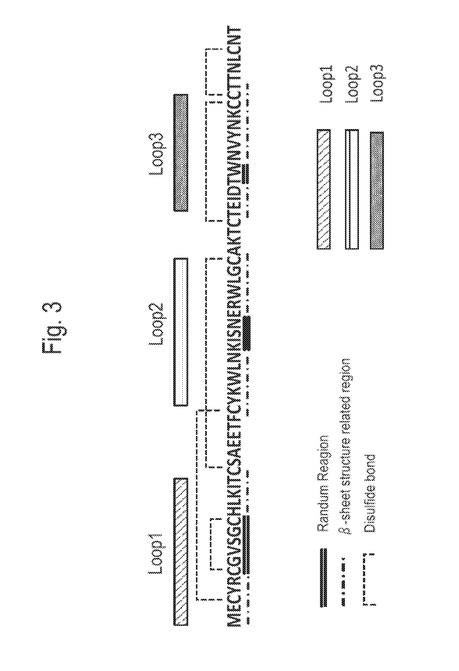
[0135]Three loop areas of bucandin shown in FIG. 3 were respectively randomized by using the following method to obtain the set of amino acid sequence of No. 1 to 13 shown in the following Table 2.
TABLE 2Loop Area (Replaceamino acid number)No.1 (X7)2 (X4)3 (X2)Round number1PTQPKRTGTRQPP62NASAVRKPETIRG63IGEVSQREALKGD64PNPADRNNPSHNR65TGLPPSDPGATHN66TNMVNRPPRRTHR67NPPTSdTPGNTTQ68PTPIQGQNLPADA79QNEPLTANTTANG710PEVDIRQKLPRKP1211ETNNGQPTIPAER1212RRSMHTVIAKNTP1213NPRTIRADLAENQ12
[0136]The 3F incorporating the amino acid sequence of No. 1 to 15 shown in Table 2, 3F (bucandin) loop 1, 3F (bucandin) loop 2, 3F ...
example 2
IVV Assay
(1) Synthesis of Puromycin-Biotin-Linker
[0143]Puromycin-biotin-linker for Puro-F-S (Geneworld) and biotin-loop (BEX Co, Ltd) were purchased. These two modified oligonucleotides were cross-linked by using bifunctional reagent (EMCS) according to the conventional method to synthesize Puromycin-biotin-linker. Puro-F-S used here was previously bound to puromycin at the one end of the spacer according to the conventional method and labelled with fluorescein. The Pyro-F-S had thiol group at another end.
[0144]Thiol group of 10 nmol of Puro-F-S was reduced by using to 100 μL of 50 mM phosphate buffer including 1 mM TCEP (tris(2-carboxyethyl)phosphine) (pH 7.0) for 6 hours at ambient temperature. Then, it was desalted just before use by using NAP-5 column (GE Healthcare). Both 10 nmol of biotin loop and 2 μmol of EMCS were added to 100 μL in total amount of sodium phosphate buffer (pH 7.0), and it was incubated at 37° C. for 30 minutes. Then, ethanol precipitation was performed at 4...
example 3
Pull Down Assay
(1) Binding of the Magnetic Beads
[0155]According to the instruction manual, the streptavidin coated magnetic beads (2.3 μm, MAGNOTEX, Takara Inc.) was washed twice with the binding buffer (10 mM Tris-HCl buffer containing 1 mM EDTA, 1 M NaCl, 0.1% Triton X-100 (pH 8.0)). 48 pmol of mRNA-puromycin linker conjugate and 1.2 mg of streptavidin beads were incubated in 120 μL of the binding buffer for 10 minutes at ambient temperature. Next, prior to add them to the cell free translation system, each bead was washed one by one by using both of the binding buffer and the translation mix buffer.
(2) Manufacture of the 3F-cDNA Display Ligand for Bait Protein Binding
[0156]After separation of the magnetic beads by using the magnetic stand, 300 μL of cell free translation extract (Ambion) was added, and incubated at 30° C. for 20 minutes. In order to increase yield of the protein-ligand fusant, the translation product was further incubated at 37° C. for 90 minutes under the hypert...
PUM
| Property | Measurement | Unit |
|---|---|---|
| temperature | aaaaa | aaaaa |
| pH | aaaaa | aaaaa |
| pH | aaaaa | aaaaa |
Abstract
Description
Claims
Application Information
 - R&D
- Intellectual Property
- Life Sciences
- Materials
- Tech Scout
- Unparalleled Data Quality
- Higher Quality Content
- 60% Fewer Hallucinations
Browse by: Latest US Patents, China's latest patents, Technical Efficacy Thesaurus, Application Domain, Technology Topic, Popular Technical Reports.
© 2025 PatSnap. All rights reserved.Legal|Privacy policy|Modern Slavery Act Transparency Statement|Sitemap|About US| Contact US: help@patsnap.com


